Anterior Cortex = HIGH RISK | Posteromedial = Low Risk | MRI Gold Standard
- Anterior cortex = HIGH RISK - tension side, poor healing, may need surgery
- Posteromedial = LOW RISK - compression side, heals well with rest
- Dreaded black line = anterior cortex stress fracture visible on XR/CT
- MRI is gold standard - shows bone marrow oedema before XR changes
- Female athlete triad - amenorrhoea, eating disorder, osteoporosis = risk factor
- Complete fracture risk if anterior cortex not treated appropriately
- “Anterior cortex stress fractures may require prophylactic IM nailing
- “XR may be normal for 2-6 weeks - order MRI if high clinical suspicion
- “Return to sport based on pain-free activity, not arbitrary timeframes
- “Ask about training errors - sudden increase in volume/intensity
Overview and Epidemiology
A stress fracture is an overuse injury: repetitive submaximal loading that exceeds the bone's ability to remodel and repair. The tibia is the most common site, carrying 50% of all stress fractures.
Who. The peak age is 17-25 years, in high-activity athletes and military recruits. Runners account for the majority of tibial stress fractures, military recruits have a 1-5% incidence during basic training, and female athletes carry a 2-10 times higher risk than males.
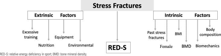
Risk factors. Intrinsic factors belong to the patient:
- Female sex
- Low BMI or an eating disorder
- Menstrual irregularity (the female athlete triad)
- Low bone mineral density
- A previous stress fracture
- Leg length discrepancy
- Pes planus or cavus foot
Extrinsic factors lie in the training, the kit and the diet:
- Too much too soon: a sudden increase in training volume
- Intensity escalation: more speed or hills without adaptation
- A change in running surface, from soft to hard
- Inadequate or worn-out footwear with poor cushioning
- Poor nutrition, with calcium or vitamin D deficiency
The female athlete triad. Energy deficiency or an eating disorder (caloric intake inadequate for the level of activity), amenorrhoea (loss of periods for over 3 months) and low bone density or osteoporosis. Always screen for it.
Anatomy and Pathophysiology
Remodelling that falls behind. Normal activity causes microdamage, and repair runs in two stages: osteoclasts resorb the damaged bone, which takes 3 weeks, then osteoblasts lay down new bone, which takes 3 months. When loading exceeds that repair capacity, a stress fracture develops.
Tension and compression. The tibia bows anteriorly, so under load the anterior cortex is the tension side and the posteromedial cortex the compression side. Tensile forces promote crack propagation; compressive forces favour healing.
Why the anterior cortex fails. It also has a poor periosteal blood supply and is cortical bone with minimal cancellous support. The posteromedial cortex has a rich periosteal blood supply and more cancellous support, and its injury typically appears as periosteal reaction.
- High Risk (Anterior)
- Anterior cortex
- Low Risk (Posteromedial)
- Posteromedial cortex
- High Risk (Anterior)
- TENSION
- Low Risk (Posteromedial)
- COMPRESSION
- High Risk (Anterior)
- Poor
- Low Risk (Posteromedial)
- Excellent
- High Risk (Anterior)
- Non-union/complete fracture
- Low Risk (Posteromedial)
- Heals well
- High Risk (Anterior)
- Dreaded black line
- Low Risk (Posteromedial)
- Periosteal reaction
- High Risk (Anterior)
- Consider surgery
- Low Risk (Posteromedial)
- Rest and activity modification
- High Risk (Anterior)
- 3-6 months
- Low Risk (Posteromedial)
- 6-8 weeks
The "dreaded black line" refers to the radiographic appearance of an anterior cortex stress fracture - a horizontal radiolucent line in the anterior tibial cortex. This represents a true cortical fracture and carries high risk of non-union or complete fracture if not managed appropriately.
Classification Systems
Two questions classify a tibial stress fracture. Where is it, which sets the risk? And how much bone is involved on MRI, which the Fredericson grade describes?
High risk: the anterior cortex. The fracture lies anteriorly in the mid-diaphysis. Treated conservatively it goes on to non-union in 30-50%, and it may require surgical fixation. The onset is insidious over weeks, the pain worse with activity and at first improving with rest, until it is eventually present at rest; the gradual onset is why it often presents late. Radiographs show a transverse lucency in the anterior cortex, and CT or MRI confirms the extent of cortical involvement.
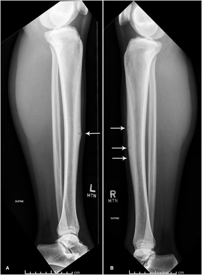
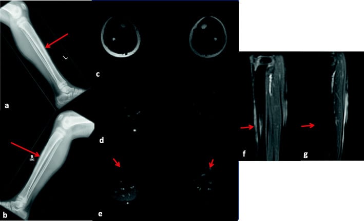
Low risk: the posteromedial cortex. This is the most common site and the majority of tibial stress fractures, usually in the distal third. It is a favourable healing environment with a low risk of complications, and it typically heals with activity modification alone. The patient has point tenderness along the posteromedial border and pain with running relieved by rest, of insidious onset. Radiographs may show periosteal reaction; MRI shows bone marrow oedema.
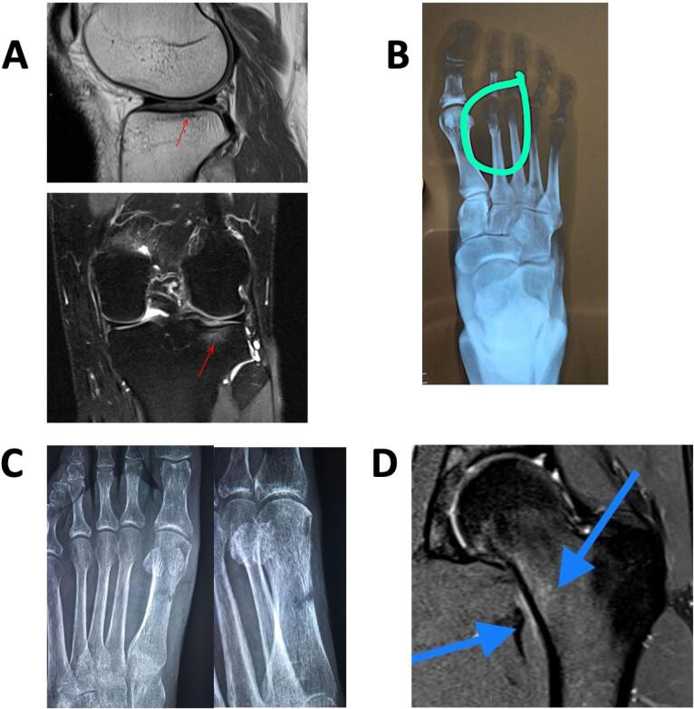
The anterior mid-diaphyseal cortex is not the only high-risk tibial stress fracture. The medial malleolus stress fracture is a second, distal high-risk site that examiners expect you to know. Seen in runners and jumping athletes (basketball, dancers), it arises from repetitive talar impingement against the medial malleolus and the tensile and torsional load there. The characteristic pattern is a vertical fracture line propagating proximally from the junction of the tibial plafond and the medial malleolus, sometimes more oblique, rather than the horizontal black line of the anterior shaft.
It is high risk for delayed union or non-union and for progression to a complete displaced medial malleolar fracture. A lesion with no visible fracture line may be tried in a boot or non-weight-bearing, but a visible fracture line, displacement, or the high-demand athlete is generally treated with internal fixation (one or two partially threaded cancellous screws across the malleolus), which gives reliable union and faster return. When you list the high-risk tibial stress fractures, name both the anterior diaphyseal cortex and the medial malleolus.
Beyond the tibia. The same high-risk label belongs to the navicular (a watershed blood supply), the lateral process of the talus (poor healing) and the proximal fifth metatarsal (the Jones fracture zone).
Severity: the Fredericson MRI grade. Each grade carries a return timeline. The grading was derived from 14 runners and has not been prospectively validated against healing time or return to sport (see the Fredericson evidence card), so the grade describes how much bone is involved, not how long a given athlete will take.
- T1 Signal
- Normal
- T2/STIR Signal
- Periosteal oedema only
- Description
- Mild stress reaction
- Return Timeline
- 2-3 weeks
- T1 Signal
- Normal
- T2/STIR Signal
- Periosteal + marrow oedema
- Description
- Moderate stress reaction
- Return Timeline
- 3-6 weeks
- T1 Signal
- Low signal marrow
- T2/STIR Signal
- Marrow oedema + cortical signal change
- Description
- Stress fracture without line
- Return Timeline
- 12-16 weeks
- T1 Signal
- Low signal marrow
- T2/STIR Signal
- Fracture line visible
- Description
- Stress fracture with line
- Return Timeline
- 14-16 weeks
- T1 Signal
- Low signal marrow
- T2/STIR Signal
- Complete fracture through cortex
- Description
- Complete stress fracture
- Return Timeline
- 16+ weeks or surgery
What the grade changes. Grades 1 and 2 are stress reactions, managed with relative rest and modified activity. Grades 3 and 4 are true stress fractures and mean complete rest from running. An anterior cortex grade 4 with a black line is a surgical consideration.
Clinical Assessment and Diagnosis
History. The story is a change in training rather than a moment of trauma. Ask about:
- Training: recent changes in volume, intensity or surface
- Onset: insidious and activity-related
- Where the pain is: anterior or posteromedial
- Night pain, which suggests a more advanced injury
- Previous stress fractures
- Diet and menstrual history, for the female athlete triad
- Medications: bisphosphonates can cause atypical fractures
Red flags. Anterior tibial pain (the high-risk location), pain at rest or at night, a history of multiple stress fractures, and signs of an eating disorder or amenorrhoea.
Examination. The leg usually looks normal, perhaps with subtle swelling. The sign is point tenderness over the fracture: on the anterior border of the mid-tibia for an anterior cortex fracture, along the posteromedial border (distal more commonly) for a posteromedial one. Three tests provoke the pain:
- Hop test - a single-leg hop reproduces it; sensitive
- Tuning fork test - vibration over the fracture site causes it; poor specificity
- Fulcrum test - bending stress reproduces it
Medial tibial stress syndrome. MTSS, the "shin splints", is periostitis without a fracture line. Radiographs are normal, with MRI if the diagnosis is uncertain. The table separates it from a stress fracture.
- MTSS
- Diffuse over 5+ cm
- Stress Fracture
- Focal point tenderness
- MTSS
- Gradual
- Stress Fracture
- Progressive worsening
- MTSS
- Early in activity, improves as the run continues
- Stress Fracture
- Worsens with activity
- MTSS
- Rare
- Stress Fracture
- Common if advanced
- MTSS
- Periosteal oedema, no marrow oedema or fracture
- Stress Fracture
- Focal bone marrow oedema, may see fracture line
The wider differential. The radiographic differential of a tibial stress lesion also includes osteoid osteoma, malignancy and chronic osteomyelitis, which can mimic a stress reaction on imaging [PMID 28343329]. The clinical differential of activity-related shin pain is summarised below.
- Key features
- Focal point tenderness, training error
- Pain pattern
- Worsens with activity, may have night pain
- Imaging / test
- MRI marrow oedema +/- fracture line
- Discriminator
- Focal lesion; positive hop test
- Key features
- Diffuse tenderness over 5cm or more
- Pain pattern
- Eases as run continues
- Imaging / test
- MRI periosteal oedema, no marrow oedema
- Discriminator
- Diffuse, no focal lesion
- Key features
- Tightness, cramping, paraesthesia
- Pain pattern
- Builds during exercise, relieved by rest
- Imaging / test
- Dynamic compartment pressure testing
- Discriminator
- Pressure rise post-exercise; neuro symptoms
- Key features
- Calf claudication in young athlete
- Pain pattern
- Exercise-induced, resolves with rest
- Imaging / test
- Ankle-brachial index, CT/MR angiography
- Discriminator
- Vascular, not bony; provocative ankle position
- Key features
- Well-localised, often nocturnal pain
- Pain pattern
- Night pain relieved by NSAIDs
- Imaging / test
- CT shows nidus with sclerosis
- Discriminator
- Classic NSAID-responsive night pain
- Key features
- Rest pain, systemic features
- Pain pattern
- Progressive, unrelated to activity
- Imaging / test
- MRI +/- biopsy, inflammatory markers
- Discriminator
- Red flags: weight loss, fever, mass
Investigations and Imaging
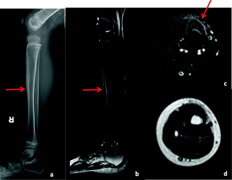
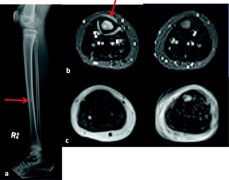
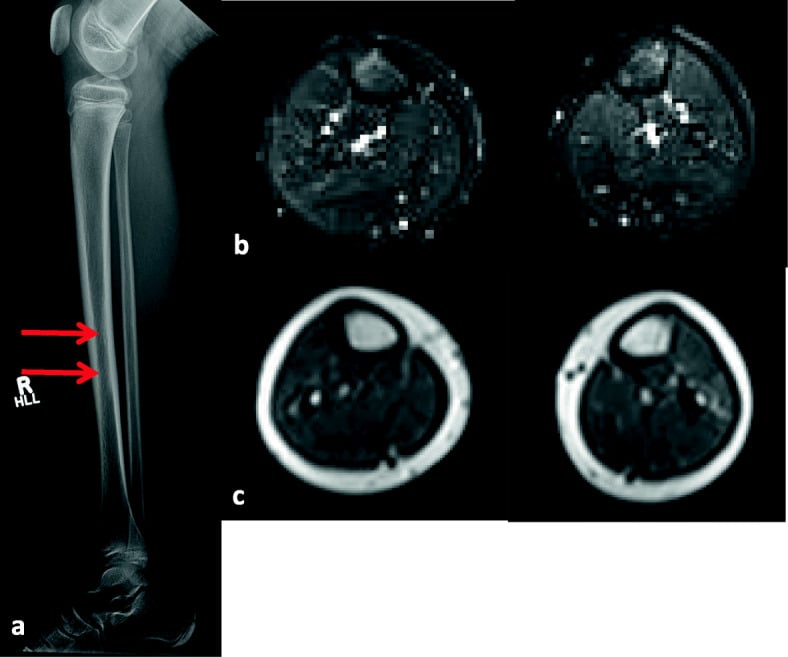
Radiographs may be normal for 2-6 weeks after symptom onset. If clinical suspicion is high, proceed directly to MRI. Do not dismiss the diagnosis based on normal XR alone.
Radiographs are first-line. They may show periosteal reaction, cortical thickening or a fracture line, but they are often negative early in the disease, and the dreaded black line of an anterior cortex fracture is a late finding.
MRI is the gold standard and the most sensitive test for early diagnosis, showing bone marrow oedema 2-6 weeks before radiographic change. The marrow shows low signal on T1 and high signal (oedema) on STIR or fat-saturated T2. MRI grades the severity (the Fredericson grade, under Classification) and so guides management.
CT is best for assessing cortical involvement. It shows the black line of an anterior cortex fracture, is useful for surgical planning, and helps when MRI is unavailable or contraindicated.
Bone scan has been largely replaced by MRI. It is very sensitive but less specific, shows a hot spot at the fracture site, and can assess several sites at once.
Management
The decision. Management depends on the risk, which is set by the location, and on the MRI grade.
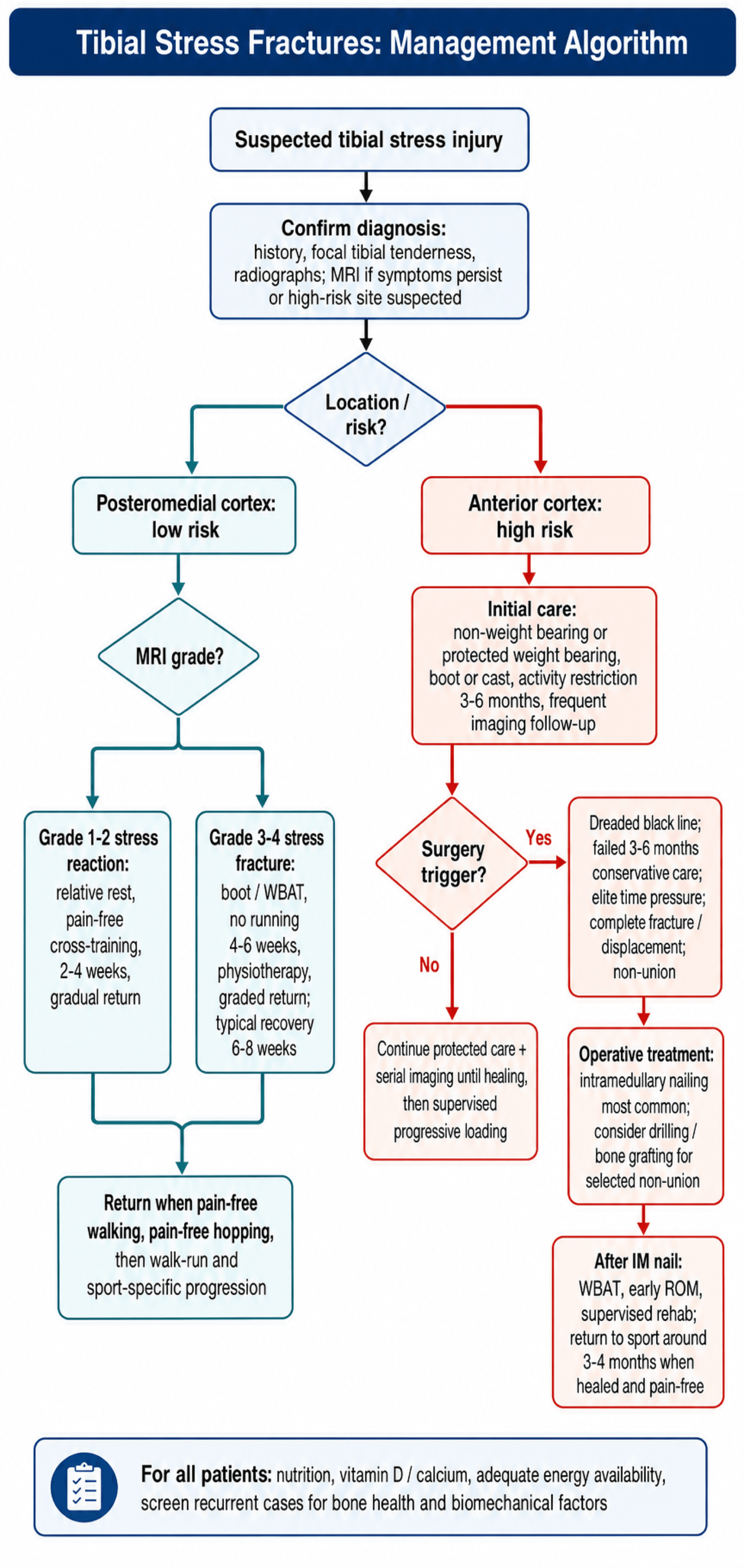
Stress reaction, grades 1-2. Relative rest, meaning pain-free activity, with cross-training (swimming, cycling) for 2-4 weeks, then a gradual return to running.
Stress fracture, grades 3-4. Weight-bearing as tolerated in a walking boot, no running for 4-6 weeks and physiotherapy for muscle conditioning, followed by a gradual return protocol over 2-4 weeks. Total recovery is typically 6-8 weeks.
That is shorter than the timelines tabled against Fredericson grades 3 and 4: the grade describes how much bone is involved, not how long this athlete will take.
Return to running is based on pain-free activity, with no arbitrary time restriction once the athlete is pain-free:
- Pain-free walking (weeks 1-2)
- Pain-free hopping and jumping (weeks 3-4)
- Walk-run programme (weeks 4-6)
- Sport-specific training (weeks 6-8)
- Full return when pain-free at sport intensity
Surgical Technique
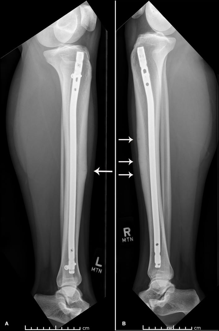
Why a nail. The intramedullary nail acts as a tension band, converting tensile to compressive forces, and allows early weight-bearing.
Relative contraindications. Active infection at the surgical site, severe osteoporosis (consider alternative fixation), and medical comorbidities that preclude surgery.
Imaging. Review the CT for cortical involvement and measure tibial length on long-leg films. Check where the fracture lies relative to the planned nail trajectory, and assess bone quality and canal diameter.
Implant. A standard tibial nail, reamed and statically locked, typically 8-10mm in diameter, its length from preoperative templating. Make sure the locking screws are available.
Counselling. Discuss the risks and benefits, consent for removal of the nail if it becomes symptomatic, set realistic expectations for return to sport and explain the rehabilitation protocol.
Intramedullary nailing is the classic operative answer for the recalcitrant anterior cortex ("dreaded black line") stress fracture, but it does not lie directly on the tension side, and the trans-tendinous or parapatellar entry causes anterior knee pain in a substantial minority. An increasingly used alternative is anterior tension-band plating: a plate applied directly to the anterior tibial cortex across the fracture.
It buttresses the tension side exactly where the fracture is, neutralising the tensile load that prevents healing. It avoids the anterior knee pain and entry-portal morbidity of the nail, does not violate the knee, and can be combined with debridement or drilling of the sclerotic edges and bone grafting. The downsides are a larger soft-tissue exposure over a poorly vascularised anterior surface (wound concerns) and the need for plate removal if prominent.
The exam point: the operative choice for the failed anterior cortex stress fracture is nailing or anterior tension-band plating, with adjunctive drilling or grafting, and you should be able to justify either by the tension-band principle.
Complications
Complete fracture. The risk factors are an anterior cortex location, delayed diagnosis, continued activity despite pain, and a black line that is ignored. It occurs in 10-20% of untreated anterior cortex stress fractures. A displaced fracture is nailed and can be treated as a typical tibial shaft fracture, with a higher risk of delayed union. It delays return to sport by 6+ months and may result in permanent activity restriction.
Non-union. More common in anterior cortex fractures, with delayed treatment, with nutritional deficiencies and in smokers. It requires salvage surgery: reamed IM nailing, bone grafting if needed, and correction of metabolic factors. Recovery is extended, 6-12 months, and non-union may limit a return to elite competition.
Recurrence. The overall rate is 10-20%, higher if the underlying risk factors are not addressed. The female athlete triad is a major risk factor, and training errors are the most common modifiable cause. Prevention addresses the risk factors: optimised nutrition, a gradual return to activity, correction of biomechanical issues and appropriate footwear.
Postoperative Care
In hospital. Weight-bearing as tolerated from the outset with the nail. Neurovascular checks every 4 hours for the first 24 hours, watching for compartment syndrome (rare but serious), DVT prophylaxis per protocol and multimodal analgesia. Discharge is typically on day 1-2.
Wounds. Keep the incisions clean and dry for 2 weeks and watch for infection. Sutures or staples come out at 14 days.
Rehabilitation runs in four phases:
- Weeks 0-2 - weight-bearing as tolerated, with crutches as needed for comfort; range of motion from the start; ankle pumps, quadriceps sets and straight-leg raises; ice and elevation; no impact activity
- Weeks 2-6 - full weight-bearing without aids; advancing range and strengthening; stationary bike when comfortable and pool exercise for cardiovascular fitness; physiotherapy 2-3 times a week
- Weeks 6-12 - progressive resistance training; light jogging once walking is pain-free; activity increased per protocol; sport-specific exercises after week 8
- Months 3-4 - full training once the criteria below are met; return to competition typically at 3-4 months
Follow-up. Review at 2 weeks for the wound and sutures, at 6 and 12 weeks with clinical examination and radiographs (considering return to sport at 12 weeks), and at 6 months for the final clinical and radiographic assessment.
Return to sport requires:
- Radiographic union, with bridging callus on radiographs
- Pain-free with all activities, including high-impact activity
- Full range of motion compared with the other side
- Functional strength restored on testing (single-leg hop, agility drills)
- Sport-specific skills without pain
Long term. Address the underlying risk factors (nutrition, training errors) and monitor for contralateral stress fractures. Most nails can remain indefinitely if asymptomatic; consider removal at 12-18 months if symptomatic.
Outcomes and Prognosis
Posteromedial fractures do well with conservative management: union over 95% with activity modification, average healing 6-8 weeks, recurrence under 5% if risk factors are addressed, excellent functional outcomes, and over 95% return to their pre-injury level. Nutrition and compliance are critical.
Anterior cortex fractures compare as follows.
- Conservative
- 50-70%
- IM Nailing
- Over 90%
- Conservative
- 4-6 months (if successful)
- IM Nailing
- 3-4 months
- Conservative
- 70-80%
- IM Nailing
- Over 90%
- Conservative
- Recurrence common with premature return; complete fracture risk if inadequately treated
- IM Nailing
- Complications low (under 5%); lower recurrence with risk factor modification
Nailing's union rate is significantly superior to conservative care, and functional outcomes after it are excellent. Weigh its low complication rate against the anterior knee pain of the entry portal, set out under Surgical Technique.
Prognostic indicators. Favourable: a posteromedial location, early diagnosis, good nutrition and bone health, compliance with activity restriction, and biomechanical issues addressed. Unfavourable: an anterior cortex location, a black line, delayed diagnosis, continued activity despite symptoms, poor bone health or the female athlete triad, and smoking.
Long term. Most patients return to full activity without restrictions, and chronic pain is rare if the fracture is appropriately treated. Recurrent stress fractures may necessitate activity modification, and the female athlete triad requires ongoing management.
Guidelines, Registries & Global Practice
Global Epidemiology
Stress fractures are a worldwide overuse problem in athletes and military recruits, and the tibia is consistently the single most common site. In a 12-month prospective study of competitive track and field athletes the overall incidence was 21.1%, with the tibia accounting for 46% of stress fractures, followed by the navicular (15%) and fibula (12%) [PMID 8775123]. Among female military recruits the reported frequency of stress fractures is in the order of 5-15% during basic training [PMID 18628731]. Female sex, low bone mineral density, menstrual disturbance and low energy availability are the dominant intrinsic risk factors across populations [PMID 8947404].
- Region
- International (US-led, ACSM/AMSSM endorsed)
- Core recommendation
- Cumulative-risk point system; clearance and return-to-play categories; multidisciplinary management of low energy availability
- Evidence basis
- Consensus / expert panel
- Region
- International (Olympic movement)
- Core recommendation
- Screen all athletes (both sexes) for low energy availability; risk-stratified return-to-sport model
- Evidence basis
- Consensus / expert panel
- Region
- International narrative standard
- Core recommendation
- Anterior tibial cortex is high risk: early imaging, non-weight-bearing immobilisation, low threshold for surgery
- Evidence basis
- Narrative review (Level V)
- Region
- International imaging standard
- Core recommendation
- Grade tibial stress injury on MRI to stage severity and guide return-to-activity timing
- Evidence basis
- Level IV correlation study
Guideline Convergence and Practice Variation
There is no single dominant national-society guideline specific to tibial stress fractures; practice is shaped by sports-medicine consensus statements rather than by AAOS, NICE or BOAST documents (which do not address this entity directly). Internationally there is strong agreement on the core principles: MRI is the imaging standard for staging, low-risk posteromedial fractures are managed with relative rest and graded return, and high-risk anterior cortex fractures (the dreaded black line) warrant aggressive management with a low threshold for intramedullary nailing [PMID 26972260] [PMID 15888719]. The main areas of genuine variation are:
- Imaging access — in high-resource settings MRI is obtained early; in limited-resource settings diagnosis often relies on serial radiographs and clinical assessment, with bone scintigraphy where MRI is unavailable.
- Timing of surgery in elite athletes — earlier nailing of anterior cortex fractures is favoured where rapid return to competition is required, whereas a longer trial of non-operative care is common elsewhere.
- Energy-availability screening — increasingly routine in high-performance and military programmes (triad/RED-S frameworks) but still under-recognised in general practice worldwide.
Registry and Preventive Evidence
Joint registries (NJR, AJRR, AOANJRR, SHAR) do not capture stress fractures, so the strongest population-level evidence comes from military and athletic cohort studies. A randomised controlled trial in 5201 female US Navy recruits showed that calcium (2000 mg) and vitamin D (800 IU) daily reduced stress fracture incidence by approximately 20% over 8 weeks [PMID 18433305], supporting nutritional optimisation as a low-cost preventive intervention across all health systems.
Viva Scenarios
Clinical Decision Scenarios
Practise clinical reasoning and management decisions out loud
“22-year-old female distance runner with 4 weeks of progressive medial shin pain. Pain worse with running, improves with rest. Point tenderness posteromedial tibia. Training for marathon, increased mileage 30% over past month.”
“25-year-old male army recruit with 6 weeks of anterior tibial pain. Initially told to 'push through'. Now has pain at rest. XR shows horizontal radiolucent line in anterior cortex.”
“19-year-old elite gymnast with third stress fracture in 2 years (metatarsal, femoral neck, now tibial). BMI 17.5, amenorrhea for 8 months. Very driven to compete at upcoming nationals.”
MCQ Practice Points
Q: Which tibial stress fracture location is HIGH RISK for non-union?
A: Anterior cortex. This is the tension side of the tibia with poor blood supply. The "dreaded black line" represents a cortical fracture with high non-union risk. Posteromedial (compression side) is LOW RISK.
Q: A runner presents with 3 weeks of tibial pain. XR is normal. What is the next step?
A: MRI. MRI is the gold standard for diagnosis, showing bone marrow edema 2-6 weeks before radiographic changes. Do not dismiss stress fracture based on normal XR.
Q: What is the treatment for an anterior tibial stress fracture with a dreaded black line?
A: Strong consideration for intramedullary nailing. Conservative treatment has 30-50% non-union rate for anterior cortex stress fractures with black line. Prophylactic nailing has over 90% union rate.
Q: What are the components of the female athlete triad?
A: Energy deficiency/eating disorder, amenorrhea, and low bone density/osteoporosis. All three components increase stress fracture risk. Must be addressed holistically - treating fracture alone will lead to recurrence.
Q: What is the primary criterion for return to running after a low-risk tibial stress fracture?
A: Pain-free activity progression - not arbitrary time limits. Patient should be pain-free with walking, then hopping, before gradual walk-run program. Typical timeline is 6-8 weeks but varies by individual.
Risk Stratification
- Anterior cortex = HIGH RISK (tension side)
- Posteromedial = LOW RISK (compression)
- Dreaded black line = surgical consideration
- MTSS = periostitis, not fracture
- Female athlete triad increases risk
Diagnosis
- XR may be normal for 2-6 weeks
- MRI is gold standard
- Fredericson grades 1-4
- CT for cortical assessment
- Point tenderness on exam
Low-Risk Management
- Rest 6-8 weeks
- Cross-training (swim, bike)
- Pain-free progression to return
- Gradual return protocol
- Address training errors
High-Risk Management
- Consider prophylactic IM nail
- Extended NWB (3-6 months) if conservative
- High non-union rate without surgery
- Complete fracture risk if ignored
- Return to sport 3-4 months post-nailing
Prevention/Nutrition
- Calcium 1000-1500mg daily
- Vitamin D over 30 ng/mL
- 10% rule for training increases
- Screen for female athlete triad
- Appropriate footwear
Evidence Base
Reamed IM Nailing for Chronic Anterior Tibial Stress Fractures
- Seven collegiate athletes with 11 chronic anterior midtibial stress fractures, all failing at least 4 months of nonoperative care, achieved clinical union at a mean of 2.7 months and radiological union at 3 months after reamed intramedullary nailing
- Mean return to sport was 4 months with a low complication rate (one insertion-site bursitis, one later traumatic distal tibial fracture)
Fredericson MRI Grading of Tibial Stress Injuries
- In 14 runners (18 symptomatic legs), MRI defined a progression from periosteal oedema through marrow involvement to frank cortical stress fracture, and correlated with an established bone-scan grading system in 14 of 18 legs
- MRI was recommended over bone scintigraphy because it more accurately correlated the degree of bone involvement with clinical symptoms while avoiding ionising radiation
Risk Factors for Stress Fractures in Track and Field Athletes
- In a 12-month prospective study of 111 athletes the stress fracture incidence was 21.1%, with the tibia the most common site
- In female athletes lower bone density, a history of menstrual disturbance, lower lean mass, leg-length discrepancy and a lower-fat diet were significant risk factors; later age of menarche and calf girth were the best independent predictors
Female Athlete Triad Coalition Consensus on Treatment and Return to Play
- The triad comprises low energy availability (with or without disordered eating), menstrual dysfunction and low bone mineral density; low energy availability is the underlying driver
- A cumulative risk-stratification point system guides clearance and return-to-play decisions, with multidisciplinary management and treatment contracts
Calcium and Vitamin D Supplementation in Female Navy Recruits (RCT)
- Randomised, double-blind, placebo-controlled trial in 5201 female Navy recruits: 2000 mg calcium plus 800 IU vitamin D daily reduced stress fracture incidence by 20% over 8 weeks (5.3% vs 6.6%, p=0.0026)
- Smoking, low physical fitness and amenorrhoea were confirmed as additional risk factors
High-Risk Stress Fractures: Diagnosis and Management
- High-risk sites (femoral neck tension side, patella, anterior tibia, medial malleolus, talus, tarsal navicular, proximal fifth metatarsal, great toe sesamoids) share a region of high tensile load and low blood flow, predisposing to delayed union and nonunion
- These require a more aggressive approach: early imaging, non-weight-bearing immobilisation, prolonged time away from sport and a careful return, with surgery often needed
IOC Consensus on Relative Energy Deficiency in Sport (RED-S): 2018 Update
- RED-S extends the female athlete triad to a broader syndrome of impaired physiological function (including bone health and stress-fracture risk) caused by relative energy deficiency, and affects male as well as female athletes
- Provides a clinical assessment and risk-stratified return-to-sport model centred on restoring energy availability