Neer Classification of Proximal Humerus Fractures
- Counting fragments, not displaced parts. The category is the number of Codman parts displaced beyond the threshold (over 1 cm OR over 45°) — a shattered-looking film with nothing past the threshold is a ONE-part fracture; a comminuted surgical neck with one displaced greater tuberosity is TWO-part.
- Using the 1 cm rule as the greater-tuberosity operation threshold. The GT fixation threshold is tighter — over 5 mm (over 3 mm in an overhead athlete/labourer) — because it retracts under the cuff and narrows the subacromial space. A "two-part by Neer" GT can still need fixation.
- Trusting the Neer label as a precise diagnosis. Category kappa is only ~0.27 — it is a treatment framework, not a reproducible radiologic label; use Hertel's criteria (short calcar under 8 mm, disrupted medial hinge) for true AVN risk.
- Sending every four-part to arthroplasty. The valgus-impacted four-part keeps its medial hinge, has a lower AVN rate, and is reasonable to fix even in older patients.
- Choosing hemi over reverse in the elderly. For a displaced complex fracture in the older patient with calcar disruption, reverse shoulder arthroplasty outperforms hemiarthroplasty (Sebastiá-Forcada) — confirm an intact deltoid and axillary nerve first.
The Neer classification system
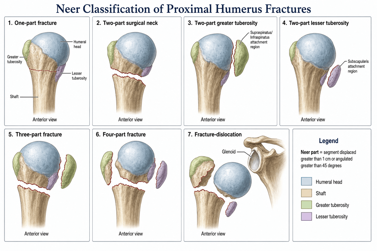
The modern classification is a marriage of two ideas. Codman (1934) showed that the proximal humerus separates into four anatomic fragments along the old epiphyseal lines — humeral head, greater tuberosity, lesser tuberosity, and shaft. Neer (1970) added a behavioural threshold: a fragment is only "displaced" if it is separated by more than 1 cm or angulated by more than 45°. The result is a four-category system built on four anatomic parts and one biomechanical rule.
- Anatomic boundary
- Anatomical neck
- Key soft-tissue attachment
- Articular surface; reliant on posteromedial vessels
- Why it matters
- Highest avascular-necrosis risk when displaced
- Anatomic boundary
- Posterolateral to the head
- Key soft-tissue attachment
- Supraspinatus, infraspinatus, teres minor
- Why it matters
- Subacromial impingement and lost external rotation if displaced superiorly
- Anatomic boundary
- Anteromedial to the head
- Key soft-tissue attachment
- Subscapularis
- Why it matters
- Lost internal rotation and risk of posterior dislocation if displaced
- Anatomic boundary
- Surgical neck region
- Key soft-tissue attachment
- Pectoralis major, deltoid, latissimus dorsi
- Why it matters
- Commonest fracture line; medial calcar comminution predicts instability
- Threshold for 'displaced'
- More than 1 cm
- How to measure
- Distance between the cortex of the displaced fragment and the parent bone
- Common pitfall
- Counting the gap in a comminuted zone as a separate part
- Threshold for 'displaced'
- More than 45 degrees
- How to measure
- Angle between the long axis of the displaced fragment and its anatomic position
- Common pitfall
- Confusing varus tilt at the surgical neck with head angulation
Head · Greater · Lesser · ShaftThe four parts
Hook:A fracture is named by which of the four parts is DISPLACED (over 1 cm or over 45°), not by how many fragments you can count.
Even a proximal humerus shattered into many fragments is a one-part fracture until at least one of the four parts is displaced more than 1 cm or angulated more than 45°. The category is a behavioural description: a four-part fracture is defined by the number of DISPLACED parts, not the number of fragments. A comminuted surgical-neck fracture with one displaced greater tuberosity is a two-part fracture.
The classification keeps invoking "AVN risk", "posteromedial vessels" and "the medial hinge" — the examiner wants the anatomy underneath it:
- The classic teaching — the anterior circumflex humeral artery (ACHA) gives an ascending/anterolateral branch that runs up the bicipital groove as the arcuate artery of Laing and was long held to supply the majority of the humeral head (hence the head's vulnerability when the fracture passes through the anatomical neck).
- The modern correction — quantitative perfusion studies (e.g. Hettrich) show the posterior circumflex humeral artery (PCHA) actually supplies the majority (around 64%) of the humeral head; both circumflex systems matter, and the head retains perfusion as long as one route survives.
- Why the calcar and medial hinge are the key — the surviving supply in a displaced fracture runs in the posteromedial vessels entering through the calcar, so a long posteromedial metaphyseal head extension (calcar 8 mm or more) with an intact medial periosteal hinge keeps the head alive — exactly the anatomical basis for Hertel's ischaemia predictors and for why the valgus-impacted four-part (hinge preserved) has a low AVN rate while calcar comminution/hinge disruption threatens the head.
Classification categories & treatment
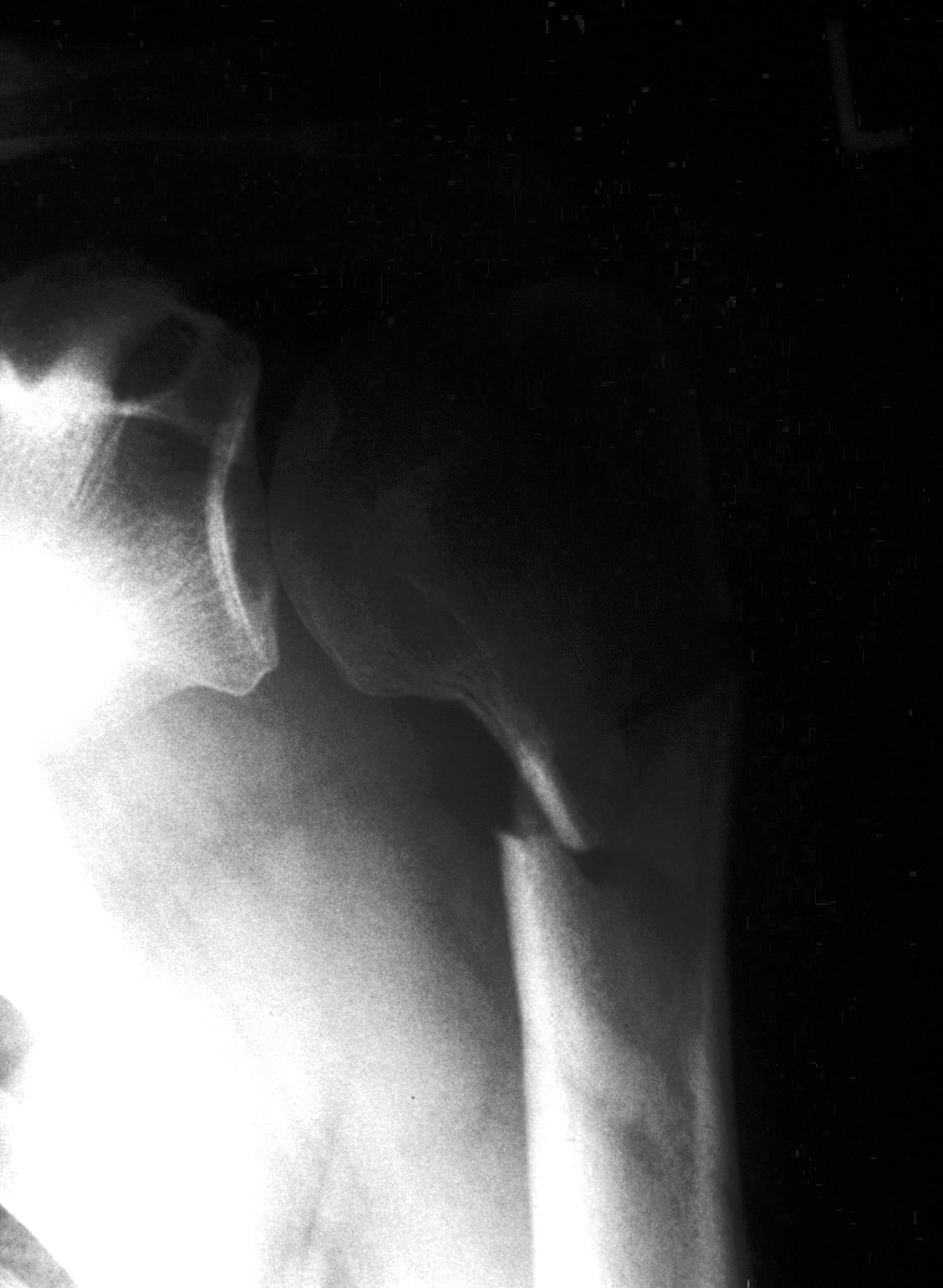
The categories progress from one-part (the commonest pattern, treated non-operatively) to four-part (often arthroplasty in the elderly). Two adjuncts sit on top: fracture-dislocation (the head is dislocated as well as fractured) and articular-surface injuries (head-splitting or impression fractures).
- Definition
- No part displaced over 1 cm or over 45° — regardless of comminution
- Typical treatment
- Sling, early pendulum mobilisation, then physiotherapy
- Key consideration
- Commonest pattern (~80% of proximal humeral fractures)
- Definition
- Exactly one part displaced
- Typical treatment
- ORIF (or closed reduction for surgical neck)
- Key consideration
- Greater tuberosity often needs fixation if displaced over 5 mm (over 3 mm in athletes/overhead workers)
- Definition
- Two parts displaced (usually surgical neck + one tuberosity)
- Typical treatment
- ORIF in younger patients; arthroplasty in older low-demand patients
- Key consideration
- AVN risk rises with each additional displaced part
- Definition
- All four parts displaced
- Typical treatment
- Arthroplasty in older patients (reverse shoulder increasingly preferred); ORIF in selected young patients
- Key consideration
- AVN risk substantial with ORIF; favours arthroplasty in the elderly
- Definition
- Head dislocated (anterior/posterior) plus a fracture
- Typical treatment
- Urgent reduction; arthroplasty for older patients with a displaced head
- Key consideration
- Two-part surgical-neck anterior dislocation in the elderly has a particularly poor prognosis
- 1Count the displaced partsApply the 1 cm / 45° rule to head, GT, LT and shaft. CT (ideally 3D) clarifies displaced parts, the medial calcar and any head-split — it changes the plan.
- 2One-part → non-operativeSling, early pendulum mobilisation, then physiotherapy — the commonest pattern and the default for minimally displaced fractures.
- 3Two/three-part → ORIF (patient-dependent)ORIF (locked plate, nail, or suture-anchor tuberosity fixation) in suitable patients; fix a greater tuberosity displaced over 5 mm (over 3 mm in athletes) even if it is under the Neer threshold.
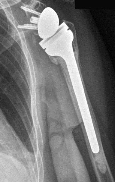
- 4Displaced four-part → arthroplasty (usually)In the elderly with calcar disruption, reverse shoulder arthroplasty is preferred over hemiarthroplasty; ORIF for selected young patients, and for the valgus-impacted variant where the medial hinge is preserved.
A two-part greater-tuberosity fracture displaced more than 5 mm (or more than 3 mm in an overhead athlete/labourer) should be fixed — the fragment retracts under the rotator cuff, the subacromial space narrows, and chronic impingement with weak external rotation is very hard to salvage later. The Neer 1 cm threshold is NOT the operation threshold for this fragment.
The variant examiners love: the humeral head is driven down into the metaphysis, the medial periosteal hinge stays intact, and the tuberosities splay around it. Because the medial perforating vessels are often preserved, the AVN rate is much lower than the classic four-part pattern, and ORIF is reasonable even in older patients — the "four-part that does not look as bad as the label suggests."
Limitations & modern context
- Inter-observer reliability is poor. The Neer-category kappa is around 0.27 (Brorson et al.), with displacement-only agreement ~0.4–0.5 even among specialists — surgeons disagree on the category in a large proportion of cases. It is a treatment framework, not a precise radiologic label.
- The category does not reliably predict outcome. Recovery depends as much on age, comorbidities, rotator-cuff status, smoking and bone quality as on the Neer category.
- Hertel's binary description (length of the posteromedial metaphyseal extension, integrity of the medial hinge, and the basic fracture type) predicts humeral-head ischaemia accurately — the combined criteria (anatomic-neck fracture, short calcar, disrupted hinge) carry positive predictive values up to ~97% — and is increasingly used alongside Neer.
- CT outperforms plain film for the number of displaced parts, the medial calcar, and the presence of a head-split — all of which change the operative plan.
- Decisions are dominated by patient factors — a frail 80-year-old with a four-part fracture may be best served by reverse shoulder arthroplasty or even non-operative care; a fit 30-year-old with the same film goes to ORIF.
- The valgus-impacted four-part behaves differently — preserved medial hinge, lower AVN risk, more fixation options.
The Neer category says nothing about the soft tissues, but the examiner expects a documented neurovascular assessment before and after any reduction or surgery:
- Axillary nerve — the most commonly injured nerve in a proximal humerus fracture or fracture-dislocation. Test regimental-badge sensation over the lateral deltoid (deltoid motor power is hard to assess acutely against a painful fracture); it is usually a neurapraxia that recovers, but documenting it matters medicolegally and because a working axillary nerve/deltoid is a prerequisite for a reverse shoulder arthroplasty.
- Axillary artery — rare but limb-threatening, and more frequent in the elderly (calcified, less elastic vessels), in high-energy injuries and in fracture-dislocations. Crucially, a palpable distal pulse does NOT exclude an axillary artery injury because of the rich collateral circulation — look for an expanding/pulsatile axillary haematoma, a bruit, pallor or pulse asymmetry, and have a low threshold for CT angiography and vascular review.
- Brachial plexus — can be injured alongside, particularly in high-energy injuries and dislocations; examine all the peripheral nerves of the limb, and check the skin for an open or tented injury.
Viva practice
- Codman's four parts (head, greater tuberosity, lesser tuberosity, shaft); a part is displaced only if over 1 cm or over 45°.
- Category = number of DISPLACED parts, not fragments — a comminuted film can still be one- or two-part.
- One-part non-operative; two/three-part ORIF; displaced four-part elderly → reverse shoulder arthroplasty (over hemi).
- Greater-tuberosity fixation threshold is tighter (over 5 mm, over 3 mm in athletes) than the Neer rule.
- Valgus-impacted four-part = preserved medial hinge, lower AVN, ORIF reasonable.
- Poor reliability (category kappa ~0.27) — use Hertel's criteria for true ischaemia risk.
1 cm or 45°The displacement rule
Hook:A part counts as displaced only if it crosses 1 cm OR 45°; if neither, it is a one-part fracture no matter how many fragments are visible.
Exam viva
Practise clinical reasoning and management decisions out loud
“A 72-year-old woman falls onto her right shoulder. Radiographs show a comminuted fracture of the surgical neck with the greater tuberosity displaced superiorly by about 8 mm, the lesser tuberosity sitting normally, and the humeral head in the glenoid. How do you classify this injury using the Neer system, and how does that guide your management?”
“A fit 78-year-old retired engineer presents after a fall. Imaging shows a displaced four-part proximal humeral fracture with disruption of the medial calcar. The deltoid and axillary nerve are intact. He wants to remain independent and will not accept a 'frozen shoulder'. Discuss your surgical reasoning and choice of implant.”
Exam cheat sheet
The four anatomic parts (Codman)
- Humeral head (anatomical neck) — highest AVN risk
- Greater tuberosity — supraspinatus, infraspinatus, teres minor
- Lesser tuberosity — subscapularis
- Humeral shaft (surgical neck) — pectoralis, deltoid, latissimus dorsi
The displacement rule (Neer)
- More than 1 cm of translation OR more than 45° of angulation
- One-part when no part meets either threshold — regardless of comminution
- Category = number of displaced parts (one → two → three → four)
- Add 'fracture-dislocation' or 'head-split' as a separate descriptor when present
Treatment by category & patient
- One-part: sling, early pendulum mobilisation, physiotherapy
- Two-part: ORIF or closed reduction (greater tuberosity if over 5 mm, over 3 mm in athletes)
- Three-part: ORIF in the young; arthroplasty in older low-demand patients
- Four-part (elderly, calcar disrupted): reverse shoulder arthroplasty preferred over hemi
- Valgus-impacted four-part: medial hinge preserved, lower AVN, ORIF often reasonable
Pitfalls to avoid
- Don't call a comminuted fracture four-part just because it looks shattered
- The greater-tuberosity fixation threshold is tighter than the Neer 1 cm rule
- Category kappa is only ~0.27 — accept disagreement on the label
- Use Hertel's criteria (calcar length, medial hinge) to gauge real AVN risk
Evidence
Displaced proximal humeral fractures. I. Classification and evaluation
- Combined Codman's four anatomic parts with a displacement threshold of 1 cm or 45° to define one-, two-, three- and four-part fractures (plus fracture-dislocations and articular injuries).
- Categorised a prospective series and linked each category to a treatment recommendation.
- Established the framework still used worldwide for surgical decision-making in proximal humeral fractures.
Displaced proximal humeral fractures. II. Treatment of three-part and four-part displacement
- Companion paper detailing the treatment of three-part and four-part displaced fractures.
- Described open reduction and internal fixation for three-part patterns and hemiarthroplasty for the older patient with a four-part fracture.
- Set out the surgical-decision framework that still anchors the classification in practice.
According to PubMed, the classification and its treatment framework come from Neer 1970 Parts I (PMID 5455339) and II (PMID 5455340); the ischaemia predictors from Hertel et al. 2004 (DOI 10.1016/j.jse.2004.01.034; combined criteria PPV up to ~97%); the poor reliability (category kappa 0.27) from Brorson et al. 2002 (DOI 10.1007/s00264-002-0369-x); and the reverse-versus-hemiarthroplasty evidence from Sebastiá-Forcada et al. 2014 (DOI 10.1016/j.jse.2014.06.035).