Three-Layer Structure | Fibre Classification | Action Potential | Injury Grading | Wallerian Degeneration
- Three-layer structure: Epineurium (outer), perineurium (surrounds fascicles), endoneurium (within fascicles)
- A-alpha fibres (largest, myelinated) conduct motor signals at 70-120 m/s; C fibres (unmyelinated) conduct pain at 0.5-2 m/s
- Action potential requires sodium influx (depolarisation) then potassium efflux (repolarisation); threshold is -55mV
- Wallerian degeneration occurs distal to injury within 24-48 hours; proximal stump regenerates at 1-3mm/day
- Sunderland Grade III (endoneurial disruption) may not recover without surgery despite intact nerve sheath
- “Nerve fibres classified by diameter (Erlanger-Gasser: A, B, C) or function (Lloyd: Ia, Ib, II, III, IV)
- “Nodes of Ranvier allow saltatory conduction - 50x faster than continuous conduction
- “Tinel's sign progression tracks regenerating axons advancing at 1mm/day
- “Second-degree injury (axonotmesis) recovers spontaneously; third-degree may require neurolysis
Overview and Introduction
Peripheral nerves transmit electrical signals between the central nervous system and the peripheral tissues. Understanding their anatomy and physiology is fundamental to managing nerve injuries and to understanding neurological deficits in orthopaedic surgery.
Scope. This page covers the structure, conduction and fibre-type basic science. The injury and repair biology that builds on it - the Seddon and Sunderland classifications, Wallerian degeneration, chromatolysis, Schwann-cell reprogramming and axonal regeneration - is covered in depth on the Nerve Injury and Regeneration page; the two are companion reads.
Why it matters. Peripheral nerve injury is an uncommon but disabling complication of limb trauma. In a large European trauma registry an associated peripheral nerve injury was recorded in approximately 3.3% of severely injured patients with upper-extremity involvement, clustering in young men after high-energy mechanisms (notably motorcycle trauma) and with humeral or forearm fractures. Accurate classification with the Seddon and Sunderland systems guides prognosis, the decision to observe versus explore, and the timing of repair.
Concepts and Mechanisms
The three connective-tissue layers
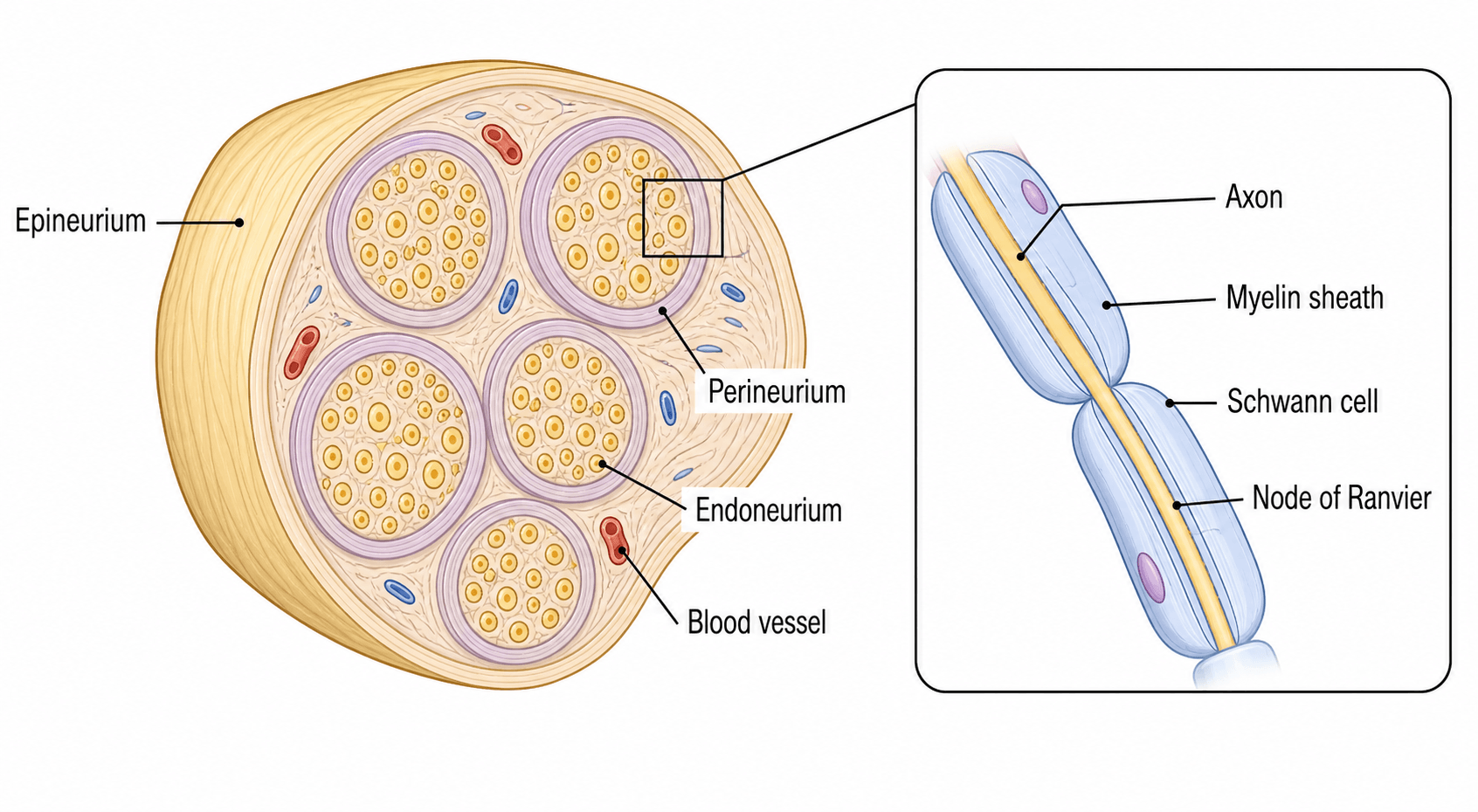
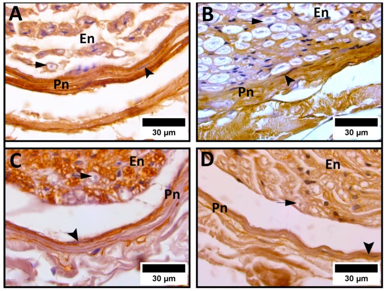
Epineurium. The outermost sheath is loose areolar connective tissue of type I and III collagen and fibroblasts, and makes up 30-75% of the nerve's cross-sectional area. It carries the vasa nervorum (longitudinal vessels) and the lymphatics, protects the fascicles mechanically, lets the nerve glide and supplies nutrients; Sunderland described it as the cushion against compression. Surgically it is the layer preserved in neurolysis and the layer that gives suture purchase in a repair.
Perineurium. Each fascicle is bounded by 7-15 concentric layers of flattened cells joined by tight junctions. This is the blood-nerve barrier: selectively permeable, it maintains the endoneurial microenvironment and its ionic composition, holds the endoneurial fluid pressure at 2-8 mmHg, and its high electrical resistance prevents current leakage. It is a tough, tension-resistant sheath, and the nerve's tensile strength and capacity to resist traction reside in it.
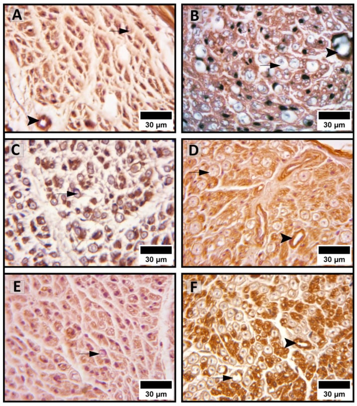
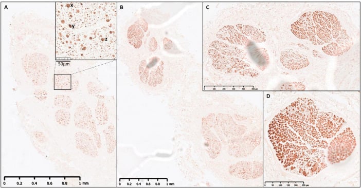
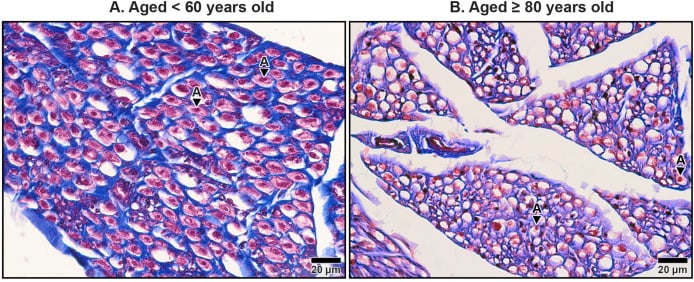
Endoneurium. Within each fascicle, type III collagen fibrils, Schwann cells and capillaries surround the individual axons, forming tubes (0.4-14 micrometres in diameter, lined by the Schwann cell basement membrane) that contain endoneurial fluid for nutrient transport. The endoneurium supports the axons mechanically, regulates their ionic environment and is the scaffold for regeneration: after injury the tubes persist and fill with Schwann cell columns, the Bands of Büngner, along which regenerating axons travel.
Why the layers matter. Injury severity is graded by how many of the sheaths are lost, and the sheaths decide the prognosis. Axons regenerating within intact endoneurial tubes (Sunderland I-II) reach their correct targets and recover spontaneously; once the endoneurium is disrupted (Sunderland III) regeneration is misdirected and recovery variable, and once the perineurium goes as well (Sunderland IV) intraneural scarring and a neuroma in continuity leave a nerve that is in continuity but recovers poorly. The layering also explains the repair options, epineurial versus fascicular, set out under fascicular organisation below.
Fascicular organisation
Fascicular patterns. Nerves differ in how their fibres are bundled:
- Monofascicular - a single large fascicle (digital nerves distally)
- Oligofascicular - a few large fascicles (2-10)
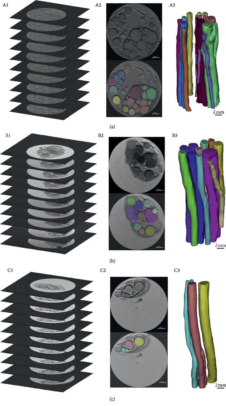
- Polyfascicular - many small fascicles (the median nerve at the wrist has 15-20)
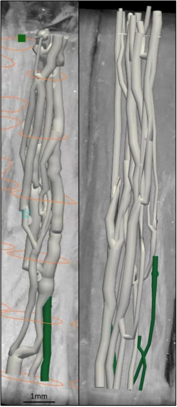
- Plexiform - fascicles branch and rejoin along the length of the nerve
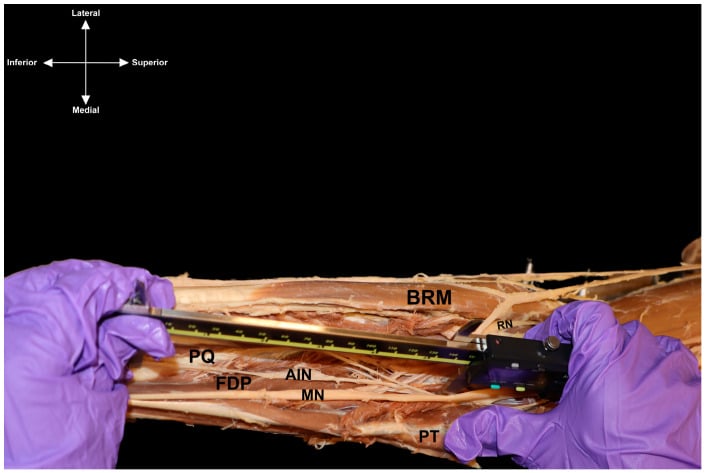
Topography. Proximally the fascicles are mixed, with motor and sensory fibres intermingled; distally they segregate, so the median nerve's motor fascicle to the recurrent branch is distinct at the wrist but mixed further up. Fascicles to particular targets also occupy consistent positions within the trunk: in the sciatic nerve the tibial division lies posteromedial and the peroneal division anterolateral, and in the ulnar nerve at the wrist the motor fascicles to the intrinsics lie dorsal. Knowing the map is what allows selective fascicular repair and internal neurolysis.
What it means for repair. Distal nerves, monofascicular or oligofascicular with segregated fibres, allow group fascicular repair with better coaptation. Proximal nerves, polyfascicular and plexiform, need an epineurial repair because fascicular matching is impossible, and attempting fascicular dissection proximally risks additional injury.
Blood supply
Extrinsic supply. Segmental regional vessels enter the epineurium at regular intervals and form extensive longitudinal anastomoses in the epineurium and perineurium: the vasa nervorum. That redundancy, together with the intrinsic supply, is why 10-15 cm of nerve can be mobilised without devascularising it.
Intrinsic supply. A rich capillary plexus lies in the perineurium, and the endoneurial capillaries are continuous capillaries with tight junctions, which is the vascular side of the blood-nerve barrier. There are watershed zones vulnerable to ischaemia, the ulnar nerve at the cubital tunnel among them.
Clinical consequences. Excessive tension impairs intraneural blood flow and impedes regeneration. Compartment syndrome raises pressure and reduces endoneurial perfusion, and diabetic microangiopathy affects the vasa nervorum and contributes to neuropathy. Respecting the vascular anatomy is what prevents iatrogenic injury during nerve repair.
Nerve Fibre Types and Classification
Fibres are classified two ways: Erlanger and Gasser by diameter and conduction speed (A, B, C, with the A group subdivided), and Lloyd by a numeric grouping of the sensory afferents (I-IV). The Lloyd groups correspond to Erlanger-Gasser types, and in both schemes size predicts speed and vulnerability.
Erlanger-Gasser classification
- Diameter (μm)
- 12-20
- Conduction Velocity (m/s)
- 70-120
- Myelination
- Heavy
- Function
- Motor, proprioception (Ia, Ib afferents)
- Diameter (μm)
- 6-12
- Conduction Velocity (m/s)
- 35-75
- Myelination
- Heavy
- Function
- Touch, pressure (mechanoreceptors)
- Diameter (μm)
- 4-8
- Conduction Velocity (m/s)
- 15-40
- Myelination
- Medium
- Function
- Motor to muscle spindles (fusimotor)
- Diameter (μm)
- 1-5
- Conduction Velocity (m/s)
- 5-30
- Myelination
- Light
- Function
- Sharp pain, temperature, crude touch
- Diameter (μm)
- 1-3
- Conduction Velocity (m/s)
- 3-15
- Myelination
- Light
- Function
- Preganglionic autonomic
- Diameter (μm)
- 0.2-1.5
- Conduction Velocity (m/s)
- 0.5-2
- Myelination
- None
- Function
- Dull pain, temperature, postganglionic autonomic
Diameter and myelin set the speed. A larger axon offers less internal resistance and conducts faster; myelination adds saltatory conduction, which is about 50 times faster than continuous conduction. The same size gradient governs regeneration, as large myelinated fibres regenerate faster than small unmyelinated ones.
Compression at two sites along a nerve causes additive impairment, the classic pairing being cervical radiculopathy with carpal tunnel syndrome. The first compression reduces axoplasmic flow and leaves the distal segment vulnerable, so treating one site may not fully resolve the symptoms.
Lloyd classification of sensory afferents
- Corresponds to
- A-alpha
- Origin
- Muscle spindle primary
- Function
- Proprioception, stretch reflex
- Clinical Test
- Deep tendon reflexes
- Corresponds to
- A-alpha
- Origin
- Golgi tendon organ
- Function
- Tension, inverse stretch reflex
- Clinical Test
- Golgi tendon reflex
- Corresponds to
- A-beta
- Origin
- Muscle spindle secondary, mechanoreceptors
- Function
- Touch, pressure, vibration
- Clinical Test
- Light touch, vibration
- Corresponds to
- A-delta
- Origin
- Nociceptors, thermoreceptors
- Function
- Sharp pain, cold
- Clinical Test
- Pinprick, cold sensation
- Corresponds to
- C
- Origin
- Nociceptors, thermoreceptors
- Function
- Dull pain, warmth
- Clinical Test
- Dull ache, warm sensation
Reading the examination. Loss of Ia and Ib fibres abolishes the deep tendon reflexes and proprioception; loss of group II removes light touch and vibration, and with them useful hand function. When groups III and IV are preserved, pain sensation is intact despite motor and proprioceptive loss. On recovery the large fibres (Ia, Ib, II) return before the small ones (III, IV).
Why fibres respond differently to injury
Compression. The large myelinated A-alpha and A-beta fibres, carrying proprioception and vibration, are the most vulnerable: the lesion is demyelination at the nodes of Ranvier with failure of saltatory conduction, a neuropraxia. The small C fibres carrying pain are relatively resistant, which is why early carpal tunnel syndrome loses vibration sense before pain.
Ischaemia. The large motor fibres, with the highest metabolic demand, fail first as ATP is depleted, the Na+/K+-ATPase stops and the membrane depolarises. Clinically, compartment syndrome causes motor loss before sensory loss.
Axonal injury. In an axonotmesis Wallerian degeneration takes every fibre type distal to the lesion. On the way back the A-alpha and A-beta fibres regenerate at 1-3 mm/day and the C fibres more slowly, so motor and large-fibre sensory recovery precede the return of pain fibres.
Preserved pain sensation does not exclude significant nerve injury, because the large proprioceptive and motor fibres are more vulnerable to compression and ischaemia. Always test vibration (128 Hz tuning fork), light touch (Semmes-Weinstein monofilaments) and two-point discrimination, not just pinprick.
The classification above is of nerve fibres; the sensory end-organs they supply are a distinct and frequently examined topic. Know the four mechanoreceptors by depth, adaptation and modality:
- Meissner corpuscles - superficial dermal papillae; fast-adapting, low-threshold; light and moving touch and flutter; dense in the fingertips for fine discrimination
- Merkel cells/discs - basal epidermis; slow-adapting, low-threshold; sustained pressure, fine spatial detail and texture; with Meissner corpuscles the main contributors to two-point discrimination in the hand
- Pacinian corpuscles - deep dermis and subcutis (onion-skin lamellae); fast-adapting, large receptive field; high-frequency vibration and deep pressure
- Ruffini endings - dermis; slow-adapting; skin stretch, sustained pressure and joint position
- Free nerve endings - pain (nociception) and temperature (A-delta and C fibres) and crude touch
Fast-adapting receptors signal change and movement and fall silent during a sustained stimulus, which is why you stop feeling your clothes; slow-adapting receptors fire throughout a maintained stimulus, which is why you keep sensing a constant grip. Two-point discrimination (Merkel plus Meissner) and vibration (Pacinian and Meissner) testing therefore assess large-fibre and end-organ function after nerve injury, and protective sensation recovers before fine discrimination.
Action Potential and Nerve Conduction
Resting potential
The resting membrane potential is -70 mV, inside negative relative to outside. It exists because the ions are distributed unevenly across the membrane and the membrane does not treat them equally:
- Sodium - high outside (140 mM), low inside (10 mM)
- Potassium - high inside (140 mM), low outside (5 mM)
- Chloride - high outside (110 mM), low inside (10 mM)
- Intracellular anions - negatively charged proteins trapped inside the cell
How it is maintained. The Na+/K+-ATPase pumps 3 Na+ out for 2 K+ in, consuming ATP, and at rest the membrane is far more permeable to K+ than to Na+ because of the potassium leak channels. K+ therefore leaks out down its gradient and leaves the inside negative. The equilibrium potentials are ENa = +60 mV and EK = -90 mV, and the resting potential sits between them, much closer to EK.
Nernst and Goldman-Hodgkin-Katz. The Nernst equation, E = (RT/zF) times ln([ion outside]/[ion inside]), gives the voltage at which the net flux of a single ion is zero; at body temperature it simplifies to E = 61 mV times log([outside]/[inside]) divided by z. The Goldman-Hodgkin-Katz equation extends this to the real membrane by weighting several ions (chiefly K+, Na+ and Cl-) by their permeability, and at rest, dominated by K+ permeability, it predicts a membrane potential approaching EK.
Hyperkalaemia (high extracellular K+) reduces the potassium gradient and makes the resting potential less negative. That partial depolarisation inactivates some of the Na+ channels, so excitability paradoxically falls and the patient becomes weak; severe hyperkalaemia can cause cardiac arrest.
The action potential
Action Potential Sequence
Membrane potential -70 mV. Voltage-gated Na+ channels closed (activation gates closed, inactivation gates open). K+ leak channels maintain the resting potential.
Stimulus reaches threshold (-55 mV). Voltage-gated Na+ channels open rapidly (activation). Na+ influx down its concentration gradient. Membrane potential reaches +40 mV. Duration 0.5-1 ms.
Na+ channels inactivate (inactivation gates close). Voltage-gated K+ channels open. K+ efflux down its concentration gradient. Membrane potential returns toward -70 mV. Duration 1-2 ms.
K+ channels remain open briefly. Membrane potential becomes more negative than resting (-80 to -90 mV). K+ channels gradually close.
Na+/K+-ATPase restores the ionic gradients. Absolute refractory period: Na+ channels inactivated, no action potential possible (0.5-1 ms). Relative refractory period: a stronger stimulus is required (2-4 ms).
All or none. A stimulus below threshold dissipates and produces nothing; at or above threshold the full action potential fires with a constant amplitude. Stimulus intensity is therefore encoded in the frequency of action potentials, rate coding, and not in their size.
The refractory periods set the ceiling. Nothing can fire during the absolute refractory period, while the Na+ channels are inactivated, and only a larger stimulus fires during the relative period. The absolute period limits the maximum firing rate to approximately 1000 Hz.
Local anaesthetics (lidocaine, bupivacaine) block voltage-gated Na+ channels and so prevent the action potential from being generated. They bind preferentially to the open and inactivated states, a use-dependent block, which is why the small C fibres carrying pain, firing more frequently, are blocked before the large A-alpha motor fibres.
Saltatory conduction
Myelin. In the peripheral nervous system a Schwann cell wraps around the axon up to 100 times; in the central nervous system one oligodendrocyte myelinates several axons. Myelin is about 70% lipid (sphingomyelin, cholesterol) and 30% protein (P0, MBP, PMP22), and its job is electrical insulation: high resistance, low capacitance.
Nodes of Ranvier. The myelin is interrupted by unmyelinated gaps of 1-2 micrometres every 1-3 mm along the axon, and the voltage-gated Na+ channels are concentrated there. Action potentials regenerate only at the nodes:
- An action potential is generated at a node
- Local current flows passively under the myelin to the next node
- Depolarisation at that node triggers a new action potential
- The process repeats, so the impulse "jumps" from node to node
The pay-off. Conduction is about 50-fold faster than in an unmyelinated fibre (120 m/s against 2 m/s), fewer Na+/K+-ATPase pumps are needed because they work only at the nodes, and the fibre can stay small: an unmyelinated axon would need a diameter 100 times larger to reach the same speed.
- Myelinated (A-alpha)
- 70-120 m/s
- Unmyelinated (C fibre)
- 0.5-2 m/s
- Ratio
- 50-100x faster
- Myelinated (A-alpha)
- 12-20 μm
- Unmyelinated (C fibre)
- 0.2-1.5 μm
- Ratio
- 10-100x larger
- Myelinated (A-alpha)
- Low (nodes only)
- Unmyelinated (C fibre)
- High (entire length)
- Ratio
- 10x more efficient
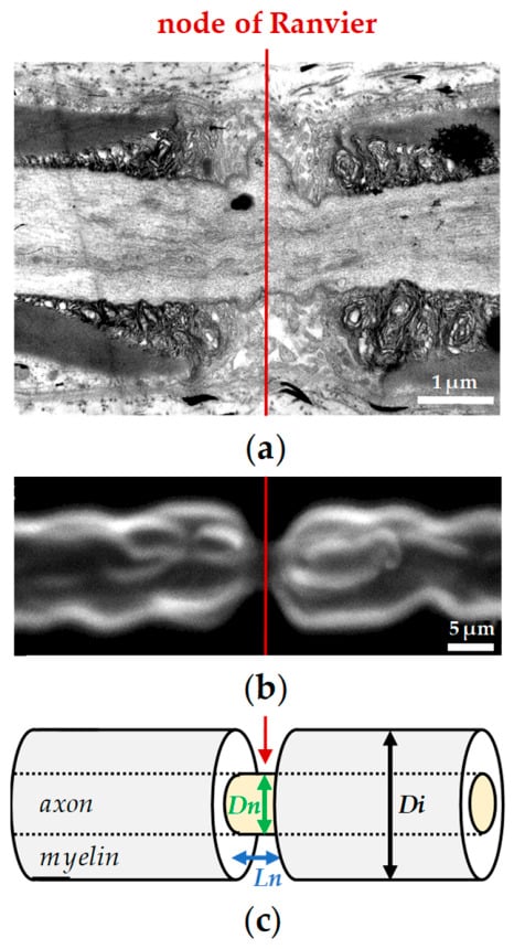
Guillain-Barré syndrome is an autoimmune attack on Schwann cells and myelin: demyelination at the nodes produces conduction block, and the large A-alpha fibres are affected, so the patient is weak. Charcot-Marie-Tooth disease type 1 is a hereditary demyelinating neuropathy (PMP22 duplication) in which repeated demyelination and remyelination build onion bulbs. Both slow conduction on nerve conduction studies.
Nerve Injury Classification
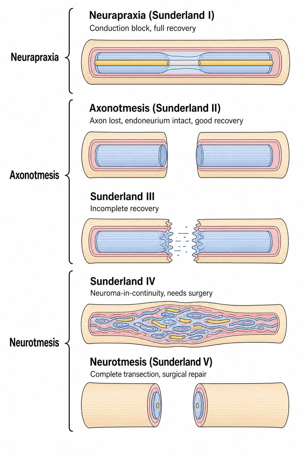
Two systems are in use and they map onto each other. Seddon's three grades are clinical, assigned from the mechanism and the examination, and guide the initial decision; Sunderland's five degrees are anatomical, defined by which sheaths are disrupted, and predict recovery and the need for surgery. Mackinnon added a sixth degree for the mixed injury.
Seddon classification
- Pathology
- Conduction block, myelin injury, axon intact
- Recovery
- Complete (100%)
- Time to Recovery
- Hours to 12 weeks
- Treatment
- Observation, expectant
- Pathology
- Axon disrupted, endoneurium intact
- Recovery
- Good to excellent (80-90%)
- Time to Recovery
- Months (1mm/day)
- Treatment
- Observation, may need neurolysis
- Pathology
- Complete nerve transection
- Recovery
- None without surgery
- Time to Recovery
- No recovery
- Treatment
- Surgical repair required
Neuropraxia. Compression, ischaemia or mild traction damages the myelin focally while the axon remains in continuity. There is a conduction block at the injury with normal conduction distal to it, and the Schwann cells remyelinate the segment over days to weeks. Saturday night palsy, a radial nerve compression, is the standard example.
Axonotmesis. A severe crush or traction tears the axon but leaves the sheath intact, so Wallerian degeneration follows distal to the injury while the endoneurium is preserved. Distal conduction is lost by 3-5 days, and recovery is by axonal regeneration at 1-3 mm/day through the intact endoneurial tubes: good, provided the target is not so distant that the muscle atrophies before the axons arrive. The closed humeral shaft fracture with a radial nerve palsy is the example.
Neurotmesis. A laceration, severe traction or high-energy trauma disrupts axons and connective tissue sheaths together. There is no recovery without surgical repair, primary or secondary, and even after repair recovery is incomplete because axons are misdirected and target muscles atrophy. An open fracture with nerve transection, or an iatrogenic nerve laceration, is the example.
Seddon's classification is clinical, made from the examination and the mechanism, not from histology. After a closed injury, distinguish the neuropraxia to be observed from the neurotmesis to be explored with serial examination, electromyography (denervation changes appear at 2-3 weeks) and Tinel's sign: advancing means axonotmesis, non-advancing means possible neurotmesis.
Sunderland classification
- Injury Level
- Myelin only (neuropraxia)
- Recovery Potential
- Complete
- Surgical Implication
- No surgery
- Injury Level
- Axon + myelin (endoneurium intact)
- Recovery Potential
- Excellent (90%)
- Surgical Implication
- Observation, rarely neurolysis
- Injury Level
- Axon + endoneurium (perineurium intact)
- Recovery Potential
- Variable (50-80%)
- Surgical Implication
- May need neurolysis
- Injury Level
- All but epineurium (perineurium disrupted)
- Recovery Potential
- Poor (less than 25%)
- Surgical Implication
- Needs surgical repair
- Injury Level
- Complete transection
- Recovery Potential
- None
- Surgical Implication
- Requires surgical repair
Grades I and II. Grade I is Seddon's neuropraxia: a conduction block with the axon intact and complete recovery. Grade II is the best case of axonotmesis: the axon is disrupted but the endoneurium intact, so the axons regenerate through their own tubes to the correct targets with minimal misdirection and excellent recovery.
Grade III. The axon and the endoneurium are disrupted but the perineurium is intact, so the nerve appears in continuity and can mislead. Intrafascicular scarring disrupts the endoneurial tubes; some axons reach their targets and others form neuromas, so recovery is variable, and the nerve may benefit from neurolysis to remove the scar, or from grafting.
Grades IV and V. In grade IV only the epineurium is intact: severe intraneural scarring produces a neuroma in continuity, the misdirected axons recover poorly without surgery, and the segment needs resection and nerve grafting. Grade V is complete transection of every layer, with no recovery without surgical repair: primary repair if the gap is under 2 cm, otherwise grafting.
Grade III is the hardest grade to diagnose and manage, because the nerve looks intact and the temptation is to observe, yet the endoneurial disruption prevents good recovery. Electrophysiology (no motor units at 3-4 months) and intra-operative nerve action potentials separate a grade III that needs resection and grafting from a grade II that will recover. This is why Sunderland added the grade: Seddon's axonotmesis contains both the good-prognosis grade II and the poor-prognosis grade III.
Mackinnon modification
Grade VI is the mixed injury, with different Sunderland grades in different fascicles. It is common in partial lacerations, stretch injuries and high-energy trauma: some fascicles are intact or grade I-II while others are transected, so on examination partial motor and sensory function is preserved. Management is selective repair of the injured fascicles while preserving the intact ones.
The surgical difficulty is telling intact from injured fascicles on the table. A nerve action potential is recorded by stimulating proximal to the injury and recording distal to it, and a fascicle that conducts one is intact; frozen section can assess fascicular injury histologically. The risk is that repairing an intact fascicle worsens the outcome by adding iatrogenic injury.
Do not assume every fascicle is injured in a partial laceration. Test for nerve action potentials across the injury and repair only the fascicles without one; preserving the intact fascicles prevents iatrogenic injury and keeps whatever function remains.
Wallerian Degeneration and Regeneration
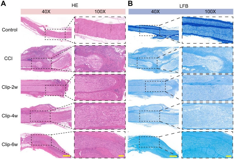
Wallerian degeneration
Wallerian Degeneration Timeline
Axon severed. Distal segment sealed. Proximal segment retracts. Initial Ca2+ influx triggers calpain activation and cytoskeletal breakdown in the distal axon.
Distal axon fragments (granular disintegration). Myelin breaks down into ovoids. Schwann cells detect the injury (lose axonal contact, upregulate the c-Jun transcription factor). Macrophages are recruited to the injury site.
Schwann cells phagocytose myelin debris (lipid-laden Schwann cells). Macrophages infiltrate and clear the remaining debris. Endoneurial tubes persist (basement membrane intact).
Schwann cells proliferate rapidly and form Bands of Büngner (columns of Schwann cells within the endoneurial tubes). They upregulate neurotrophic factors (NGF, BDNF, GDNF) and express adhesion molecules (N-CAM, L1) to guide regenerating axons.
Debris clearance complete. Endoneurial tubes contain Schwann cell columns ready to support regeneration. If no regenerating axon arrives, the Schwann cells eventually atrophy and the tubes collapse (after 12-18 months).
The molecular programme. Calcium influx activates calpains, the proteases that break the axon down, and the ubiquitin-proteasome system degrades the cytoskeletal proteins, neurofilaments and tubulin. The Schwann cells dedifferentiate, losing the myelin phenotype for a repair phenotype under the control of c-Jun, the master transcription factor of the repair programme, and their neurotrophic factors (NGF, BDNF, GDNF) support the regenerating axons.
What the clinician sees. The distal axon goes on conducting for 24-48 hours after the injury, which is useful for intra-operative nerve stimulation. Denervation changes on EMG, fibrillation potentials and positive sharp waves, appear at 2-3 weeks: the time it takes for Wallerian degeneration to complete and for the muscle fibre membrane to become unstable as acetylcholine receptors are upregulated. Do not expect EMG changes in the first week.
Axonal regeneration
The proximal stump. The neuron's response to losing its axon runs in sequence:
- Chromatolysis (24-48 hours): the cell body swells, the Nissl substance disperses and the nucleus moves to the periphery as protein synthesis increases
- Gene expression changes: growth-associated proteins (GAP-43), tubulin and actin are upregulated
- Growth cone formation: the axon tip forms a motile growth cone with filopodia
- Sprouting: multiple sprouts, 5-50 per axon, emerge from the proximal stump
Finding the way. The growth cone follows a chemotactic gradient of neurotrophic factors (NGF, BDNF), and by contact guidance follows the Schwann cells of the Bands of Büngner within the endoneurial tubes, adhering to N-CAM, L1 and laminin on their surface. Guidance is partially selective: motor axons prefer motor pathways and sensory axons sensory pathways.
The rate. Axons advance at 1-3 mm/day, on average 1 mm/day clinically, after an initial delay of 3-4 weeks for growth cone formation and crossing the scar at the repair site. Percussion over the regenerating axons produces tingling, Tinel's sign, which tracks the advancing front; over long distances, brachial plexus to hand, regeneration may take 12-18 months.
The regenerated fibre is not the original. Schwann cells remyelinate the regenerated axons, but the internodes are shorter than before (10-20% of normal) and the myelin thinner, so conduction velocity recovers to only 60-80% of the original.
Early repair (within 3 months) beats delayed repair because (1) Schwann cell support is optimal, (2) the muscle endplates remain receptive for 12-18 months, (3) the target organs are closer, with less distance to regenerate, and (4) there is less scar. Delayed repair beyond 12-18 months has poor outcomes because of muscle atrophy, endplate loss and endoneurial tube collapse.
Factors affecting recovery
Patient factors. Children regenerate better than adults, with a faster rate and more plasticity. Diabetes impairs regeneration through microangiopathy and neuropathy, nicotine reduces blood flow to the nerve, and vitamin deficiencies (B12, folate) impair regeneration.
Injury factors. A clean laceration does better than a crush or avulsion, a pure sensory nerve better than a pure motor one and a motor nerve better than a mixed one, and an associated vascular injury or soft-tissue loss worsens the prognosis. Distal injuries do better than proximal ones, for the reasons in the pearl below.
Surgical factors. Early repair, within 3 months, beats delayed repair. The repair must be tension-free, because tension impairs blood flow and regeneration; precise fascicular alignment reduces misdirection; and a direct repair beats a graft, as an autograft beats a conduit.
Biology. Short distances (digital nerves) recover excellently and long distances (brachial plexus) give poor motor recovery; motor endplates degenerate irreversibly after 12-18 months of denervation. Axons may reach the wrong targets, motor to sensory and vice versa, and axons blocked by scar form painful neuromas.
- Good Prognosis
- Child (less than 10 years)
- Poor Prognosis
- Adult (greater than 40 years)
- Good Prognosis
- Sharp laceration
- Poor Prognosis
- Crush, avulsion, traction
- Good Prognosis
- Distal (digital nerve)
- Poor Prognosis
- Proximal (brachial plexus)
- Good Prognosis
- Early repair (less than 3 months)
- Poor Prognosis
- Delayed repair (greater than 12 months)
- Good Prognosis
- Pure sensory
- Poor Prognosis
- Mixed motor-sensory
Distal injuries (the median nerve at the wrist) recover excellently because the regeneration distance is short (the hand muscles are 5-10 cm away), the muscle has less time to atrophy, and the fascicles are better organised, with motor and sensory segregated. Proximal injuries (a brachial plexus root avulsion) recover poorly because the distance may be 40-60 cm to the hand, the muscles atrophy before reinnervation, and the mixed fascicles cause misdirection. This is why nerve transfers, moving a distal donor nerve to a proximal target, have revolutionised brachial plexus surgery.
The painful neuroma is the clinical face of failed regeneration. It is a disorganised tangle of regenerating axons, Schwann cells and fibrous tissue that forms when regrowing axons fail to enter a distal endoneurial tube.
- End (terminal) neuroma - at the cut end of a transected nerve with no distal target: the classic painful amputation-stump neuroma
- Neuroma-in-continuity - a fusiform swelling within an injured but non-transected nerve (Sunderland III-IV) where intrafascicular scar blocks or misdirects axons: the lesion that needs intra-operative nerve action potentials to decide resect-and-graft versus neurolysis
Clinically it is a tender, often Tinel-positive nodule with shooting or burning pain in the nerve distribution, hypersensitivity and, in amputees, prosthesis intolerance.
- Primary prevention - a tension-free, well-aligned repair gives the axons a distal target; at amputation, traction neurectomy lets the cut end retract into healthy soft tissue away from the wound and the weight-bearing surfaces
- Established treatment - excise the neuroma and transpose or bury the proximal stump into muscle or bone, away from scar and load
- Modern reconstruction - targeted muscle reinnervation (TMR) coapts the cut nerve to a nearby motor nerve or muscle so the axons have somewhere to go, and the regenerative peripheral nerve interface (RPNI) wraps the nerve end in a free muscle graft; both markedly reduce neuroma and phantom or residual-limb pain and are now standard in amputation surgery
Differential Diagnosis of Acute Limb Weakness or Sensory Loss
- Distribution / Pattern
- Single named-nerve territory (e.g. wrist drop with radial palsy)
- Key Distinguishing Feature
- Maps to one peripheral nerve; mechanism (fracture, laceration, compression)
- Confirmatory Test
- Nerve conduction studies / EMG; advancing Tinel's sign
- Distribution / Pattern
- Dermatomal/myotomal (single root)
- Key Distinguishing Feature
- Neck pain, radiation in a root pattern, positive Spurling test; reflex loss for that root
- Confirmatory Test
- MRI cervical spine; EMG showing paraspinal involvement
- Distribution / Pattern
- Multiple nerves / whole limb, may be pan-plexal
- Key Distinguishing Feature
- High-energy traction (motorcycle); Horner sign suggests root avulsion
- Confirmatory Test
- MRI / CT myelography; EMG; clinical pattern
- Distribution / Pattern
- Regional, pain out of proportion, passive-stretch pain
- Key Distinguishing Feature
- Tense compartment, pain dominates early; sensory then motor loss late
- Confirmatory Test
- Compartment pressure measurement (clinical diagnosis)
- Distribution / Pattern
- Whole limb distal to occlusion
- Key Distinguishing Feature
- Pulseless, pale, cold, paraesthesia and paralysis (the 6 Ps)
- Confirmatory Test
- Doppler / angiography
- Distribution / Pattern
- Hemibody, often face involved, upper motor neuron signs
- Key Distinguishing Feature
- Upper motor neuron pattern, hyperreflexia, no peripheral nerve mapping
- Confirmatory Test
- CT/MRI brain
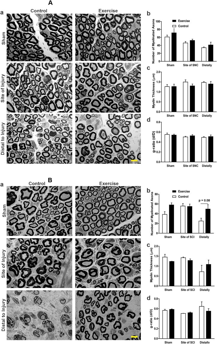
A focal peripheral nerve lesion maps to a single named-nerve territory and is confirmed electrodiagnostically; the critical step is to exclude compartment syndrome and acute arterial ischaemia, which are limb-threatening and demand emergency intervention rather than expectant nerve management.
Management Algorithm
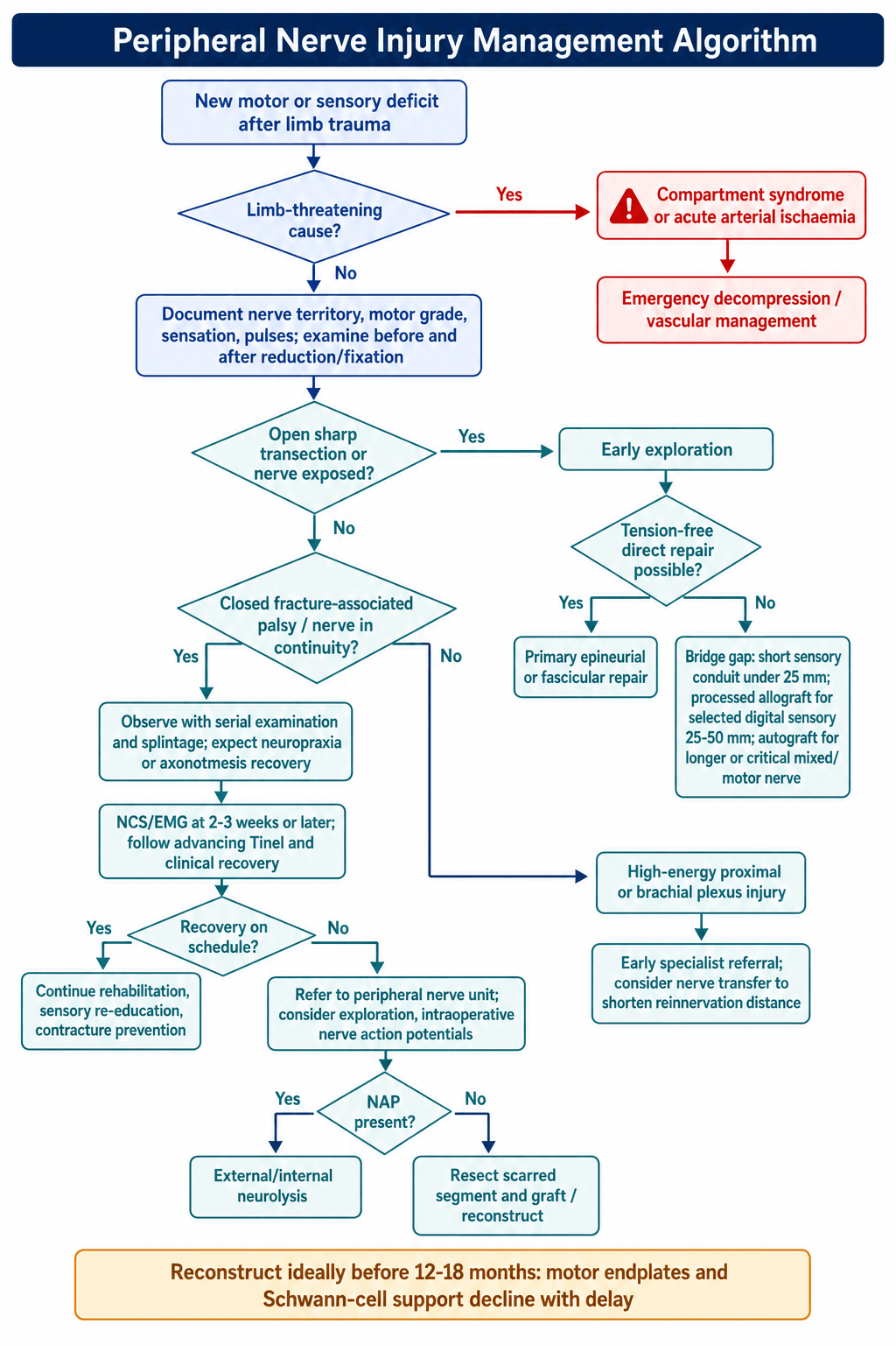
Guidelines, Registries & Global Practice
Global Epidemiology
- Peripheral nerve injury (PNI) is an uncommon complication of limb trauma. In the European TraumaRegister DGU cohort of 49,382 severely injured patients with upper-extremity involvement, an associated nerve injury was present in approximately 3.3%.
- PNI clusters in young males (mean age approximately 40 years, around 79% male) and after high-energy mechanisms, with motorcycle accidents the single most common cause in that registry.
- Typical concomitant lesions are humeral (approximately 37%) and ulnar (approximately 20%) fractures, vascular laceration, and extensive soft-tissue damage; PNI roughly doubles inpatient rehabilitation need and lengthens hospital stay.
- In penetrating and ballistic trauma, nerve deficits are common (around 30% of upper-extremity gunshot injuries in one Level 1 trauma series) but are frequently neuropraxic or axonotmetic, with more than half improving spontaneously, supporting initial expectant management of nerve-in-continuity ballistic injuries.
- In limited-resource and conflict settings, blast and gunshot mechanisms predominate and the ulnar nerve is the most frequently injured peripheral nerve.
Classification and Decision Frameworks (Side by Side)
- What it standardises
- Severity grading of nerve injury
- Key recommendation
- Use Seddon for clinical/initial decisions; Sunderland five-degree (plus Mackinnon degree VI mixed) to predict recovery and need for surgery
- Evidence basis
- Foundational anatomical-pathological description; widely adopted worldwide
- What it standardises
- Diagnostic and repair principles
- Key recommendation
- Tension-free coaptation; early exploration of sharp/open transections; autograft or processed allograft for gaps not amenable to direct repair (conduits reserved for short sensory gaps under 25 mm)
- Evidence basis
- Expert consensus and cohort/registry data (e.g. RANGER)
- What it standardises
- Open injuries and nerve repair pathways
- Key recommendation
- Repair clean transections primarily within recommended timeframes; refer complex/closed lesions to specialist nerve units
- Evidence basis
- Standards for trauma and society guidance
- What it standardises
- Nerve in the context of fracture care
- Key recommendation
- Document neurology before and after reduction/fixation; observe most closed fracture-associated palsies; explore if iatrogenic or no recovery by expected window
- Evidence basis
- Educational consensus, observational evidence
- What it standardises
- Brachial plexus and reconstruction
- Key recommendation
- Early referral for traumatic plexus injury; favour nerve transfers for proximal injuries to shorten reinnervation distance
- Evidence basis
- Consensus plus cohort outcome data
Evidence-Based Reconstruction Principles (Global Consensus)
- Timing: clean, sharp transections are repaired primarily and early; closed fracture-associated palsies (e.g. radial nerve palsy with humeral shaft fracture) are usually observed because most are neuropraxia or axonotmesis that recover. Reconstruction should ideally occur within the 12-18 month window before motor endplates and repair Schwann cells degenerate.
- Repair ladder by gap: direct tension-free coaptation is preferred; hollow conduits are reserved for short sensory gaps (typically under 25 mm) in low-demand nerves; processed nerve allograft has registry support for digital nerve gaps of 25-50 mm (around 86% meaningful sensory recovery); autograft (sural nerve) remains the reference standard for longer or critical motor/mixed nerves.
- Nerve transfers (e.g. double fascicular Oberlin-type transfer for elbow flexion, spinal accessory to suprascapular nerve) shorten the distance from coaptation to target muscle and have improved outcomes in proximal and brachial plexus injuries.
- Electrodiagnostics: nerve conduction studies and needle EMG are timed at 2-3 weeks or later after injury, when denervation changes (fibrillations, positive sharp waves) appear; intraoperative nerve action potentials help decide between neurolysis and resection-grafting for a nerve-in-continuity.
Practice Variation and Resource Setting
- High-resource settings: ready access to microsurgery, intraoperative neuromonitoring, processed allograft and conduits, and specialist peripheral-nerve and brachial-plexus units; increasing use of nerve transfers.
- Limited-resource and conflict settings: predominance of blast/gunshot injuries, delayed presentation, reliance on autograft and tendon transfers rather than commercial conduits/allograft, and emphasis on rehabilitation and protective sensory retraining.
- Across all settings, hand therapy and structured rehabilitation (sensory re-education, prevention of contracture, protective sensation education) are integral to functional outcome.
Exam Relevance (All Boards)
- Nerve anatomy and physiology is a core basic-science topic for fellowship candidates worldwide.
- Examiners commonly test the three-layer architecture, the ionic basis of the action potential and saltatory conduction, and the Seddon/Sunderland classifications with their clinical implications.
- The highest-yield distinction is Sunderland degree II (good prognosis) versus degree III (poor prognosis) in a nerve that appears in continuity, and the use of intraoperative nerve action potentials to resolve it.
MCQ Practice Points
Q: What are the five grades of the Sunderland nerve injury classification?
A: Grade I (neurapraxia): Local conduction block, myelin injury, full recovery weeks. Grade II (axonotmesis): Axon damage, endoneurium intact, full recovery months. Grade III: Endoneurium damaged. Grade IV: Perineurium damaged. Grade V (neurotmesis): Complete transection. Grades III-V require surgical intervention.
Q: What is the rate of nerve regeneration after injury?
A: Peripheral nerves regenerate at approximately 1mm/day or 1 inch/month. This guides timing expectations for motor recovery. Sunderland Grade II injuries recover at this rate once Wallerian degeneration completes (~3 weeks). More proximal injuries take longer due to greater distance to end organs.
Q: What is Wallerian degeneration?
A: Wallerian degeneration is the organized process of distal nerve segment breakdown following axonal injury. Begins within 24-48 hours, completes by 3 weeks. Involves axon fragmentation, myelin breakdown, and Schwann cell proliferation forming Bands of Büngner to guide regenerating axons. Essential for successful regeneration.
Q: What are the structural layers of a peripheral nerve from inside to outside?
A: From inside out: Axon (nerve fiber), Endoneurium (surrounds individual axons), Perineurium (surrounds fascicles - creates blood-nerve barrier), Epineurium (outermost layer surrounding nerve trunk). The internal epineurium fills space between fascicles. Understanding crucial for nerve repair technique.
Q: What determines nerve conduction velocity?
A: Myelination and axon diameter are primary determinants. Large myelinated fibers (Aα) conduct at 70-120 m/s (motor, proprioception). Small unmyelinated C fibers conduct at 0.5-2 m/s (pain, temperature). Saltatory conduction between nodes of Ranvier enables rapid transmission in myelinated fibers.
Basic Science Viva Scenarios
Practise clinical reasoning and management decisions out loud
“Describe the anatomical structure of a peripheral nerve. What are the three connective tissue layers and their functions?”
“Explain the generation and propagation of an action potential in a myelinated nerve fiber. What is saltatory conduction?”
“A patient has a closed humeral shaft fracture with radial nerve palsy noted immediately after injury. Classify nerve injuries and describe the process of Wallerian degeneration.”
Three-Layer Structure
- Epineurium: outer loose connective tissue, vasa nervorum, 30-75% cross-section, mechanical protection
- Perineurium: 7-15 cell layers with tight junctions, blood-nerve barrier, maintains ionic environment
- Endoneurium: collagen tubes around axons within fascicles, forms Bands of Büngner
- Endoneurial tubes critical: intact (Sunderland I-II) = good recovery; endoneurium disrupted (III) = variable recovery; perineurium disrupted (IV) = poor recovery
Nerve Fiber Classification
- A-alpha: largest (12-20 μm), fastest (70-120 m/s), motor and proprioception
- A-beta: medium (6-12 μm), 35-75 m/s, touch and vibration
- A-delta: small (1-5 μm), 5-30 m/s, sharp pain and temperature
- C fibers: smallest (0.2-1.5 μm), slowest (0.5-2 m/s), dull pain (unmyelinated)
- Large fibers most vulnerable to compression; small fibers most resistant
Action Potential
- Resting potential: -70 mV (Na+/K+-ATPase maintains gradients)
- Threshold: -55 mV triggers voltage-gated Na+ channels
- Depolarization: Na+ influx to +40 mV (0.5-1 ms)
- Repolarization: K+ efflux returns to -70 mV (1-2 ms)
- Absolute refractory: 0.5-1 ms (Na+ channels inactivated)
- Saltatory conduction: action potential jumps node to node (50x faster than continuous)
Seddon Classification
- Neuropraxia: conduction block, myelin injury, axon intact, full recovery (hours to 12 weeks)
- Axonotmesis: axon disrupted, endoneurium intact, good recovery at 1 mm/day
- Neurotmesis: complete transection, no recovery without surgery
Sunderland Classification
- Grade I: myelin only (neuropraxia), complete recovery
- Grade II: axon + myelin, endoneurium intact, excellent recovery (90%)
- Grade III: axon + endoneurium disrupted, perineurium intact, variable recovery (50-80%), may need neurolysis
- Grade IV: only epineurium intact, poor recovery (less than 25%), needs grafting
- Grade V: complete transection, requires surgical repair
- Mackinnon Grade VI: mixed injury (different grades in different fascicles)
Wallerian Degeneration
- Distal axon degenerates 24-48 hours after injury
- Schwann cells phagocytose debris, form Bands of Büngner (1-4 weeks)
- Bands of Büngner: Schwann cell columns guide regenerating axons
- Neurotrophic factors: NGF, BDNF, GDNF support regeneration
- EMG denervation changes (fibrillations) appear at 2-3 weeks
- Window for repair: 12-18 months (optimal Schwann support and muscle endplate viability)
Axonal Regeneration
- Regeneration rate: 1-3 mm/day (average 1 mm/day clinically)
- Initial delay: 3-4 weeks (growth cone formation, crossing scar)
- Tinel's sign: tracks advancing regeneration (percussion causes tingling)
- Factors for good recovery: young age, distal injury, sharp laceration, early repair (less than 3 months)
- Factors for poor recovery: proximal injury, crush/avulsion, delayed repair (greater than 12 months)
Evidence Base
Anatomy and Physiology of Nerve Injury (Sunderland's Five-Degree System)
- Microstructure of nerve trunks underpins a five-degree classification of injury severity, with partial and mixed types each having defined pathology and clinical features
- Tensile strength and capacity to resist traction reside in the fascicular (perineurial) tissue; the epineurium provides a cushion against compression
- Fascicular plexuses redistribute and mix branch fibres along the nerve, complicating fascicular matching at repair
- After transection, changes in axons, endoneurial tubes, fasciculi and the nerve trunk together determine the outcome of regeneration
Double Crush in Nerve Entrapment Syndromes
- Original description of the double crush hypothesis: a single axon compressed at two sites is more vulnerable than at either site alone
- Proposed that proximal impairment of axoplasmic flow renders distal segments susceptible to a second compression
- Frequently invoked for coexisting cervical radiculopathy and carpal tunnel syndrome
- Predicts that treating only one compression site may not fully resolve symptoms
Repair Schwann Cell, c-Jun and Bands of Büngner
- Nerve injury reprograms myelin and Remak Schwann cells into a dedicated repair phenotype that supports neuronal survival and axon regrowth
- Repair cells up-regulate trophic factors and cytokines, drive myelin clearance (autophagy and macrophage recruitment), and form Bands of Büngner to guide axons
- This repair programme is controlled transcriptionally by c-Jun, rapidly up-regulated after injury
- Without c-Jun, a dysfunctional repair cell forms, leading to neuronal death and failure of functional recovery
Double Nerve Transfer for Elbow Flexion (Oberlin Plus)
- Double fascicular transfer (ulnar fascicle to biceps motor branch plus median fascicle to brachialis branch) for upper-trunk brachial plexus palsy
- Grade 4 (MRC) elbow flexion restored in all 10 patients with adequate follow-up
- Patients could lift 1-5 kg with no secondary loss of grip strength or sensation
- Distal donor-to-target coaptation shortens reinnervation distance compared with proximal repair or grafting
Processed Nerve Allograft for Digital Nerve Gaps (RANGER Registry)
- Fifty digital nerve repairs with gaps of 25-50 mm (mean 35 mm) reconstructed with processed nerve allograft
- Meaningful recovery (MRC S3 or greater) achieved in 86% of repairs
- Results compared favourably with historical autograft recovery (reported 60-88%) without donor-site morbidity
- Processed allograft extends the gap range previously reserved for autograft, beyond the under-25 mm limit of hollow conduits
Peripheral Nerve Injury in Severe Upper-Limb Trauma (TraumaRegister DGU)
- Among 49,382 severely injured patients with upper-extremity involvement, 3.3% had an associated peripheral nerve injury
- Nerve-injured patients were younger (mean 40.6 versus 47.2 years) and more often male (78.6%)
- Motorcycle accidents were the single most common mechanism (32.5%); humeral (37.2%) and ulnar (20.3%) fractures were the typical concomitant lesions
- Nerve injury was associated with longer hospital stay (30.6 versus 24.2 days) and greater need for inpatient rehabilitation