Forestier Disease | Mechanical Dysphagia from Anterior Osteophytes
- DISH (Forestier's Disease) is a systemic condition causing 'flowing wax' calcification of the ALL.
- OALL is the cervical manifestation, which can mechanically compress the esophagus (Dysphagia) or Airway.
- The airway can be 'Difficult' to intubate due to osteophytes preventing visualization.
- Surgery (Osteophytectomy) is reserved for severe dysphagia or airway compromise.
- Unlike OPLL, OALL does NOT typically cause myelopathy directly.
- “Look for 'Candle Wax' dripping appearance on X-ray.
- “Ask about 'Aspiration Pneumonia' history in elderly patients with neck stiffness.
- “Differentiation from Ankylosing Spondylitis (AS): DISH spares the SI joints and facets.
- “Surgery should preserve the disc space (Osteophytectomy ONLY) unless instability exists.
At a Glance
| Feature | DISH (Forestier) | Ankylosing Spondylitis (AS) |
|---|---|---|
| Age Onset | Old (over 50) | Young (under 40) |
| SI Joints | Normal (Spared) | Fused (Sacroiliitis) |
| Disc Space | Preserved | Calcified/Narrowed |
| HLA-B27 | Normal Prevalence | Highly Associated (+) |
Mnemonics
DISHResnick Criteria for DISH
Hook:Diagnostic criteria.
DATESymptoms of OALL
Hook:Clinical presentation.
HORSESurgical Risks
Hook:Complications of osteophytectomy.
Overview and Epidemiology
Definition
- DISH (Forestier disease) is a systemic, non-inflammatory enthesopathy causing "flowing" ossification of spinal ligaments and peripheral entheses, sparing the disc and synovial joints.
- OALL (ossification of the anterior longitudinal ligament) is the cervical expression that grows anteriorly, compressing the pharynx, oesophagus and airway rather than the cord.
Key epidemiology (global)
- Radiographic prevalence rises with age: roughly 10 percent over 50 years and up to 25-30 percent over 70 years in autopsy and CT cohorts.
- Male predominance (about 2:1); rare under 40 years.
- Cervical osteophytes are most marked at C3-C6; symptomatic dysphagia affects a minority (often quoted under 20 percent of those with cervical DISH).
- Strongly linked to metabolic syndrome (type 2 diabetes, obesity, hypertension) — DISH is increasingly viewed as a metabolic bone phenotype, not simple "wear and tear".
- The rigid, brittle "bamboo-like" spine fractures easily after low-energy trauma (chalk-stick fractures) with a high rate of neurological injury.
Pathophysiology and Anatomy
The Anterior Longitudinal Ligament (ALL)
- Broad ligament covering the anterior vertebral bodies.
- Prevents hyperextension.
- In DISH, it ossifies but spares the annulus fibrosus and nucleus pulposus (unlike Ankylosing Spondylitis).
The Swallowing Mechanism
- Phase 1 (Oral): Bolus prep.
- Phase 2 (Pharyngeal): Pharynx constricts, Hyoid elevates, Epiglottis inverts.
- Phase 3 (Esophageal): Peristalsis. OALL disrupts Phase 2 by physically blocking epiglottic inversion or narrowing the pharyngeal space.
Classification Systems
Resnick Criteria (Gold Standard)
Used to diagnose DISH and distinguish from normal spondylosis.
- Flowing calcification along the anterolateral aspect of at least 4 contiguous vertebral bodies.
- Preservation of disc height in the involved segments, and absence of extensive radiographic changes of degenerative disc disease (vacuum sign, marginal sclerosis).
- Absence of apophyseal joint ankylosis or sacroiliac joint erosion/sclerosis/fusion.
Clinical Assessment
Presentation
- Dysphagia: Most common presenting symptom of cervical OALL (a minority of all cervical DISH). "Food sticking".
- Globus Sensation: Feeling of a lump in the throat.
- Dysphonia: Hoarseness (RLN compression or vocal cord edema).
- Stiffness: Decreased cervical ROM.
- Dyspnea: Rare, caused by laryngeal edema or massive C3 osteophyte compressing glottis.
Examination
- Palpation: Hard, bony mass palpable in the posterior pharynx (beware gag reflex).
- Neck: Stiffness, loss of extension.
- Neuro: Usually normal (OALL grows OUT, not IN to the canal).
Imaging and Investigations
Workup Protocol
- "Flowing Candle Wax" appearance anterior to bodies.
- Radiolucent line: Between the ossified ALL and the vertebral body (unossified deep layer).
- Check disc heights (spared).
- Video Fluoroscopic Swallowing Study (VFSS).
- Critical to demonstrate the mechanical cause of dysphagia.
- Shows the bolus hitting the osteophyte and spiraling or causing aspiration.
- CT: Defines bony anatomy for resection.
- ENT Nasendoscopy: Mandatory to rule out intrinsic malignancy (cancer) or vocal cord palsy BEFORE surgery.
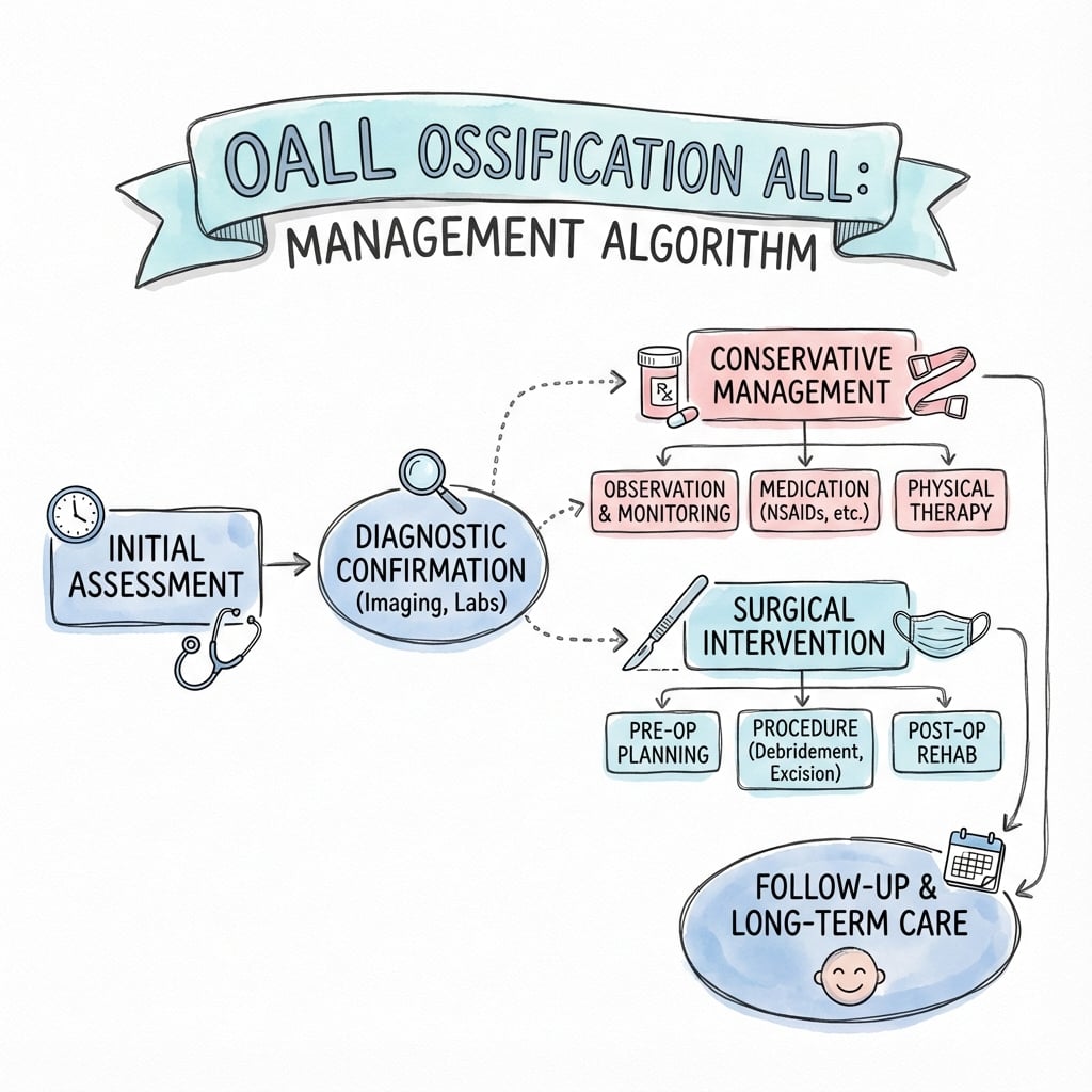
Management Algorithm
Non-Operative Management
First Line Treatment
- Dietary Modification: Soft foods, thicken fluids (Speech Pathology involvement).
- NSAIDs: Reduce soft tissue inflammation around the osteophytes.
- Steroids: Short course for acute flare of dysphagia.
- Review: Monitor weight and aspiration signs.
Surgical Technique
Anterior Cervical Osteophytectomy
- Goal: Resect the anterior bony mass to decompress the esophagus.
- Position: Supine, neck slightly extended (careful! hyperextension can fracture the fused spine). Mayfield head clamp or horseshoe.
- Approach: Standard Smith-Robinson (Anterior Cervical).
Step-by-Step:
- Incision: Transverse skin incision at the level of the osteophyte (fluoroscopy to confirm).
- Dissection: Deep dissection medial to the Carotid sheath and lateral to the Trachea/Esophagus.
- Exposure:
- The prevertebral fascia will be tight over the mass.
- Identification of the "valley" between the osteophyte and the disc space is crucial.
- Adhesions: The esophagus is often adherent to the tip of the osteophytes due to chronic inflammation. Use a peanut dissector or wet Raytec to gently peel.
- Resection:
- Use a high speed Diamond Burr or Leksell Rongeur.
- Resect the "Peaks" (Over the vertebral bodies).
- Be careful at the "Valleys" (Over the discs) not to violate the annulus.
- Limits: Resect flush with the anterior margin of the vertebral body. Do not chase lateral osteophytes near the Foramen Transversarium (Vertebral Artery risk).
- Disc Sparing: Do not enter the disc space.
- Bone Wax: Apply bone wax to the bleeding cancellous surface.
- Closure: Deep drain is essential. Close Platysma and Skin.
Fusion?
- Generally NOT indicated unless there is pre-existing instability.
- Fusion increases adjacent segment disease.
Complications
| Complication | Risk Level | Management |
|---|---|---|
| Recurrent Laryngeal Nerve Palsy | High | Retraction injury. Observe. Vocal cord medialization if permanent. |
| Esophageal Perforation | High (Adhesions) | Direct repair + muscle flap. NGT feeding. Antibiotics. |
| Regrowth | Moderate (years) | Use bone wax. Ensure complete resection. |
| Hematoma | Moderate | Place drain. Watch airway. |
Radiographic osteophyte regrowth is well recognised because the underlying metabolic driver persists, but symptomatic recurrence requiring re-operation is uncommon — about 4 percent in the largest pooled surgical series at a mean follow-up of nearly 4 years. Counsel patients that regrowth is slow and most stay symptom-free.
Postoperative Care
- Airway: Monitor for retropharyngeal hematoma / edema (Stridor).
- Feeding: Nasogastric feeding may be needed if esophageal repair done or severe edema.
- Swallow Study: Repeat contrast swallow before oral intake if dura/esophagus concerns.
Outcomes and Prognosis
- Success Rate: High for dysphagia resolution — improvement in about 95 percent of operated patients in the largest pooled series.
- Aspiration: May not resolve if the cause was neurological or permanent muscle damage.
- QoL: Significant improvement in eating ability.
Evidence Base
Radiographic & pathological criteria for DISH
- Evaluated 215 cadaveric spines and 100 patients with the disease.
- Defined the radiographic hallmark: flowing anterolateral ossification with a subjacent radiolucent line and preserved disc height.
- Established the three-part criteria distinguishing DISH from ankylosing spondylitis and degenerative spondylosis.
Surgical outcomes for cervical DISH dysphagia/airway obstruction
- 138 articles describing 419 patients (mean age 67 years, 85% male).
- Surgery (most often anterolateral approach) in 66 percent; dysphagia improved in 95.5 percent of operated patients.
- Total post-operative complication rate 22.1 percent (12.7 percent within 1 month).
- Dysphagia recurred in only 4 percent (12 patients) at mean 3.7-year follow-up.
Airway management in OALL of the cervical spine
- Two OALL patients with unexpectedly difficult tracheal intubation from anterior bridging osteophytes.
- Osteophytes displace the posterior hypopharyngeal wall, defeating both direct and fiberoptic-assisted awake intubation.
- OALL found in roughly 12 percent of autopsies and progresses with age.
DISH, metabolic disease and bone quality (Camargo cohort)
- 1545 postmenopausal women; DISH prevalence 8.2 percent.
- DISH associated with significantly higher obesity, metabolic syndrome, hypertension and type 2 diabetes.
- Despite higher lumbar BMD, DISH carried lower trabecular bone score and more vertebral fractures (28.6 vs 15.1 percent).
Vertebral fracture prevalence in DISH vs AS
- 7 DISH studies (n=1193): pooled vertebral fracture prevalence 22.6 percent (95% CI 13.4-33.4).
- 26 AS studies (n=2875): pooled prevalence 15.2 percent.
- DISH fractures cluster at the thoracolumbar junction; AS at the mid-thoracic spine.
CT vs MRI in ankylosing spine trauma
- 124 DISH/AS trauma patients imaged with both CT and MRI.
- MRI revealed additional injuries in 4.8 percent and changed management in 3.2 percent — mostly disco-ligamentous hyperextension injuries.
- Authors advise selective MRI for non-ankylosed levels or neurological deficit.
Clinical Decision Scenarios
Practise clinical reasoning and management decisions out loud
“75M presents with 6 months of progressive difficulty swallowing solids. He has lost 5kg. He has a history of 'stiff neck'. Lateral X-ray shows massive anterior bone formation C3-C6. What is the diagnosis and workup?”
“You are called to ED for a DISH patient who has fallen and hit his head. GCS 8. The ED registrar cannot intubate. Why?”
“During osteophytectomy, you notice a Bubble in the wound field. What has happened and what do you do?”
MCQ Practice Points
Q: Which feature distinguishes DISH from Ankylosing Spondylitis? A: Preservation of SI joints and Facet joints (No ankylosis).
Q: What structure ossifies in DISH? A: Anterior Longitudinal Ligament (ALL).
Q: Post-operative hoarseness after osteophytectomy is most likely due to? A: Recurrent Laryngeal Nerve (RLN) neurapraxia (from retraction).
Q: DISH is strongly associated with which metabolic disorder? A: Type 2 Diabetes Mellitus / Obesity.
Q: When is fusion indicated in OALL surgery? A: Only if instability is present. (E.g. fracture or iatrogenic disc violation). Routine fusion is unnecessary.
Guidelines, Registries & Global Practice
Global epidemiology
- Radiographic DISH is common worldwide in older adults: roughly 10 percent over 50 and 25-30 percent over 70, with consistent male predominance across European, North American and Asian CT/autopsy cohorts.
- Population studies repeatedly tie DISH to metabolic syndrome, so prevalence tracks with regional obesity and type 2 diabetes burden.
- Symptomatic cervical OALL (dysphagia or airway obstruction) remains uncommon, and the published surgical literature is dominated by case reports and small series — no randomised data exist.
| Body | Relevant stance |
|---|---|
| AO Spine | Treats the ankylosed (DISH/AS) spine as a high-risk fracture pattern; low-energy trauma needs full-length CT and a high index of suspicion for unstable three-column injury. |
| NICE / BOA (UK) | No DISH-specific guidance; managed under general dysphagia (2-week-wait cancer pathway to exclude malignancy first) and spinal trauma standards. |
| AAOS / NASS (US) | No dedicated guideline; surgical decompression reserved for refractory mechanical dysphagia or airway compromise after ENT/swallow workup. |
| Difficult Airway Society / ASA | Cervical OALL is a recognised predictor of difficult intubation — plan awake fiberoptic technique and avoid forced hyperextension. |
Registry & practice variation
- There is no implant registry relevant to OALL because osteophytectomy is usually fusion-free; registry data (NJR, AOANJRR, AJRR) only apply if an instrumented fusion is added for instability or a chalk-stick fracture.
- Well-resourced settings: video fluoroscopic swallow study, CT planning, ENT nasendoscopy and awake fiberoptic intubation are standard before surgery.
- Limited-resource settings: diagnosis often rests on plain lateral radiographs and barium swallow; dietary modification and speech-pathology input carry most of the load, with surgery reserved for severe weight loss or aspiration.
Controversies & Areas of Uncertainty
- No high-level evidence: the entire surgical literature is case reports and small series; there are no randomised trials or formal guidelines, so recommendations are consensus-based (Harlianto/Verlaan systematic review).
- Fusion vs osteophytectomy alone: most authors avoid routine fusion to spare adjacent-segment disease and operative time, reserving it for instability or iatrogenic disc violation — but the threshold is not standardised.
- Defining "mechanical" dysphagia: dysphagia in elderly DISH patients is often multifactorial (neurological, presbyphagia, reflux). A positive swallow study does not guarantee the osteophyte is the sole cause, and a subset have persistent symptoms after technically successful surgery.
- Predicting recurrence: radiographic regrowth is common yet symptomatic recurrence is rare (~4 percent); no reliable predictor or proven preventive (bisphosphonates, metabolic control) exists.
- Imaging in trauma: whether every ankylosed-spine trauma patient needs MRI in addition to CT remains debated — current data suggest selective use for neurological deficit or suspected disco-ligamentous injury.
Exam Day Cheat Sheet
Key Concepts
- Flowing Anterolateral Ossification
- Disc Height Preserved
- SI Joints Normal
- Dysphagia over Dyspnea
Criteria (Resnick)
- 4+ Contiguous bodies
- Discs spared
- No Facet/SI fusion
- Flowing Candles
Surgery
- Indication: Severe Dysphagia/Wt Loss
- Procedure: Anterior Osteophytectomy
- Protect Esophagus!
- No Fusion usually
Risks
- Esophageal Perforation
- RLN Palsy
- Recurrence
- Hematoma (Airway)
Image Manifest
- [3-preoperative-computed-tomography-ct-a-ct-of-the-ce.png]: Sagittal CT showing OALL and disc preservation
- [5-preoperative-esophagram-revealed-extrinsic-compres.png]: Barium swallow showing esophageal compression