Direct Bone-Implant Contact | Surface-Dependent | Biomechanical Fixation | Time-Dependent Process
- Osseointegration = direct structural and functional connection between bone and implant surface
- Titanium is gold standard due to oxide layer and biocompatibility
- Surface roughness (1-10 micrometers) enhances bone apposition and integration
- Primary stability (press-fit) essential for successful secondary biological fixation
- Micromotion greater than 150 micrometers inhibits osseointegration and promotes fibrous tissue
- “Distance osteogenesis (bone grows from host bed) requires gap less than 500 micrometers
- “Contact osteogenesis (bone forms on implant) requires rough surface and biocompatible material
- “Porous coatings allow bone ingrowth (50-400 micrometer pores optimal)
- “Hydroxyapatite coating accelerates early integration but may degrade over time
Overview and Definition
Definition. Brånemark defined osseointegration as the "direct structural and functional connection between ordered, living bone and the surface of a load-carrying implant", with no intervening fibrous tissue. At the ultrastructural level that means bone mineral in contact with the titanium oxide layer; clinically it means stable, non-mobile fixation that can withstand functional loading.
History. Per-Ingvar Brånemark first described the phenomenon in 1952, when he observed titanium chambers becoming permanently incorporated into rabbit bone. He coined the term in 1981 and applied the principle to dental implants, which revolutionised implant dentistry and later orthopaedic surgery.
What it needs. A biocompatible material, stable fixation and appropriate healing time. Titanium is the gold standard material because of its oxide layer and biocompatibility; tantalum and hydroxyapatite are also biocompatible. Fixation is mechanical at first, a press-fit at surgery, and biological later, as bone forms on and around the implant. Whether bone or fibrous tissue ends up at the interface is determined by primary stability and the control of micromotion during healing.
Why it matters. Osseointegration gives durable biological fixation to joint replacements, dental implants and bone-anchored prostheses, and it provides stable, load-bearing fixation for decades. Direct bone contact allows physiological load transfer without the interface breaking down. Fibrous fixation is mechanically weak and painful under load and loosens progressively under cyclic loading, so an implant that fails to osseointegrate ends in aseptic loosening and revision surgery.
Osseointegration specifically refers to bone-implant contact. Biointegration is a broader term that includes soft-tissue integration (e.g. tendon-bone, ligament-bone). In exams, use "osseointegration" for bone-implant interfaces and be specific about the tissue type involved.
Anatomy of the Bone-Implant Interface
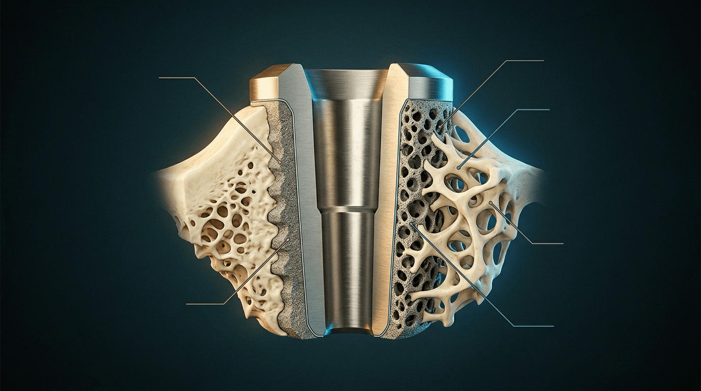
From the implant surface outward. In successful osseointegration, bone mineral is in direct contact with the oxide layer at the molecular level:
- Titanium oxide (TiO₂) - a 2-10 nm passivation layer
- Proteoglycan layer - 20-50 nm of adsorbed proteins, including fibronectin and vitronectin
- Mineralised bone - directly apposed, with no fibrous tissue
- Osteocyte network - canaliculi connecting to the implant surface
The oxide layer is what bone contacts. Titanium forms its TiO₂ layer spontaneously, within milliseconds, in air and in body fluids. The layer is 2-10 nm thick, amorphous or crystalline, highly biocompatible and osteoconductive. It is chemically inert and not cytotoxic, prevents corrosion and metal-ion release, and allows direct bone apposition with minimal inflammatory response and no foreign-body rejection.
Its surface chemistry. The oxide surface carries hydroxyl groups (-OH), protein adsorption sites and calcium phosphate precipitation sites. It is negatively charged, which attracts proteins and cells, and its low surface energy keeps the foreign-body response low.
If the titanium oxide layer is damaged (e.g. by scratching during surgery), it regenerates within milliseconds when exposed to oxygen or water. This "self-healing" property maintains biocompatibility even after surface damage. The oxide layer is not a coating that can be worn off - it is an intrinsic property of titanium in oxidising environments.
The protein layer. Before any cell arrives, proteins from blood and tissue fluid coat the oxide within seconds: fibronectin (cell adhesion), vitronectin (osteoblast binding), albumin (a passivating protein) and complement proteins. This conditioning layer, not the bare metal, is what osteoblasts actually "see". Cells attach to it in sequence:
- Proteins adsorb to the TiO₂ surface
- Osteoblast integrins bind the adsorbed proteins
- The osteoblast adheres and spreads, and adhesion triggers osteogenic differentiation signals
- Matrix is deposited and mineralises
The Vroman effect. The composition of the protein layer changes over time through competitive, sequential adsorption. The most abundant and highly mobile proteins, such as albumin and fibrinogen, reach the surface fastest and adsorb first. They are progressively displaced by less abundant but higher-affinity proteins, such as high-molecular-weight kininogen and the cell-adhesive proteins fibronectin and vitronectin, which bind the surface more tightly and are adsorbed to titanium within minutes.
Why the sequence matters. Osteoblast integrins recognise fibronectin and vitronectin in the final conditioning layer, so the Vroman sequence determines whether the surface becomes osteoconductive, supporting osteoblast attachment, spreading and matrix deposition. The surface's chemistry, charge, energy and wettability (e.g. the hydroxylated TiO₂ surface) modulate which proteins ultimately dominate. Bioactive and hydrophilic treatments aim to bias the process toward retaining cell-adhesive proteins and so accelerate early osteoblast attachment. That is the rationale behind hydrophilic and nanotextured implant surfaces, and the reason surface design targets protein adsorption, not just roughness.
Histological zones. The zones of bone around an osseointegrated implant:
- Interface zone, 0-50 μm - newly formed woven bone, high osteocyte density, active remodelling
- Transition zone, 50-500 μm - mixed woven and lamellar bone, maturing gradually
- Host bone, beyond 500 μm - native cortical or cancellous bone of normal architecture
- Osseointegration
- Bone directly on implant
- Fibrous Encapsulation
- Fibrous tissue layer
- Osseointegration
- Less than 50 nm
- Fibrous Encapsulation
- 50-500 μm fibrous membrane
- Osseointegration
- Less than 150 μm
- Fibrous Encapsulation
- High micromotion present
- Osseointegration
- Excellent
- Fibrous Encapsulation
- Poor (loose implant)
- Osseointegration
- Stable fixation
- Fibrous Encapsulation
- Implant loosening
- Osseointegration
- Direct bone contact (BIC%)
- Fibrous Encapsulation
- Fibrous tissue, inflammation
Biological Mechanisms of Osseointegration
Two routes to the surface. Bone reaches an implant either by forming on the implant itself (contact osteogenesis) or by growing out from the host bed across a gap (distance osteogenesis). Contact osteogenesis is faster and more robust and gives better bone-implant contact, which is why it is preferred and why most modern surface treatments aim to promote it.
Contact osteogenesis. Osteogenic cells migrate to the implant, mesenchymal stem cells differentiate directly on its surface, and osteoblasts deposit bone matrix onto it with no intervening cartilage or fibrous tissue. It depends on biocompatible surface chemistry and is the primary mechanism for cementless implants with rough surfaces. Its requirements:
- A biocompatible material - titanium, tantalum or hydroxyapatite
- Surface roughness of Ra 1-10 μm
- Micromotion under 150 μm
- No bacterial contamination
Its timeline. From haematoma to mature lamellar bone on the implant:
- Days 0-7 - haematoma formation and the inflammatory phase
- Weeks 1-2 - mesenchymal stem cells recruited to the surface
- Weeks 2-4 - osteoblast differentiation and woven bone deposition
- Weeks 4-12 - woven bone remodels to lamellar bone
- Months 3-6 - mature lamellar bone in direct contact with the implant
Distance osteogenesis. Bone formation begins at the host bone surface, and osteoblasts migrate across the gap toward the implant until new bone bridges it, much as a fracture heals across a gap. It occurs around smooth-surfaced implants and in small gaps around press-fit components. It depends on osteoblasts migrating from host bone, so it is slower than contact osteogenesis, and it needs a gap under 500 μm (ideally under 200 μm), stable fixation with no micromotion, adequate vascularity in the gap, and time for the bone to migrate.
- Weeks 0-2 - blood clot fills the gap and angiogenesis begins
- Weeks 2-4 - woven bone forms from the host bed outward
- Weeks 4-8 - bone bridges the gap to reach the implant surface
- Weeks 8-16 - consolidation and remodelling
- Months 4-12 - mature lamellar bone fills the gap
Gap size decides the outcome. The narrower the gap, the more reliably it fills with bone:
- Under 50 μm - direct apposition, optimal
- 50-200 μm - reliable osseointegration
- 200-500 μm - possible, but integration is delayed
- Over 500 μm - fibrous tissue interposes and integration fails
Gaps over 500 μm do not reliably osseointegrate: fibrous tissue forms instead of bone, and fixation is unstable and eventually loosens. The aim at surgery is a tight press-fit, with a gap under 200 μm.
Remodelling for the life of the implant. After initial integration at 3-6 months, bone keeps remodelling according to the mechanical stress it carries (Wolff's law). Density increases where the bone is well loaded and bone resorbs where it is stress-shielded, so the pattern of load transfer determines the bone architecture around the implant. Osseointegration remains a dynamic process throughout the life of the implant:
- Months 6-12 - initial adaptation to loading
- Years 1-5 - progressive remodelling toward a density equilibrium
- Beyond 5 years - a stable bone-implant interface, if loading is appropriate
When adaptation fails. Stress shielding causes proximal bone loss around stiff femoral stems (see Complications). Overload causes peri-implant fracture or bone resorption, and micromotion from inadequate fixation causes late loosening.
Cells and signals. The cellular sequence at the interface:
- Timing
- Day 0-1
- Function
- Release growth factors (PDGF, TGF-β)
- Significance
- Initiate healing cascade
- Timing
- Day 1-3
- Function
- Debris removal, inflammatory signals
- Significance
- Acute inflammation (normal)
- Timing
- Day 3-7
- Function
- Phagocytosis, release cytokines
- Significance
- Transition to repair phase
- Timing
- Day 7-14
- Function
- Differentiate to osteoblasts
- Significance
- Key cells for bone formation
- Timing
- Day 14+
- Function
- Deposit bone matrix on implant
- Significance
- Create osseointegration
- Timing
- Week 4+
- Function
- Remodel woven to lamellar bone
- Significance
- Mature bone formation
The growth factors involved:
- BMP-2 and BMP-7 - osteoblast differentiation
- VEGF - angiogenesis, essential for bone formation
- PDGF and TGF-β - mesenchymal stem cell recruitment
- FGF - osteoblast proliferation
Materials and Surface Modifications
Pure titanium (grades 1-4). Excellent biocompatibility, moderate strength and good corrosion resistance. It is used in dental implants and some bone screws.
Ti-6Al-4V. The most common orthopaedic titanium alloy: stronger than pure titanium, lower in modulus than cobalt-chrome, and it still forms the protective TiO₂ layer. It is used for femoral stems, acetabular cups and fracture fixation.
Titanium's other advantages. It has a high strength-to-weight ratio and resists fatigue, its osseointegration is predictable and reliable, and its surface can be roughened, coated or treated to enhance integration.
Titanium (modulus 110 GPa) reduces stress shielding compared to cobalt-chrome (modulus 210 GPa) but is softer and more prone to scratching. Cobalt-chrome has better wear properties for bearing surfaces, titanium is better for stems and fixation.
Primary and Secondary Stability
Two kinds of stability. Primary stability is mechanical, the press-fit achieved at surgery. Secondary stability is biological, the fixation that bone formation provides over 3-6 months. Primary stability is essential: if it is lost before secondary stability develops, the implant fails.
Micromotion. Micromotion greater than 150 μm at the bone-implant interface prevents osseointegration and promotes fibrous tissue formation instead, which is why absolute stability is required during the healing period. The press-fit is what limits micromotion until secondary biological stability develops.
Implant Stability Over Time
Primary (mechanical) stability: press-fit fixation and friction at the bone-implant interface. It depends on implant geometry, bone quality and surgical technique.
Bone at the interface resorbs because of surgical trauma, and primary stability declines before secondary stability develops. This is the risk period for early loosening if initial fixation is inadequate or loading excessive.
Secondary stability emerges: woven bone forms at the interface and begins to provide biological fixation, with a gradual increase in bone-implant contact. The stability minimum occurs around 6-8 weeks, then stability increases.
Secondary (biological) stability: lamellar bone remodelling and mature osseointegration, with 60-90% bone-implant contact. Fixation can now tolerate full physiological loads.
Ongoing remodelling: bone adapts to its loading pattern by Wolff's law and reaches a stable equilibrium if loading is appropriate.
Weeks 6-8 post-op are the critical period, when primary stability is declining but secondary stability is not yet established. This "stability valley" is when implants are most vulnerable to failure. Protected weight-bearing through it is essential unless the press-fit is good enough to keep micromotion below 150 μm (see Postoperative Care).
What sets primary stability. On the implant side, a tapered geometry is better than a straight one, a larger diameter gives more contact and a longer implant more fixation area; thread design matters for screws, as does the surface's coefficient of friction. On the bone side, cortical bone holds better than trabecular and young bone better than osteoporotic, and the fixation site (metaphysis or diaphysis) and the surgical technique (undersizing or oversizing the preparation) both matter. There must be sufficient trabecular and cortical bone for fixation.
Poor bone quality (osteoporosis, revision surgery, elderly patients) reduces primary stability. Consider cement augmentation, longer stems, metaphyseal fixation, or protected weight-bearing for a longer period (12 weeks vs 6 weeks).
Investigations
Clinical assessment. A well-osseointegrated implant gives pain-free function with no start-up pain (pain on the first few steps), is stable on examination and allows a normal gait. A loose implant causes activity-related pain, especially with loading; start-up pain is the classic sign of femoral loosening, and thigh pain points to femoral stem loosening. A loose implant may be stable at first, held by fibrous fixation, but the process is progressive.
Radiographs. Baseline films, taken immediately after surgery or at 6 weeks, are essential: every later film is compared with them and with prior films. Signs of successful osseointegration:
- Stable implant position on serial films, with no subsidence or migration after initial settling
- No progressive radiolucent lines
- Trabecular bone incorporated into the porous surface
- Bone densification adjacent to the implant (spot welds)
- Endosteal bone formation (calcar remodelling in the hip)
Signs of failure and loosening:
- New or progressive radiolucent lines (greater than 2 mm = definite loosening)
- Implant migration or subsidence, component rotation or angular change
- Periprosthetic osteolysis (scalloped lesions)
- Reactive sclerosis or proximal bone loss (stress shielding pattern)
Reading a radiolucent line. A well-osseointegrated implant should have no radiolucent line at the bone-implant interface, because bone is in direct contact with the porous surface. A partial line under 1 mm may represent normal trabecular remodelling, but a complete radiolucent line indicates fibrous encapsulation rather than osseointegration.
- Zones Involved
- Partial (1-2 zones)
- Progression
- Stable
- Interpretation
- Likely normal healing or fibrous tissue at interface
- Zones Involved
- Multiple zones
- Progression
- Stable
- Interpretation
- Fibrous fixation, monitor closely
- Zones Involved
- Complete (all zones)
- Progression
- Progressive
- Interpretation
- Definite loosening, failure of osseointegration
- Zones Involved
- Any
- Progression
- Increasing over time
- Interpretation
- Active loosening process, intervention required
Zones. Radiolucent lines are reported systematically by zone, width and progression, and examiners expect them described that way:
- Gruen zones (femoral stem, THA) - 7 zones on the AP view, from zone 1 at the greater trochanter to zone 7 at the calcar, and 7 on the lateral; complete involvement of all zones is definite loosening
- DeLee-Charnley zones (acetabular cup) - 3 zones on the AP radiograph: zone 1 superolateral, zone 2 superior, zone 3 inferomedial; progressive lucencies in all 3 zones indicate cup loosening
- Knee - 7 zones for the tibial component and 7 for the femoral component
The painful cementless implant. Failed osseointegration has to be separated from its mimics:
- Key Clinical Clue
- Start-up and activity-related pain, progressive
- Best Discriminating Test
- Serial radiographs (progressive radiolucent lines, migration)
- Distinguishing Feature
- Normal CRP/ESR, no organism on aspiration
- Key Clinical Clue
- Rest pain, warmth, effusion, sinus
- Best Discriminating Test
- Joint aspiration (cell count, culture, alpha-defensin)
- Distinguishing Feature
- Raised CRP/ESR, positive culture/biofilm
- Key Clinical Clue
- Acute pain after fall or load
- Best Discriminating Test
- Radiographs (Vancouver classification)
- Distinguishing Feature
- Cortical breach; stem may be well-fixed (B1) or loose (B2)
- Key Clinical Clue
- Often asymptomatic, proximal
- Best Discriminating Test
- Radiographs (proximal cortical thinning, Engh grade)
- Distinguishing Feature
- Stable implant, no migration, no lucent line at tip
- Key Clinical Clue
- Late new pain after pain-free interval
- Best Discriminating Test
- Radiographs / CT (scalloped lytic lesions)
- Distinguishing Feature
- Focal lysis around well-fixed implant, often eccentric liner wear
- Key Clinical Clue
- Pain not load-related, no startup pain
- Best Discriminating Test
- Spinal/vascular work-up, normal implant imaging
- Distinguishing Feature
- Implant radiographically and clinically well-fixed
Management
Choosing the fixation. Cementless fixation is preferred in younger patients (under 60-65 years) with good bone quality: it allows bone ingrowth and leaves no cement mantle to fail. Cemented fixation is preferred in the elderly, in osteoporotic bone, and when immediate fixation is required without 6-12 weeks of protected weight-bearing.
General guidance on fixation by age:
- Under 65 - cementless preferred (excellent long-term outcomes)
- 65-75 - either option acceptable, surgeon/patient preference
- Over 75 - cemented often preferred, especially if osteoporotic
- Femoral neck fracture over 70 - cemented has lower fracture and mortality risk
These are general guidelines - individual patient factors (bone quality, expected activity, life expectancy) should guide decision-making.
Cementless osseointegration may not be appropriate in:
- Severe osteoporosis (cannot achieve primary stability)
- Elderly patients with limited life expectancy
- Revision surgery with massive bone loss
- A patient who cannot comply with weight-bearing restrictions
In these cases, cemented fixation may provide more reliable immediate stability without relying on osseointegration.
Optimising the patient. The modifiable risk factors to address before surgery:
- Smoking impairs angiogenesis and osteoblast function and reduces osseointegration success by 10-20%; recommend cessation 4-6 weeks before surgery
- Diabetes - an HbA1c greater than 8% is associated with poorer outcomes; optimise glycaemic control preoperatively, targeting an HbA1c under 7.5% if possible
- Osteoporosis reduces primary stability; consider bisphosphonates, and a longer period of protected weight-bearing may be needed
Medications. Bisphosphonates do not impair osseointegration and may enhance it, so they need not be stopped, and calcium and vitamin D supplements continue; any vitamin D deficiency should be addressed. Anticoagulation follows the VTE protocol. Corticosteroids may impair bone healing and chemotherapy reduces bone formation, so both are avoided if possible in the first 6-12 weeks. Teriparatide, a PTH analogue, may enhance bone formation (off-label).
The evidence on NSAIDs and osseointegration is nuanced. Animal studies suggest NSAIDs may impair bone formation around implants. However, clinical studies have not consistently shown worse outcomes. Most surgeons avoid prolonged NSAID use in the early postoperative period (6-12 weeks) as a precaution, but short-term use is likely acceptable. This is a common viva question where examiners want to see you acknowledge the controversy.
Biological enhancement. Bone marrow aspirate supplies autologous cells; growth factors such as BMP are not routinely used on implants; and platelet-rich plasma has limited evidence for implants.
Surgical Technique
The intraoperative goals. The operation has to achieve:
- Primary stability - a tight press-fit with axial and rotational stability and no visible toggling of the implant
- Maximum bone-implant contact - accurate reaming and broaching, a minimal gap, and no excessive bone removal; preserve cancellous bone where possible, because excessive reaming gives a poor press-fit
- A protected interface - no thermal injury, careful handling that does not scratch the surface, and no contamination of the porous surface
Implant choice and position. Size the implant for a tight press-fit, with a rough or porous surface, in titanium or Ti-6Al-4V alloy. Place it in the correct alignment and at the correct depth; malalignment increases stress and loosening.
Press-fit. The prepared cavity is undersized by 0.5-1 mm relative to the implant, creating an interference fit in which the bone is slightly compressed around it. Too tight risks fracture; too loose results in micromotion and failure of osseointegration. Confirm stability before closure, and assess bone quality so that the technique can be adjusted if the bone is osteoporotic.
Cementless femoral stem.
- Start with the smallest broach and progress by 1-2 sizes until the broach has axial and rotational stability
- Confirm leg length and offset, and assess stability through the range of motion
- Insert the implant with axial impaction; a stable stem should require the extraction instrument to remove it
- The implant should not move with manipulation. If it is unstable, upsize or consider cement
Cementless acetabular cup.
- Ream with sequential hemispherical reamers to bleeding subchondral bone, the final reamer 1-2 mm undersized to the cup
- Impact the cup into the prepared acetabulum, targeting 40-45° inclination and 15-25° anteversion
- Confirm stability: the cup should require the extractor to remove it
- Screws are optional: they provide additional primary stability, go in the safe zone (posterosuperior quadrant), and may not be required with a good press-fit
Heat. Friction from reaming generates heat, and dead bone at the implant interface cannot participate in osseointegration: it is replaced by fibrous tissue and the implant loosens. This is a common cause of "unexplained" early loosening. Prevent it with cool saline irrigation during reaming, sharp instruments (replace dull reamers), an intermittent technique that pauses to cool, and no excessive force or speed. The risk is highest in dense sclerotic bone at revision surgery, with power reaming without irrigation, and with prolonged reaming by dull instruments.
Bone temperatures greater than 47°C for 1 minute cause osteocyte death.
Handling porous and coated implants. Contamination (fibres, debris) impairs bone contact, and scratching can damage the oxide layer or coating; either kind of surface damage reduces the potential for osseointegration.
- Handle the implant by its smooth, non-porous areas, with the dedicated inserter designed for it
- Keep it in its protective packaging until use, and insert it carefully with controlled impaction
- Never touch the porous surface with gloves or instruments, use metal instruments on it, or let it contact drapes or gowns
- Do not drop or scratch the implant
Complications
How integration fails. Any of these failure modes can occur early, as a failure to integrate, or late, as the loss of established integration:
- Aseptic loosening - fibrous encapsulation instead of bone integration; the most common
- Periprosthetic infection - bacteria prevent osseointegration and promote fibrous tissue
- Periprosthetic fracture - disrupts the bone-implant interface
- Stress shielding - bone resorption from unloading, the proximal femur in THA
Aseptic loosening. The most common cause of THA and TKA revision. Its mechanisms are failure of primary stability (micromotion), fibrous encapsulation instead of bone formation, progressive osteolysis from wear debris, and stress shielding with bone resorption. The patient has pain with activity, especially loading, and start-up pain, classic for a loose femoral stem, with symptoms progressing over months to years. The radiographic signs are described under Investigations.
Wear debris (polyethylene, metal, ceramic) triggers a macrophage inflammatory response at the bone-implant interface. This releases osteoclast-activating cytokines (IL-1, IL-6, TNF-α), leading to bone resorption around the implant. This is why reducing wear (XLPE, ceramic bearings) is critical for long-term implant survival.
- Early Failure (less than 2 years)
- Failed primary stability, infection
- Late Failure (greater than 2 years)
- Osteolysis, stress shielding, late infection
- Early Failure (less than 2 years)
- Persistent pain from surgery
- Late Failure (greater than 2 years)
- New onset pain after pain-free interval
- Early Failure (less than 2 years)
- Fibrous tissue at interface
- Late Failure (greater than 2 years)
- Osteolysis, granuloma, bone resorption
- Early Failure (less than 2 years)
- Early radiolucent lines, migration
- Late Failure (greater than 2 years)
- Progressive osteolysis, stress shielding
- Early Failure (less than 2 years)
- Revision with optimised fixation
- Late Failure (greater than 2 years)
- Revision +/- bone grafting, address osteolysis
Stress shielding. A stiff implant shields bone from its normal stress, and the bone resorbs by Wolff's law ("use it or lose it"). The proximal femur is most affected in THA: stiff implants such as cobalt-chrome femoral stems transfer load distally and shield the proximal bone, and the effect is worse with stiff, fully coated stems. The Engh classification grades its severity:
- Grade 1 - minimal (cortical thinning only)
- Grade 2 - moderate (calcar rounding, cortical thinning)
- Grade 3 - severe (absent calcar, marked thinning)
Prevention strategies: (1) use tapered, proximally coated stems, which have less distal fixation and transfer load proximally; (2) choose titanium over cobalt-chrome (lower modulus, more load to bone); (3) consider shorter stems, which preserve proximal loading, in appropriate patients. Once established, stress shielding is irreversible but often not clinically significant if the implant remains well fixed.
Periprosthetic infection. Bacterial biofilm on the implant surface and the inflammatory response destroy bone, and direct bone-implant contact cannot be achieved. The risk factors are diabetes, smoking and obesity, immunosuppression, prolonged operative time and prior surgery at the site. Treatment often requires implant removal with staged revision (spacer, then reimplantation), and osseointegration cannot be achieved until the infection is cleared.
Periprosthetic fracture. The Vancouver classification for THA:
- Type A - trochanteric (A-G, A-L)
- Type B - around or below the stem (B1, B2, B3)
- Type C - well distal to the stem
B1 and B2 differ in the state of osseointegration. A B1 fracture has a well-fixed stem with intact osseointegration and is treated by ORIF with the stable stem preserved, maintaining integration. A B2 fracture combines the fracture with a loose stem and lost osseointegration; the loose stem must be revised, and achieving new integration is challenging.
Postoperative Care
Loading. Immediate full weight-bearing is now the modern standard for most cementless THA with a good press-fit, excellent bone quality and a standard approach without osteotomies. The evidence supports early mobilisation, and modern cementless implants can often tolerate immediate full loading because an adequate press-fit keeps micromotion below the 150 μm threshold. Protected weight-bearing for 6-12 weeks remains appropriate when primary stability is suboptimal, the bone is osteoporotic, bone graft has been placed around the implant, or the operation is a complex reconstruction or a revision. Progression runs: weeks 0-6 per the surgeon's protocol, weeks 6-12 as tolerated, and full activities usually permitted from 3 months.
- Weight-Bearing
- Full WB immediately
- Rationale
- Primary stability achieved, accelerates recovery
- Weight-Bearing
- PWB 6-12 weeks
- Rationale
- Reduced primary stability, higher micromotion risk
- Weight-Bearing
- PWB 6-12 weeks
- Rationale
- Allow graft incorporation and integration
- Weight-Bearing
- PWB 6-12 weeks
- Rationale
- Protect reconstruction until healed
- Weight-Bearing
- Gradual loading over months
- Rationale
- Skin-implant interface needs protection
Early studies recommended protected weight-bearing after cementless THA, but this was based on cemented implants. With modern porous-coated implants achieving good primary stability, early loading actually stimulates bone formation (Wolff's law) without compromising osseointegration.
Follow-up. The early reviews and annual surveillance:
- 2 weeks - wound check
- 6 weeks - clinical review and radiograph
- 3 months - functional assessment
- Annually - clinical examination for pain, function and stability, and a radiograph for radiolucent lines, osteolysis and implant position
When to review early. New onset pain after a pain-free interval, start-up pain or thigh pain, and signs of infection all need early review.
Thigh pain after cementless THA is common in the first 12-18 months and often represents bone remodelling around the implant. It is more common with stiff, fully coated stems that engage the diaphysis. Most cases resolve spontaneously. However, progressive thigh pain with start-up symptoms or new radiolucent lines indicates failing osseointegration and requires closer monitoring.
Activity. Walking (unlimited), swimming, cycling, golf and other low-impact activities are generally permitted after an osseointegrated implant, with return to sedentary work at 2-6 weeks and to physical work at 3-6 months. Caution is advised with running and jumping (high impact), contact sports (fracture risk) and heavy lifting, which should be limited to functional needs. Activity level does not clearly affect long-term survival; higher activity may increase wear, which is less relevant with XLPE, and patient satisfaction is often higher with fewer restrictions.
Outcomes
Cementless hip. AOANJRR 2023 survival for cementless THA with osseointegrated implants:
- 10 years - 95-97%
- 15 years - 92-95%
- 20 years - 85-90%
Against cemented fixation. For THA, cementless fixation is now standard in most patients under 70 years, with equivalent or superior long-term outcomes to cemented fixation. At 15 years the AOANJRR cumulative revision rate is 5.8% for cementless and 6.4% for cemented THA, with hybrid fixation similar to cementless at most time points, and revision for aseptic loosening is less than 5%. By age, the registry finds cementless fixation clearly superior under 55, both excellent with cementless slightly better from 55 to 75, and cemented fixation possibly preferred over 75 because of bone quality. XLPE has dramatically reduced wear-related failure.
What predicts a good hip. Good primary stability at surgery, a porous-coated titanium surface, and adequate liner thickness (XLPE).
Cementless knee. Survival is 95-96% at 10 years and 90-94% at 15. Early studies showed higher loosening with cementless components, and modern designs with trabecular metal show equivalent outcomes; the AOANJRR still finds cemented fixation slightly better at 15 years, with revision rates of 5.8% cemented against 6.9% cementless. Cemented fixation remains the gold standard for TKA, though modern cementless designs are closing the gap, and cementless fixation is used selectively in younger patients and for metaphyseal fixation. For hip and knee alike, the choice depends on patient age, bone quality and surgeon experience.
- Success Rate
- Over 95% at 15 years
- Time to Integration
- 3-6 months
- Key Factors
- Surface, press-fit, bone quality
- Success Rate
- Over 95% at 15 years
- Time to Integration
- 3-6 months
- Key Factors
- Press-fit, screw augmentation
- Success Rate
- 90-94% at 15 years
- Time to Integration
- 3-6 months
- Key Factors
- Less proven than cemented
- Success Rate
- 90-95% at 10 years
- Time to Integration
- 3-6 months
- Key Factors
- Bone density, smoking, diabetes
- Success Rate
- 90-95%
- Time to Integration
- 6-12 months
- Key Factors
- Staged protocol, skin care
Bone-anchored prostheses. After transfemoral amputation, implant survival is 90-95% at 5 years, the infection rate at the skin-implant interface 10-20%, and mechanical complications 5-10%, and patient satisfaction is high, better than with a socket. Compared with a socket prosthesis, patients walk better, gain enhanced proprioception (osseoperception), have fewer skin problems and don and doff the limb faster. The limitations are staged surgery (6-12 months), an ongoing risk of infection at the abutment, availability limited to high-volume centres, and the careful patient selection required.
Clinical Applications in Orthopaedics
Hip. Cementless femoral stems carry proximal or full porous coating, and acetabular cups are hemispherical, press-fit and porous coated. Modern designs have a success rate greater than 95% at 10 years, and osseointegration is evident on radiographs by 3-6 months.
Knee. Cementless components are less common than in the hip, cemented fixation being standard. Porous-coated tibial baseplates and femoral components are available, and younger patients (under 55 years) may benefit from cementless fixation, which requires good bone quality and precise surgical technique.
Shoulder. Cemented versus cementless fixation of the glenoid component is controversial. Cementless humeral stems with press-fit metaphyseal fixation are common, and in a reverse shoulder osseointegration of the glenoid baseplate is critical for longevity.
Factors Affecting Osseointegration Success
Each requirement for osseointegration has a favourable and an unfavourable form, and a clinical strategy for it. Early overload disrupts healing, so loading is protected until secondary stability develops unless the press-fit allows otherwise.
- Favourable
- Young, dense, healthy bone
- Unfavourable
- Osteoporotic, irradiated bone
- Clinical Strategy
- Augment poor bone with cement or biologics; address osteoporosis if present
- Favourable
- Press-fit, less than 150 μm motion
- Unfavourable
- Loose fit, excessive motion
- Clinical Strategy
- Undersize preparation, larger implant, screw fixation
- Favourable
- Less than 200 μm
- Unfavourable
- Greater than 500 μm
- Clinical Strategy
- Line-to-line or undersized preparation (see Surgical Technique)
- Favourable
- Protected 6-12 weeks, gradual increase
- Unfavourable
- Immediate full weight-bearing without an adequate press-fit
- Clinical Strategy
- Crutches, walker, graduated progression
- Favourable
- Rough (Ra 1-10 μm), clean
- Unfavourable
- Smooth, contaminated
- Clinical Strategy
- Grit-blast and acid-etch, ultrasonic clean
- Favourable
- Good blood supply, young
- Unfavourable
- Avascular, smoker, diabetic
- Clinical Strategy
- Optimise medical conditions, smoking cessation
- Favourable
- Sterile technique, prophylaxis
- Unfavourable
- Bacterial contamination
- Clinical Strategy
- Antibiotics, debridement if infected
- Favourable
- Healthy, well nourished, compliant
- Unfavourable
- Diabetes, smoking, steroids
- Clinical Strategy
- Medical optimisation, patient education
The scenarios that put osseointegration at highest risk:
- Revision surgery (poor bone stock, scar tissue)
- Osteoporotic bone (reduced primary stability)
- Smoking (impairs angiogenesis and bone formation)
- Diabetes (poor glycaemic control reduces healing)
- Infection (promotes fibrous tissue, prevents bone apposition)
- Excessive early loading (micromotion greater than 150 μm prevents integration)
Guidelines, Registries & Global Practice
Global picture. Osseointegration is the biological basis of all cementless fixation. Worldwide, cementless fixation now dominates primary total hip arthroplasty in most high-income registries, while cemented fixation remains the predominant approach for total knee arthroplasty and for hip fracture hemiarthroplasty/THA in older patients. The major national joint registries — NJR (England, Wales, NI), AJRR (USA), AOANJRR (Australia), Swedish (SHAR), Norwegian and NZJR — provide the principal real-world evidence on how different fixation methods and implant surfaces osseointegrate and survive. There is no single global guideline for fixation choice; practice is registry- and evidence-led rather than codified.
- THA fixation stance
- Cemented or hybrid favoured, especially over 65; cementless acceptable in younger bone
- TKA fixation stance
- Cemented standard
- Basis
- NJR survivorship and cost-effectiveness analysis
- THA fixation stance
- Cementless dominant for primary THA across most ages
- TKA fixation stance
- Cemented majority; cementless growing
- Basis
- AJRR registry trends
- THA fixation stance
- Cementless dominant; cemented preferred in elderly/osteoporotic and neck-of-femur fracture
- TKA fixation stance
- Cemented standard (over 85%)
- Basis
- AOANJRR cumulative revision data
- THA fixation stance
- Historically strong cemented tradition; cementless rising in younger patients
- TKA fixation stance
- Cemented standard
- Basis
- Long-term Swedish/Norwegian registry data
Consistent cross-registry signals:
- Modern porous-coated/cementless THA achieves excellent long-term survival (broadly 90% or better at 15 years)
- Cementless and cemented THA give broadly comparable mid-term revision rates in most cohorts
- Cementless fixation in the elderly with osteoporotic bone carries a higher early periprosthetic fracture risk
- For hip fracture, cemented stems reduce periprosthetic fracture vs cementless in older patients
- For TKA, cemented fixation remains the registry benchmark; modern cementless designs are approaching parity
Why practice differs by region and resource setting:
- Surgeon training/tradition: Scandinavian cemented heritage vs North American cementless preference in high-income settings
- Patient demographics: Younger, higher-demand populations favour cementless osseointegration
- Bone quality: Higher osteoporosis prevalence shifts choice toward cement for reliable immediate fixation
- Resource/cost: Cement is cheaper and avoids dependence on press-fit; cementless implants and instrumentation cost more
- Bearing trends: Ceramic-on-highly-cross-linked-polyethylene increasingly standard worldwide to limit wear-related osteolysis
National joint registries (NJR, AJRR, AOANJRR, Swedish, Norwegian, NZJR) are the world's largest real-world datasets on how implants osseointegrate and survive. They track revision by fixation method, surface technology and specific brand, allowing early identification of poorly performing implants. Quoting registry evidence — not just one country's — demonstrates a global, evidence-based understanding in any orthopaedic exam.
MCQ Practice Points
Q: What is the definition of osseointegration as described by Brånemark?
A: "Direct structural and functional connection between living bone and the surface of a load-bearing implant." Key features: no fibrous tissue interposition, bone-implant contact, and functional load transfer. Histologically defined as direct bone-to-implant contact without intervening soft tissue. Contrast with fibrous integration (fibrous capsule around implant = failure).
Q: What surface modifications improve osseointegration of titanium implants?
A: (1) Macro-texture: porous coating, plasma spray, sintered beads (allows bone ingrowth). (2) Micro-texture: grit-blasting, acid-etching (increases surface area). (3) Nano-texture: hydroxyapatite coating (osteoconductive, accelerates osseointegration). Rougher surfaces (Ra 1-2 μm) have better bone-implant contact than smooth surfaces. Hydroxyapatite coating accelerates initial osseointegration but may delaminate long-term.
Q: What is the optimal initial stability (primary fixation) for cementless implants to achieve osseointegration?
A: Micromotion under 150 μm (ideally under 50 μm). Micromotion greater than 150 μm leads to fibrous tissue formation instead of bone ingrowth. Press-fit interference (typically 1-2mm larger than prepared cavity) creates initial stability. Bone ingrowth occurs over 6-12 weeks. Early full weight-bearing may be permitted if press-fit is adequate.
Q: How does osseointegration differ between cemented and cementless implant fixation?
A: Cemented: PMMA cement fills gap between implant and bone; no direct bone-implant contact; relies on mechanical interlock (cement-bone and cement-implant interfaces). Cementless: Requires direct bone growth onto/into implant surface; no intermediate material; relies on biological fixation through bone ingrowth/ongrowth. Both can achieve excellent long-term fixation.
Q: What factors impair osseointegration of cementless implants?
A: (1) Excessive micromotion (greater than 150 μm), (2) Inadequate initial stability (poor press-fit), (3) Gap greater than 2mm between implant and bone, (4) Infection, (5) Patient factors: smoking, diabetes, bisphosphonates (controversial), radiation. NSAIDs may impair early bone healing but effect on osseointegration is controversial. Hydroxyapatite coating can bridge gaps up to 2mm.
Exam Viva Scenarios
Practise clinical reasoning and management decisions out loud
“An examiner asks you to describe what happens at the bone-implant interface at the molecular level during successful osseointegration.”
“What surface characteristics optimize osseointegration in cementless total hip arthroplasty?”
“A 65-year-old patient is 5 years post-cementless THA and presents with thigh pain. X-rays show a radiolucent line at the bone-implant interface. How do you assess whether the implant is osseointegrated or loose?”
“A 55-year-old diabetic smoker with osteoporosis requires primary THA. How do you optimize osseointegration in this high-risk patient?”
“During cementless THA, you insert the final broach but it feels slightly loose with some toggle. What do you do?”
“A patient is 8 years post-cementless THA with progressive thigh pain. X-rays show radiolucent lines in all Gruen zones and proximal femoral osteolysis. What is your diagnosis and management?”
“A patient is 6 months post-cementless THA and reports mild thigh pain with weight-bearing. X-rays show stable implant position with no radiolucent lines. How do you manage this?”
“A 72-year-old patient with moderate osteoporosis requires primary THA. Would you use cemented or cementless fixation?”
“An examiner asks you how the AOANJRR helps monitor osseointegration outcomes in Australia.”
Core Definition and History
- Direct bone-implant contact without fibrous tissue layer
- Brånemark 1952 discovery (titanium in rabbit bone), coined term 1981
- First applied to dental implants, then orthopaedic surgery
- Titanium gold standard due to TiO2 oxide layer (2-10 nm, biocompatible)
Requirements for Success
- Biocompatible material: Titanium, tantalum, hydroxyapatite
- Primary stability: Micromotion LESS than 150 micrometers (critical threshold)
- Gap distance: Less than 500 micrometers (ideally less than 200 micrometers)
- Surface roughness: Ra 1-10 micrometers optimal (moderate roughness)
- Protected loading: 6-12 weeks weight-bearing restriction
- Adequate bone quality, vascularity, no infection
Biological Mechanisms
- Contact osteogenesis: Bone forms ON implant surface (rough surface required)
- Distance osteogenesis: Bone grows FROM host bed across gap (less than 500 micrometers)
- Timeline: Week 2-4 woven bone, Week 4-12 remodeling, Month 3-6 mature lamellar
- Bone-implant contact: 60-90% in successful integration
Primary vs Secondary Stability
- Primary = mechanical press-fit at surgery (friction, geometry)
- Secondary = biological fixation via bone formation (3-6 months)
- Stability valley at 6-8 weeks (primary declining, secondary developing)
- This is most critical period - protected weight-bearing essential
- Excessive micromotion during this period → fibrous tissue → failure
Surface Modifications
- Grit-blast + acid-etch: Creates Ra 1-5 micrometers (standard)
- Porous coatings: 100-200 micrometer pores, 30-50% porosity (bone ingrowth)
- Hydroxyapatite coating: Accelerates early integration but may resorb
- Smooth surface (Ra less than 0.5 micrometers): Poor integration, fibrous tissue
Critical Numbers
- Micromotion threshold: Less than 150 micrometers (above = fibrous tissue)
- Gap limit: Less than 500 micrometers for integration
- Optimal roughness: Ra 1-10 micrometers
- Optimal pore size: 100-200 micrometers for ingrowth
- Integration timeline: 3-6 months for mature lamellar bone
- Protected weight-bearing: 6-12 weeks minimum
Clinical Applications
- Cementless THA/TKA: Porous-coated stems and cups
- Dental implants: Original Brånemark application
- Spinal implants: Pedicle screws, titanium cages (not PEEK)
- Bone-anchored prostheses: Transcutaneous implants for limb loss
- Indications: Young patients, good bone quality vs cemented in elderly/osteoporotic
Exam Tips and Traps
- Always mention Brånemark when defining osseointegration
- Know micromotion threshold (150 micrometers) - frequently asked
- Explain stability valley concept (6-8 weeks critical)
- Titanium oxide layer (TiO2) essential for biocompatibility
- PEEK does NOT osseointegrate (bioinert, not bioactive)
- HA coating accelerates early but no long-term advantage proven
Evidence Base
Brånemark - Osseointegrated Titanium Fixtures (Foundational Clinical Series)
- 91% positive 5-9 year result in approximately 400 consecutive edentulous patients restored with titanium implants
- Good results attributed to anchorage in living bone without interposing soft-tissue layer
- SEM and TEM of removed implants gave direct structural evidence of osseointegration
- Established the foundational clinical principle later adopted in orthopaedic cementless fixation
Surface Topography and Bone Integration (Systematic Review)
- Systematic review of 100 in-vivo studies of titanium surface topography and bone response
- Smooth (Sa less than 0.5 micrometers) and minimally rough (Sa 0.5-1 micrometers) surfaces showed weaker bone responses than rougher surfaces
- Moderately rough surfaces (Sa greater than 1-2 micrometers) gave the strongest bone-to-implant contact and removal-torque results
- Most published studies used inadequate surface characterisation, highlighting need for standardised measurement
Micromotion Threshold for Bone Ingrowth (Landmark)
- Bone ingrowth into porous-surfaced implants occurred despite small relative movement (up to approximately 28 micrometers)
- Movement of 150 micrometers or more produced fibrous connective-tissue attachment instead of bone
- Canine femoral implant studies defined the movement window separating bone from fibrous fixation
- Initial implant micromotion is the key determinant of bone versus fibrous-tissue interface
Optimum Pore Size for Bone Ingrowth Fixation (Landmark)
- Cobalt-base alloy implants with four pore-size ranges placed in canine femora for 4, 8 and 12 weeks
- Pore size of approximately 50-400 micrometers gave the maximum fixation shear strength (17 MPa)
- This optimum range achieved peak fixation strength in the shortest time (by 8 weeks)
- Defined the design window for porous-surfaced cementless implant fixation by bone ingrowth
Radiographic Signs of Biologic Fixation (Engh Score)
- Defined and validated radiographic signs predicting osseointegration versus mechanical stability of cementless femoral stems
- Signs were correlated with histological fixation in retrieved implants and stability confirmed at reoperation
- A two-year fixation/stability score predicted durable stability through five years in 1005 cases
- Low scores correlated strongly with symptomatic loosening
Bone-Anchored (Osseointegrated) Limb Prosthesis - Prospective Safety
- Prospective cohort of 90 lower-limb amputees treated with press-fit titanium osseointegration implants
- Soft-tissue infections were common but mostly treated successfully with antibiotics; only one septic implant failure and no aseptic loosening at one year
- Prosthesis-use score improved from 52 to 88 and global quality-of-life score from 40 to 71 at one year
- Confirmed safety and functional benefit of transcutaneous osseointegration over socket suspension at short-term follow-up