MPFL | Trochlear Dysplasia | TT-TG Distance | Surgical Options
- MPFL is the PRIMARY soft tissue restraint to lateral patellar translation (50-60%)
- MPFL ruptures in almost ALL acute patellar dislocations
- TT-TG greater than 20mm indicates lateral malalignment needing tibial tubercle osteotomy
- Trochlear dysplasia is the most common anatomical risk factor
- First-time dislocators: conservative unless loose body or osteochondral fragment
- “MPFL attaches just distal to adductor tubercle on femur
- “Patella alta (Insall-Salvati greater than 1.2) increases instability
- “Recurrence rate 50% after first dislocation, 80% after second
- “Combined procedures often needed for significant anatomical abnormalities
Overview and Epidemiology
Lateral dislocation is the near-universal direction in patellofemoral instability, and the condition is strongly associated with anatomical abnormalities.
Who. The incidence is 7-77 per 100,000 a year, with a peak at 15-19 years. Females outnumber males in some studies, and dislocation often happens during sport or pivoting activities.
Mechanism. A twist on a planted foot with the knee flexed, or a valgus force with external rotation. A direct blow to the medial patella is a rare cause. In a dysplastic knee, dislocation may occur with minimal trauma.
After a first dislocation treated conservatively the recurrence risk is 50%; after a second it is 80%+. That jump after the second dislocation justifies earlier surgery in recurrent cases, and anatomical risk factors predict higher recurrence.
Predicting Recurrence: Instability Risk Scores
The first-dislocation figure is an average. Validated composite scores stratify the individual risk and help decide who to stabilise early, rather than relying on the dislocation count alone.
Patellar Instability Severity Score (Balcarek). A points-based score combining skeletal immaturity or young age, bilateral instability, trochlear dysplasia grade (Dejour), patellar height (Caton-Deschamps) and TT-TG distance. Higher totals predict markedly higher redislocation risk and support earlier stabilisation in first-time dislocators with high-risk anatomy.
Recurrent Instability of the Patella (RIP) score (Hevesi/Krych). It stratifies first-time dislocators using skeletal immaturity (open physes), trochlear dysplasia (sulcus depth) and tubercle lateralisation / patellar height. A high-risk profile carries a substantially elevated recurrence rate.
Both scores make the same point: it is the bundle of anatomical risk factors (dysplasia, alta, a raised TT-TG, immaturity), not simply the number of dislocations, that drives recurrence.
Pathophysiology and Anatomy
The MPFL. The medial patellofemoral ligament is the primary soft-tissue restraint to lateral patellar translation, providing 50-60% of the restraint in the first 30° of flexion. It matters most in that early arc, before the patella has entered the trochlea. It ruptures in almost all acute dislocations and is the target of surgical reconstruction.
Its attachments. On the femur it attaches between the adductor tubercle and the medial epicondyle, just distal and anterior to the adductor tubercle: the Schoettle point, the landmark for the femoral tunnel. On the patella it attaches to the proximal two-thirds of the medial border and blends with the VMO. It is about 55mm long.
The trochlea. The normal trochlea is a concave groove that accepts the patella during flexion and engages it by 20-30°. A dysplastic trochlea is flat or convex, and the patella then has no bony constraint.
Alignment. Patella alta, a high-riding patella, delays that engagement. The Q angle is the quadriceps vector; valgus and femoral anteversion increase it, and femoral anteversion, an internal rotation of the femur, increases the lateral patellar vector. The TT-TG distance, from the tibial tubercle to the trochlear groove on CT or MRI, measures the lateral offset of the tubercle.
Every factor counts. Trochlear dysplasia, patella alta, a raised TT-TG, femoral anteversion, genu valgum and rotational malalignment must all be assessed. Failure to address them leads to recurrence.
PATELLAAnatomical Risk Factors
Hook:The PATELLA itself tells you all the risk factors!
Classification Systems
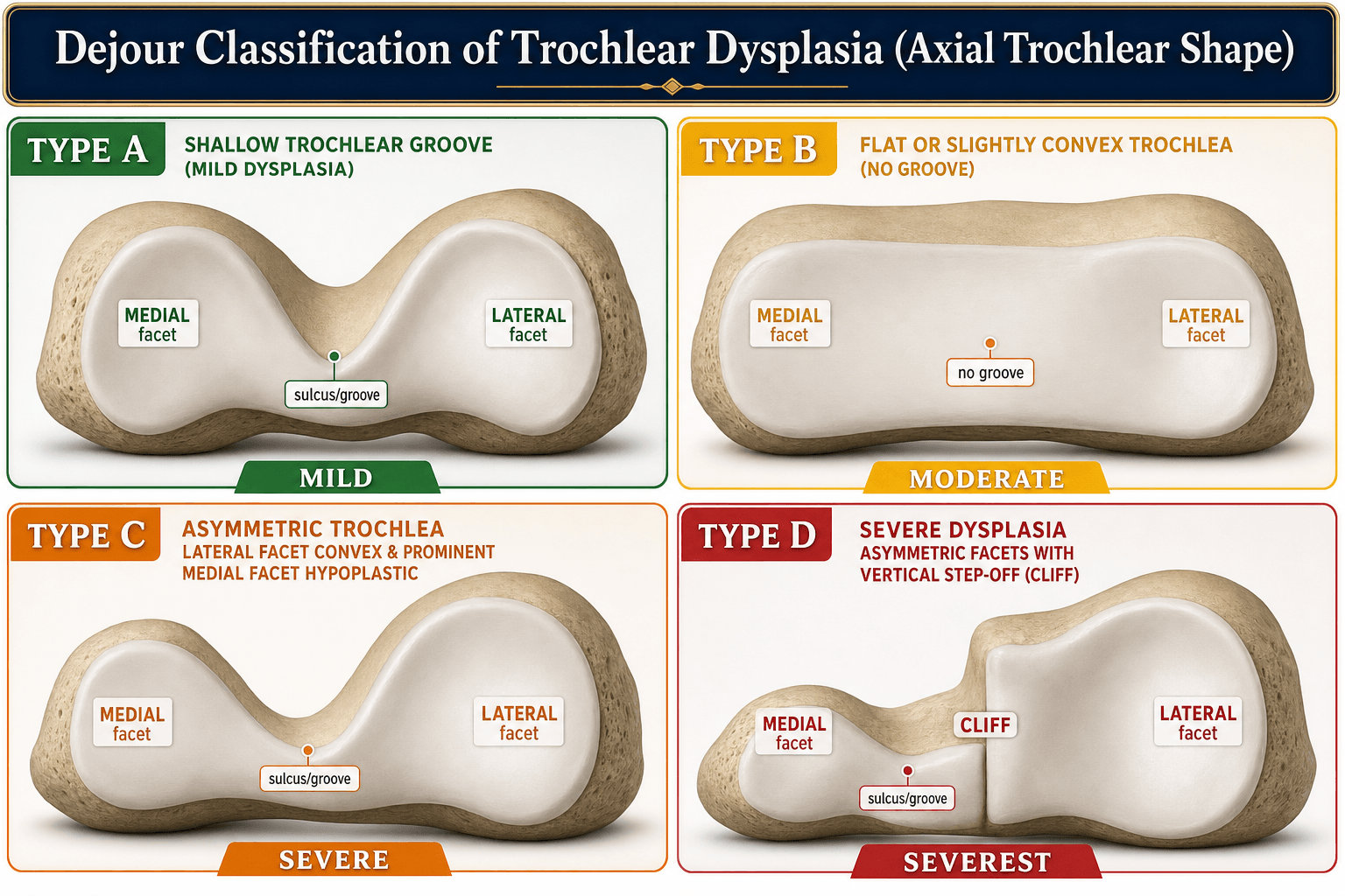
Trochlear dysplasia. Dejour grades it into four types from the radiograph and CT, by the shape of the trochlea and the signs that accompany it.
- Radiograph/CT Finding
- Crossing sign only
- Trochlear Morphology
- Shallow trochlea
- Surgical Implication
- MPFL may suffice
- Radiograph/CT Finding
- Crossing sign + supratrochlear spur
- Trochlear Morphology
- Flat trochlea
- Surgical Implication
- Consider bony procedure
- Radiograph/CT Finding
- Crossing sign + double contour
- Trochlear Morphology
- Asymmetric facets
- Surgical Implication
- Bony procedure likely needed
- Radiograph/CT Finding
- All above + cliff pattern
- Trochlear Morphology
- Convex trochlea
- Surgical Implication
- Trochleoplasty may be needed
The crossing sign, where the trochlear groove line crosses the anterior femoral cortex on the lateral radiograph, is the most sensitive finding and is present in every type.
Patellar height. Both indices are ratios measured on the lateral knee.
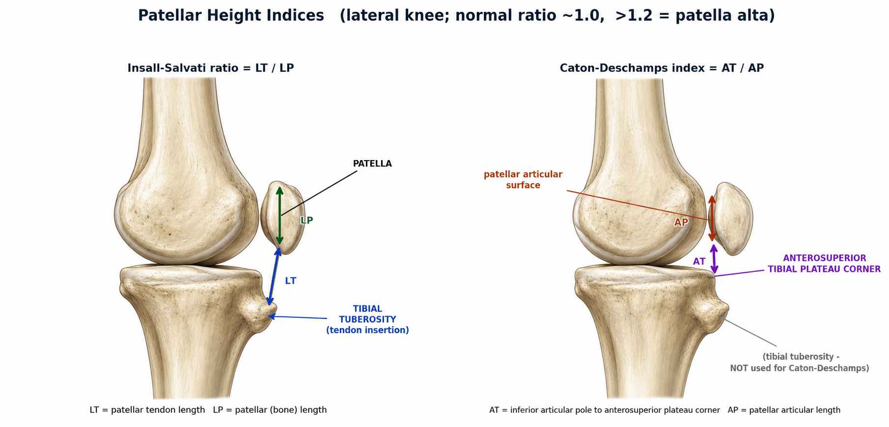
Each measurement has a threshold to recall:
- TT-TG distance, on axial CT or MRI - normal less than 20mm; greater than 20mm is a pathological lateral offset
- Insall-Salvati ratio - greater than 1.2 is alta, greater than 1.4 severe
- Caton-Deschamps index - greater than 1.2 is alta
- Sulcus angle, the angle between the trochlear facets - normal less than 145°; greater than 145° indicates dysplasia
- Lateral trochlear inclination, the angle of the lateral facet - less than 11° indicates dysplasia
MPFL tear pattern. Tears are most common on the femoral side (50-65%), often as a bony avulsion. Patellar-side tears are less common and may be an avulsion from the proximal patella; mid-substance tears are the least common. More than one site can tear, and MRI determines the pattern for surgical planning.
Clinical Assessment
History. Patients often describe the patella moving laterally, and it usually reduces as the knee extends. An immediate haemarthrosis is common. Ask about previous dislocations.
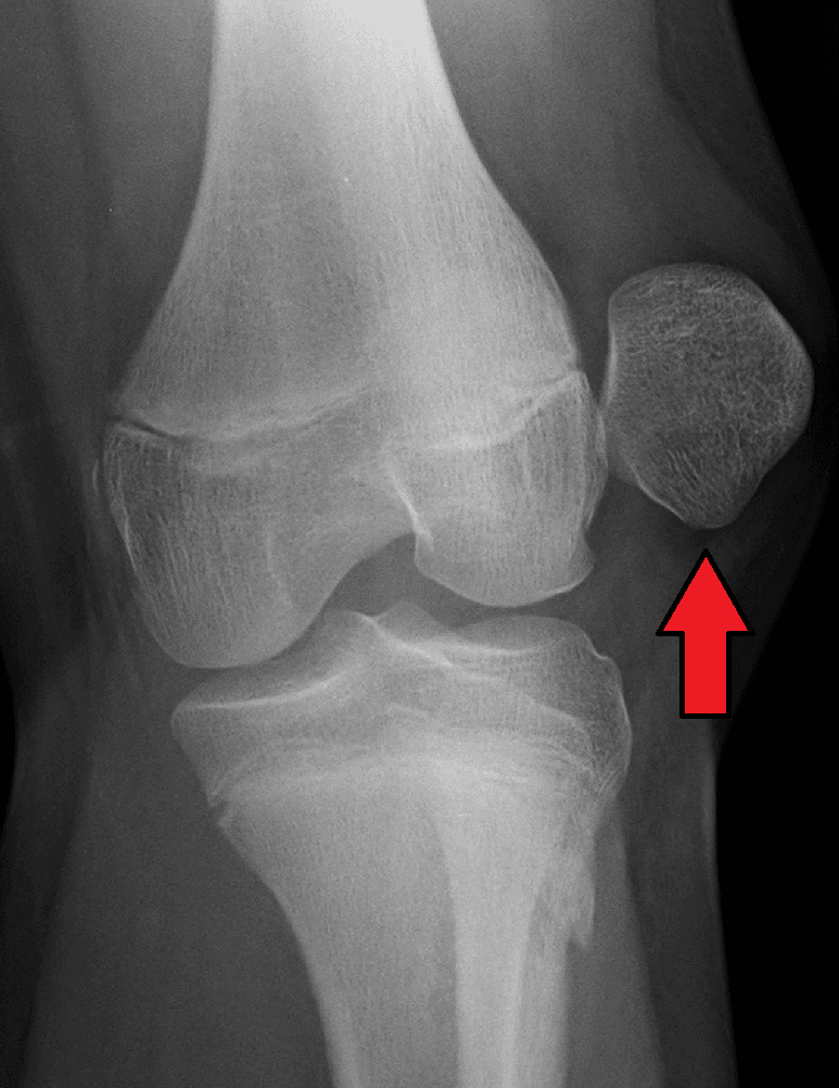
Examination. In an acute injury the effusion is a haemarthrosis. Beyond that, examine for the factors that predispose to instability:
- Alignment - valgus, femoral anteversion, tibial torsion; an increased Q angle suggests malalignment
- Tracking - the J-sign (lateral tracking in terminal extension), lateral tilt, subluxation
- Laxity - the Beighton score for generalised hypermobility
- The other knee - the predisposition is often bilateral
The apprehension test is the most reliable clinical sign of patellofemoral instability. With the knee in 30° of flexion, apply a lateral force to the patella. The test is positive if the patient resists or shows apprehension, a fear of dislocation.
Investigations
Radiographs. The standard views:
- AP weight-bearing - valgus alignment
- Lateral - trochlear dysplasia (crossing sign, supratrochlear spur) and patella alta (Insall-Salvati)
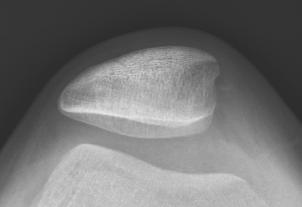
- Skyline/Merchant - patellar tilt, subluxation and trochlear morphology
- Long-leg alignment - if an osteotomy for valgus is being considered
MRI locates the MPFL tear and its extent, shows osteochondral injury to the medial patella and lateral femoral condyle and any loose cartilage or bone fragments in the joint, and assesses trochlear morphology.
The acute dislocation shows an MPFL tear (usually femoral side), bone marrow oedema in both the medial patella and the lateral femoral condyle (the kissing contusion), possibly an osteochondral fragment, and a haemarthrosis. This pattern is pathognomonic.
CT is the most accurate test for the TT-TG, measured on axial images from the deepest point of the trochlear groove to the tibial tubercle. The TT-PCL distance is an alternative less affected by trochlear dysplasia. CT also gives the rotational profile (femoral anteversion, tibial torsion), and 3D reconstruction of the trochlea helps surgical planning.
Differential Diagnosis
- Key History
- Lateral give-way on pivot, visible/felt patellar shift
- Examination
- Positive apprehension, J-sign
- Discriminator
- Reduces on extension; medial patella/lateral condyle bone bruise on MRI
- Key History
- Anterior pain with stairs/sitting, no true dislocation
- Examination
- Pain on patellar compression, no apprehension
- Discriminator
- No instability event or apprehension
- Key History
- Pivot injury, audible pop, rapid effusion
- Examination
- Positive Lachman/pivot shift
- Discriminator
- Anterior tibial translation, not patellar shift
- Key History
- Medial click/snapping, no frank dislocation
- Examination
- Palpable tender medial band
- Discriminator
- Mechanical snapping without apprehension
- Key History
- Sudden inability to extend, palpable gap
- Examination
- Extensor lag, patella alta/baja
- Discriminator
- Loss of active extension
- Key History
- Locking, catching after injury
- Examination
- Effusion, locking
- Discriminator
- Loose body on imaging - may coexist with dislocation
Management Algorithm
The decision. Recurrent instability is an indication for surgery, and the operation is built around the anatomy: MPFL reconstruction for the soft tissue, with bony procedures added for the abnormalities the measurements reveal.
First Dislocation
The acute knee. A tense effusion is aspirated. Radiographs rule out a fracture or loose body, and MRI assesses the MPFL and any osteochondral injury.
Non-operative management is appropriate for most first-time dislocators without a loose body: a brace in extension for 2-4 weeks, then progressive range of motion and quadriceps strengthening. Full recovery takes 6-12 weeks.
Early surgery. An osteochondral loose body needs fixation or removal. With a loose body, a large chondral or MPFL avulsion, high-risk anatomy or a professional athlete, early MPFL repair or reconstruction may be warranted.
FLAPIndications for Surgery After First Dislocation
Hook:Fix the FLAP - surgical indications after first dislocation!
Recurrent Instability
MPFL reconstruction is the standard operation. It may be sufficient alone if the anatomy is favourable, and lateral retinacular tightness is addressed if present. Before surgery, identify every anatomical risk factor: CT for the TT-TG, MRI for the soft tissue and radiographs for dysplasia.
Adding bony procedures. The measurements decide what goes with the MPFL reconstruction:
- TT-TG less than 20mm, no dysplasia - MPFL reconstruction alone
- TT-TG greater than 20mm - tibial tubercle medialisation
- Patella alta - tibial tubercle distalisation, or combined with medialisation
- High-grade dysplasia (Dejour B-D) - consider trochleoplasty, which is controversial and technically demanding
- Combined abnormalities - may need MPFL reconstruction, TTO and other procedures
The 20mm threshold is a guide. Borderline values (15-20mm) are weighed together with the TT-PCL, patellar height and dysplasia (see Controversies).
Surgical Technique
Graft. Gracilis or semitendinosus autograft is the most common choice. Allograft is an option, and a quadriceps tendon strip is gaining popularity.
Patellar fixation. Two tunnels in the proximal two-thirds of the medial patella, with suture anchors or interference screws. Avoid full-thickness tunnels.
Femoral tunnel. Its placement is the most important technical factor. The tunnel goes at the Schoettle point, always confirmed with intraoperative fluoroscopy. Schöttle defined the point on a true lateral view as 1mm anterior to the posterior cortex extension line, 2.5mm distal to the posterior origin of the medial femoral condyle, and proximal to the most posterior point of Blumensaat's line.
Tensioning. Tension the graft with the knee at 30-60° of flexion, and confirm full flexion and patellar tracking before final fixation.
Overtensioning the graft increases patellofemoral contact pressures and accelerates cartilage wear. Once tensioned, the patella should still translate one quadrant laterally.
Complications
- Cause
- Missed anatomical factor, tunnel malposition
- Prevention
- Address all risk factors, correct tunnel placement
- Management
- Revision surgery addressing missed factors
- Cause
- Overtensioned graft, tunnel malposition
- Prevention
- Proper tensioning, correct tunnel position
- Management
- Graft release, revision if severe
- Cause
- Overtensioning, prolonged immobilisation
- Prevention
- Early ROM, proper tensioning
- Management
- Physical therapy, MUA if needed
- Cause
- Full-thickness patellar tunnels
- Prevention
- Partial thickness tunnels or anchors
- Management
- ORIF
- Cause
- Tunnel malposition, early return to sport
- Prevention
- Correct technique, appropriate rehab
- Management
- Revision reconstruction
Femoral tunnel malposition is the most common cause of MPFL reconstruction failure. Anterior or distal placement changes graft isometry, leading to graft laxity in flexion or patellofemoral overload.
Postoperative Care
Rehabilitation Protocol
Brace locked in extension for ambulation. ROM exercises 0-90° out of brace. WBAT with crutches. CPM if available.
Progressive ROM to full. Unlock brace. Wean crutches. Quadriceps strengthening, VMO focus. No resisted knee extension 0-45°.
Full weight-bearing, no brace. Progressive closed chain strengthening. Stationary bike, pool exercises.
Running at 3-4 months if strength adequate. Sport-specific training. Full return 6-9 months.
After a TTO, weight-bearing is protected for 6-8 weeks until the osteotomy has healed. Confirm radiographic healing before advancing to full weight-bearing. The osteotomy may delay return to sport.
Outcomes and Prognosis
Good prognosis goes with an isolated MPFL rupture without dysplasia, a normal TT-TG distance, a first or second dislocation, compliance with rehabilitation and correct surgical technique.
Poor prognosis goes with severe trochlear dysplasia (Dejour C-D), multiple dislocations with cartilage damage, uncorrected anatomical abnormalities, generalised ligamentous laxity and tunnel malposition.
Guidelines, Registries & Global Practice
Global Epidemiology
- Incidence of first-time patellar dislocation approximately 5.8-7 per 100,000 overall, rising to 29-77 per 100,000 in adolescents aged 10-17.
- Peak incidence 15-19 years; sex distribution roughly equal, with the highest single-group risk in females aged 10-17 (Fithian cohort).
- The majority of patients carry 2 or more anatomical risk factors; trochlear dysplasia is present in around 85% of recurrent cases.
Society Guidance, Side by Side
- First Dislocation
- Non-operative unless osteochondral fragment or loose body
- Recurrent Instability
- MPFL reconstruction favoured over repair
- Bony Correction
- TTO when TT-TG elevated; trochleoplasty selectively
- First Dislocation
- Rehabilitation first-line; image to exclude fragment
- Recurrent Instability
- Reconstruction with anatomical femoral tunnel
- Bony Correction
- Address dysplasia/alta at high-volume centres
- First Dislocation
- Non-operative default; early surgery if APIFs plus fragment
- Recurrent Instability
- Reconstruction; a-la-carte philosophy
- Bony Correction
- Trochleoplasty established for high-grade (Dejour B-D)
- First Dislocation
- Individualised risk stratification
- Recurrent Instability
- Correct ALL contributing factors
- Bony Correction
- Bony procedures driven by measurements, not reflex
The unifying global theme is an "a-la-carte" or menu-based philosophy (popularised by the Lyon/Dejour school and endorsed across ESSKA): identify every contributing factor and correct each one, rather than applying a single operation to all patients.
- No dedicated arthroplasty-style registry exists for patellar instability; evidence is driven by RCTs, cohorts and meta-analyses.
- Pooled MPFL reconstruction redislocation approximately 1-2% (Schneider meta-analysis).
- Trochleoplasty recurrence approximately 2.4% but with 17-27% rates of stiffness/OA/reoperation (Leclerc meta-analysis).
- High-resource: routine MRI plus CT for TT-TG, intra-operative fluoroscopy, trochleoplasty at specialist centres, allograft availability.
- Limited-resource: diagnosis on plain radiographs and clinical exam; gracilis/semitendinosus autograft preferred (no allograft cost); trochleoplasty rarely available - reconstruction plus TTO is the workhorse.
- Adjustable-loop or anchor fixation chosen by availability; the anatomical Schöttle target remains universal regardless of fixation device.
Patellofemoral instability is a common viva topic. Know the MPFL anatomy, TT-TG threshold (20mm), Dejour classification, and be able to articulate a systematic "a-la-carte" approach to surgical planning that addresses every anatomical abnormality.
Controversies and Areas of Uncertainty
Acute repair reduces redislocation in high-risk children (Askenberger RCT) but most data favour reconstruction for established recurrent instability. The role of repair in adults remains debated.
TT-TG of 20mm is a guide, not an absolute cut-off. Borderline values (15-20mm), TT-PCL, patellar height and dysplasia must be weighed together. Over-medialisation risks medial overload and arthrosis.
Highly effective for dislocation control in Dejour B-D, but the 17-27% rates of stiffness, OA and reoperation, plus a steep learning curve, keep it confined to specialist centres. The threshold for offering it is not standardised.
In open physes, femoral tunnels risk physeal injury and growth disturbance. Physeal-sparing or soft-tissue techniques are preferred, and TTO is generally deferred until skeletal maturity.
MCQ Practice Points
Q: What percent of restraint to lateral patellar translation does the MPFL provide? A: 50-60% of restraint in the first 30° of flexion. It is the primary soft tissue restraint and ruptures in almost all acute dislocations.
Q: What TT-TG distance is considered pathological? A: Greater than 20mm. This indicates need for tibial tubercle medialization in addition to MPFL reconstruction.
Q: What is the recurrence rate after a first patellar dislocation treated conservatively? A: Approximately 50%. After a second dislocation, the rate increases to 80%+.
Q: Where does the MPFL attach on the femur? A: Between the adductor tubercle and medial epicondyle at the Schoettle point. This is just distal and anterior to the adductor tubercle.
Q: What Insall-Salvati ratio indicates patella alta? A: Greater than 1.2. Normal is 0.8-1.2. Patella alta delays patellar engagement in the trochlea.
Q: What radiographic finding is common to all Dejour types of trochlear dysplasia? A: The crossing sign - where the trochlear groove line crosses the anterior femoral cortex on lateral radiograph.
Exam Viva Scenarios
Practise clinical reasoning and management decisions out loud
“A 16-year-old netball player presents after her knee gave way while pivoting. She felt the patella move laterally and it reduced when she extended her knee. There is a moderate effusion. Radiographs show no fracture. How would you manage this?”
“A 22-year-old woman has had 4 patellar dislocations in the past 2 years. Examination shows positive apprehension, no significant valgus. CT shows TT-TG of 24mm. Radiographs show Dejour Type A trochlear dysplasia. What would you recommend?”
“A 19-year-old male had MPFL reconstruction 18 months ago but has had 2 further dislocations since. Examination shows persistent apprehension. CT shows TT-TG of 16mm. Review of operative notes shows the femoral tunnel was placed at the medial epicondyle. What is your assessment?”
MPFL Anatomy
- Primary restraint 50-60% (first 30° flexion)
- Femoral attachment: Schoettle point
- Between adductor tubercle and medial epicondyle
- Ruptures in almost all acute dislocations
Risk Factors (PATELLA)
- Patella alta (Insall-Salvati greater than 1.2)
- Anteversion (femoral)
- Trochlear dysplasia (Dejour A-D)
- Excessive TT-TG (greater than 20mm)
Measurements
- TT-TG: Normal less than 20mm
- Insall-Salvati: Normal 0.8-1.2
- Crossing sign: Trochlear dysplasia marker
- Sulcus angle: greater than 145° = dysplasia
Treatment Algorithm
- First dislocation no loose body: Conservative
- Recurrent, normal anatomy: MPFL reconstruction
- TT-TG greater than 20mm: MPFL + TTO medialization
- Patella alta: MPFL + TTO distalization
MPFL Reconstruction Keys
- Femoral tunnel at Schoettle point (CRITICAL)
- Tension at 30-60° flexion
- Avoid overtensioning
- Confirm full flexion before fixation
Prognosis
- 50% recurrence after 1st dislocation
- 80%+ recurrence after 2nd dislocation
- 80-95% success with MPFL reconstruction
- Tunnel malposition = most common failure cause
Evidence Base and Key Studies
Epidemiology and Natural History of Acute Patellar Dislocation
- Prospective cohort of 189 patients followed 2-5 years
- First-time dislocators had a 17% recurrent instability rate during follow-up
- Patients with a prior history of instability had a 49% recurrence rate
- Prior instability conferred 6.6x higher odds of further episodes; risk highest in females aged 10-17
Outcomes After Isolated MPFL Reconstruction (Meta-analysis)
- Systematic review and meta-analysis of 14 studies
- Pooled recurrent instability after isolated MPFL reconstruction only 1.2% (95% CI 0.3-2.1%)
- 84.1% of patients returned to sport; mean postoperative Kujala 85.8
- Reoperation risk 3.1%; positive apprehension sign in 3.6%
MPFL Repair vs Brace After First-Time Dislocation (RCT)
- Randomised controlled trial of 74 skeletally immature children (9-14 years)
- Redislocation rate 22% after arthroscopic MPFL repair vs 43% with knee brace (P = 0.047)
- Repair did NOT improve subjective or objective knee function over bracing
- 81% of patients had 2 or more anatomical patellar instability risk factors
Factors of Patellar Instability (Landmark Classification Paper)
- Analysed 143 unstable knees vs control radiographs/CT
- Trochlear dysplasia present in 85% (crossing sign in 96%) - the dominant factor
- TT-TG distance pathological (20mm or more) in 56% of unstable knees
- Patella alta (Caton-Deschamps 1.2 or more) in 24%; factors seen in only 3-6.5% of controls
Radiographic Landmark for the MPFL Femoral Tunnel (Schöttle Point)
- Cadaveric study of 8 knees defining the radiographic femoral insertion
- Point lies 1mm anterior to the posterior cortex extension line
- 2.5mm distal to the posterior origin of the medial femoral condyle
- Proximal to the most posterior point of the Blumensaat line on a true lateral
Trochleoplasty Outcomes and Complications (Meta-analysis)
- Meta-analysis of 28 studies and 1000 trochleoplasty procedures
- Overall recurrent dislocation rate only 2.4%
- Dejour deepening trochleoplasty most effective (1 recurrence in 349 knees, 0.28%)
- Patellofemoral osteoarthritis 27%, stiffness 7%, further surgery 17%
TT-PCL Distance Threshold (Systematic Review)
- Systematic review of 4 studies (300 unstable knees, 144 control knees)
- Mean TT-PCL 21.1mm in instability vs 18.8mm in controls (P less than 0.0001)
- Moderate-to-strong correlation between TT-PCL and TT-TG on MRI
- Excellent inter- and intra-observer reliability; pathological threshold of 21mm advocated