Eccentric Loading | Axillary Fold Loss | Surgical Repair Best for Active | Tietjen Classification
TIETJEN CLASSIFICATION (BY RUPTURE LOCATION)
Critical Must-Knows
- Mechanism is eccentric loading in abduction and external rotation (bench press lowering phase)
- Sternal head is most commonly injured (inserts more distally on humerus)
- Clinical triad: ecchymosis, loss of anterior axillary fold, palpable defect
- MRI confirms diagnosis, shows location of tear and degree of retraction
- Surgical repair produces superior strength and cosmesis in active individuals
- Bone anchors or transosseous tunnels to lateral bicipital groove of humerus
Clinical Pearls
- "Pectoralis major has two heads: clavicular (upper) and sternal (lower, larger)
- "Sternal head inserts more distally on humerus - experiences greater tension
- "Injury occurs during eccentric phase of bench press (lowering weight)
- "Complete ruptures show loss of anterior axillary fold and palpable defect
- "Surgery within 2-8 weeks produces best outcomes for active individuals
Critical Pectoralis Major Rupture Exam Points
Mechanism Recognition
Eccentric contraction during bench press - injury occurs when lowering a heavy weight with shoulder in abduction and external rotation. The sternal head experiences maximal tension at this position and fails first.
Clinical Diagnosis
Classic triad: anterior chest ecchymosis, loss of anterior axillary fold (asymmetric), and palpable defect with humeral insertion ruptures. Resisted adduction reproduces pain and weakness.
Surgical Timing
Optimal window is 2-8 weeks. Acute (under 2 weeks) has tissue friability. After 8 weeks, tissue retraction and scarring make repair difficult. Chronic cases may need reconstruction.
Operative Indications
Active individuals benefit most from surgical repair. Type III (tendon ruptures) produce best surgical outcomes. Surgery restores 95-100% of strength versus 60-70% with nonoperative management.
Quick Decision Guide - Treatment Selection
| Tear Type | Patient Profile | Treatment | Expected Outcome |
|---|---|---|---|
| Type I (muscle belly) | Any activity level | Nonoperative (sling, early motion) | Good functional outcome, some weakness acceptable |
| Type II (MTJ) | Active, athletic | Consider operative | Variable - better with surgery in high demand |
| Type III (tendon) | Active, athletic, young | Operative (bone anchors/tunnels) | 95-100% strength return, excellent cosmesis |
| Type III (tendon) | Sedentary, elderly | Discuss options (may choose nonop) | 60-70% strength, cosmetic deficit acceptable to some |
| Chronic (over 3 months) | Symptomatic weakness | Reconstruction (allograft augmentation) | Inferior to acute repair, reasonable improvement |
BENCH - PBENCH - Pectoralis Major Rupture Features
| B | Bench press mechanism Eccentric loading during lowering phase |
| E | Ecchymosis anterior chest Bruising visible over chest wall |
| N | No anterior axillary fold Loss of fold with complete rupture |
| C | Clavicular and sternal heads Sternal head most commonly injured |
| H | Humeral insertion site Lateral bicipital groove - surgical repair site |
| B | Bench press mechanism Eccentric loading during lowering phase | C | Clavicular and sternal heads Sternal head most commonly injured |
| E | Ecchymosis anterior chest Bruising visible over chest wall | H | Humeral insertion site Lateral bicipital groove - surgical repair site |
| N | No anterior axillary fold Loss of fold with complete rupture |
Hook:BENCH reminds you of the classic injury mechanism and the key clinical findings
TIETJEN - CTIETJEN - Classification by Location
| T | Type I = muscle belly Tear Intramuscular rupture - nonoperative |
| I | Type II = In between (MTJ) Musculotendinous junction - consider operative |
| E | Type III = End (tEndon) Tendon rupture - best surgical results |
| T | Timing matters (2-8 weeks) Optimal surgical window |
| J | Junction to humerus repair Bone anchors or transosseous tunnels |
| E | Eccentric contraction causes Lowering phase of bench press |
| N | Need MRI for diagnosis Confirms location and degree of retraction |
| T | Type I = muscle belly Tear Intramuscular rupture - nonoperative | T | Timing matters (2-8 weeks) Optimal surgical window | N | Need MRI for diagnosis Confirms location and degree of retraction |
| I | Type II = In between (MTJ) Musculotendinous junction - consider operative | J | Junction to humerus repair Bone anchors or transosseous tunnels | ||
| E | Type III = End (tEndon) Tendon rupture - best surgical results | E | Eccentric contraction causes Lowering phase of bench press |
Hook:TIETJEN classification helps guide operative versus nonoperative decision
STERNAL - ASTERNAL - Anatomy and Function
| S | Sternal head larger Lower, larger portion of muscle |
| T | Twists 180 degrees Fibers twist to insert on humerus |
| E | External rotation vulnerable Combined with abduction increases risk |
| R | Rib attachments (ribs 2-6) Origin of sternal head |
| N | Narrow tendon insertion All fibers converge to 5cm insertion |
| A | Adduction primary action Horizontal adduction and internal rotation |
| L | Lateral bicipital groove Insertion site on humerus |
| S | Sternal head larger Lower, larger portion of muscle | R | Rib attachments (ribs 2-6) Origin of sternal head | L | Lateral bicipital groove Insertion site on humerus |
| T | Twists 180 degrees Fibers twist to insert on humerus | N | Narrow tendon insertion All fibers converge to 5cm insertion | ||
| E | External rotation vulnerable Combined with abduction increases risk | A | Adduction primary action Horizontal adduction and internal rotation |
Hook:STERNAL anatomy explains why the sternal head is most vulnerable to rupture
REPAIR - SREPAIR - Surgical Principles
| R | Recognize early (2-8 weeks best) Optimal tissue quality window |
| E | Expose deltopectoral interval Standard surgical approach |
| P | Prepare humeral footprint Decorticate lateral bicipital groove |
| A | Anchors or tunnels Bone anchors (easier) or transosseous |
| I | Insert sutures through tendon Locking Krackow or whipstitch pattern |
| R | Repair with arm in adduction/IR Decreases tension on repair |
| R | Recognize early (2-8 weeks best) Optimal tissue quality window | P | Prepare humeral footprint Decorticate lateral bicipital groove | I | Insert sutures through tendon Locking Krackow or whipstitch pattern |
| E | Expose deltopectoral interval Standard surgical approach | A | Anchors or tunnels Bone anchors (easier) or transosseous | R | Repair with arm in adduction/IR Decreases tension on repair |
Hook:REPAIR outlines the key steps in surgical management
Overview and Epidemiology
Pectoralis major rupture is an uncommon injury that predominantly affects young, athletic males during weightlifting activities. The incidence has increased over the past 20 years due to the popularity of resistance training and heavy bench press exercises.
Mechanism of injury:
- Eccentric contraction during bench press lowering phase (80% of cases)
- Shoulder positioned in abduction and external rotation
- Sudden excessive load or loss of control during lowering
- Sternal head experiences greatest tension and fails first
- Other mechanisms: water skiing, wrestling, football tackles, rock climbing
Why Bench Press?
The bench press places the pectoralis major at maximal tension when the shoulder is in abduction and external rotation during the eccentric (lowering) phase. The sternal head inserts more distally on the humerus and experiences greater tensile forces than the clavicular head.
Epidemiology:
- Age: predominantly 20-40 years (peak athletic activity)
- Gender: male predominance (over 95% of cases)
- Sport: weightlifting (especially bench press), football, wrestling, rugby
- Side: right side more common in right-handed individuals (stronger side)
- Increasing incidence: related to popularity of gym training and heavier weights
Risk factors:
- Anabolic steroid use (tendon strength not matched to muscle hypertrophy)
- Previous pectoralis injury
- Inadequate warm-up
- Excessive weight/poor technique
- Sudden increase in training intensity
Pathophysiology and Mechanisms
Pectoralis major anatomy:
The pectoralis major is a large, fan-shaped muscle of the anterior chest wall with complex fiber orientation.
Two heads:
-
Clavicular head (upper portion)
- Origin: medial half of anterior clavicle
- Smaller, more horizontal fibers
- Inserts more proximally on humerus
-
Sternal head (lower portion)
- Origin: anterior sternum and costal cartilages of ribs 2-6
- Larger, represents about 80% of muscle mass
- Fibers twist 180 degrees before insertion
- Inserts more distally on humerus (experiences greater tension)
Insertion:
- Both heads converge to form a flat tendon approximately 5cm wide
- Inserts on lateral lip of bicipital groove of humerus
- Fibers twist so that sternal head inserts superior to clavicular head
- Insertion is 2-3cm long on humeral shaft
Function:
- Primary: adduction and internal rotation of shoulder
- Horizontal adduction (bringing arm across chest)
- Clavicular head: flexion of shoulder
- Sternal head: extension from flexed position
- Important for pushing, throwing, and climbing activities
Fiber Twist Anatomy
The pectoralis major fibers undergo a 180-degree twist before insertion. The lowest sternal fibers (from rib 6) insert most superiorly on the humerus, while the clavicular fibers insert inferiorly. This creates a mechanical disadvantage for the sternal fibers during eccentric loading.
Biomechanics of injury:
When the shoulder is in abduction and external rotation (bottom of bench press):
- Pectoralis major is maximally stretched
- Eccentric contraction occurs as weight is lowered (muscle lengthening under load)
- Sternal head experiences greatest tension (distal insertion = longer moment arm)
- Failure typically occurs at musculotendinous junction or tendinous insertion
- Complete rupture more common than partial
Blood supply:
- Pectoral branch of thoracoacromial artery (main supply)
- Lateral thoracic artery
- Perforating branches from internal mammary
- Rich blood supply - rarely a concern for healing
Nerve supply:
- Lateral pectoral nerve (C5-C7) - clavicular head
- Medial pectoral nerve (C8-T1) - sternal head
- Nerves enter from deep surface - generally not at risk during repair
Classification Systems
Tietjen Classification (by anatomic location - most commonly used)
| Type | Location | Characteristics | Treatment |
|---|---|---|---|
| Type I | Muscle belly | Intramuscular tear | Usually nonoperative |
| Type II | Musculotendinous junction | MTJ disruption | Variable - consider operative in active |
| Type III | Tendon rupture | Insertion avulsion or tendon failure | Operative preferred (best results) |
Clinical relevance:
- Type I: least common, typically incomplete, good nonoperative outcomes
- Type II: intermediate zone, may have both muscle and tendon involvement
- Type III: most common (60-70%), best surgical results, complete rupture typical
Type III Best for Surgery
Type III (tendon) ruptures have the best surgical outcomes because healthy tendon can be securely repaired to bone. Type I (muscle) ruptures are difficult to repair and do reasonably well nonoperatively.
Clinical Presentation and Assessment
Acute presentation:
Patients typically present shortly after injury with a characteristic history and clinical findings.
History:
- Mechanism: bench press (80%), other weightlifting, contact sports, or trauma
- Sensation: "pop" or "tearing" felt in anterior chest/shoulder
- Pain: immediate, severe anterior chest pain
- Swelling: rapid onset of chest wall swelling
- Weakness: inability to continue exercise, difficulty with pushing activities
- Previous injuries: prior pectoralis strain or partial tears
The Pop
Patients frequently report hearing or feeling a "pop" during the injury (70-80% of cases). This occurs at the moment of tendon failure and is a strong indicator of complete rupture.
Physical examination:
Clinical Examination Findings
| Finding | Significance | Timing |
|---|---|---|
| Ecchymosis over anterior chest | Bleeding from torn muscle/tendon | Appears within 24-48 hours, peaks at 3-5 days |
| Loss of anterior axillary fold | Complete rupture of tendon (asymmetry when comparing sides) | Immediate if complete; more obvious after swelling subsides |
| Palpable defect | Tendon retraction from humeral insertion | Easier to palpate after acute swelling resolves (1-2 weeks) |
| Asymmetric muscle contour | Medial bunching of muscle belly | Progressive as swelling decreases |
| Weakness with resisted adduction | Loss of primary pectoralis function | Immediate; quantifiable with strength testing |
| Pain with resisted testing | Active inflammation and tissue injury | Acute phase; improves over weeks |
Specific examination maneuvers:
-
Visual inspection
- Patient upright with arms by sides
- Compare anterior axillary folds bilaterally
- Asymmetry indicates complete rupture
- May see medial muscle bunching
-
Palpation
- Palpate along course of pectoralis major
- Feel for defect at humeral insertion
- Compare to contralateral side
- Easier after acute swelling subsides (1-2 weeks)
-
Strength testing
- Horizontal adduction against resistance (primary test)
- Internal rotation against resistance
- Forward elevation (clavicular head function)
- Compare to contralateral side
-
Special tests
- Press test: patient presses palms together in front of chest - look for asymmetric contraction
- Wall push: observe anterior axillary fold during wall push-up
Functional deficits:
- Difficulty with pushing activities (push-ups, bench press)
- Weakness with throwing or striking
- Cosmetic deformity may be primary concern for some patients
- Daily activities usually not significantly affected
Differential diagnosis:
Differential Diagnosis of Acute Anterior Chest/Shoulder Pain After Lifting
| Condition | Distinguishing features | Key investigation |
|---|---|---|
| Pectoralis major rupture | Bench-press 'pop', loss of anterior axillary fold, ecchymosis, weak resisted adduction | MRI (location, retraction) |
| Pectoralis major strain/contusion | Pain and tenderness but axillary fold preserved, no palpable defect, strength relatively intact | Clinical; MRI shows oedema without discontinuity |
| Proximal long head of biceps rupture | 'Popeye' deformity distally, ecchymosis along arm, antecubital pain, supination weakness | Clinical; ultrasound/MRI |
| Subpectoral (proximal) biceps or subscapularis tear | Anterior shoulder pain, positive lift-off/belly-press, internal rotation weakness | MRI of shoulder |
| Anterior shoulder dislocation/subluxation | Mechanism of forced abduction-external rotation, apprehension, possible Bankart | Radiographs, MRI arthrogram |
| Rib fracture / costochondral injury | Focal bony/cartilage tenderness, pain on chest compression, no fold loss | Radiographs, clinical |
Investigations
Imaging is essential for:
- Confirming diagnosis
- Determining location of tear (Tietjen classification)
- Assessing completeness of tear
- Identifying degree of tendon retraction
- Surgical planning
Plain radiographs:
Usually normal in isolated pectoralis rupture.
May show:
- Soft tissue swelling over anterior chest
- Rarely: avulsion fracture of humeral insertion (very uncommon)
Limitations: cannot visualize muscle or tendon pathology
Ultrasound:
Can be useful in experienced hands but operator-dependent.
Advantages:
- Dynamic examination
- Can assess degree of retraction
- Lower cost than MRI
Disadvantages:
- Operator-dependent
- Limited by body habitus and acute swelling
- Less detailed than MRI
Not first-line imaging in most centers.
MRI (gold standard):
MRI is Diagnostic Standard
MRI is the investigation of choice for pectoralis major rupture. It confirms the diagnosis, localizes the tear (Tietjen type), assesses completeness, measures retraction, and guides surgical planning.
MRI protocol:
- Axial, coronal, and sagittal sequences
- STIR or fat-suppressed sequences show edema
- Focus on humeral insertion and muscle-tendon junction
Key MRI findings:
| Finding | Significance |
|---|---|
| Location of tear | Type I (muscle), II (MTJ), or III (tendon) |
| Completeness | Partial versus complete disruption |
| Tendon retraction | Distance from humeral insertion (affects surgical difficulty) |
| Muscle edema/hematoma | Acute injury findings |
| Muscle atrophy | Chronic injury indicator |
| Tendon quality | Important for surgical planning |
Retraction Distance
The degree of tendon retraction on MRI is important for surgical planning. Less than 2cm retraction is easily repaired directly to bone. Greater than 5cm retraction may require allograft augmentation in chronic cases.
CT scan:
Rarely indicated. May be useful if:
- Avulsion fracture suspected
- MRI contraindicated
- Combined bony injury present
When to image:
Timing of imaging:
- Acute: MRI can be performed immediately if diagnosis uncertain
- Preferred timing: 5-7 days after injury allows acute swelling/edema to decrease, improving image quality
- Chronic: MRI shows atrophy and retraction for reconstruction planning
Management Algorithm
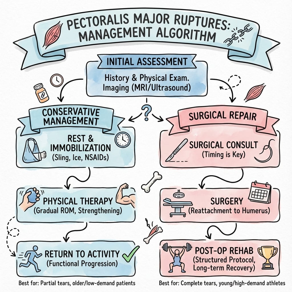
Emergency/acute management:
All patients with suspected pectoralis major rupture:
- Ice and analgesia for comfort
- Sling for comfort (not immobilization requirement)
- Avoid active adduction/internal rotation to prevent further injury
- Clinical examination once acute pain controlled
- MRI to confirm diagnosis and classify injury (can wait 5-7 days for better quality)
Not an Emergency
Pectoralis major rupture is not a surgical emergency. Optimal surgical timing is 2-8 weeks, allowing time for thorough assessment, patient counseling, and surgical planning.
Initial counseling:
- Explain diagnosis and Tietjen classification
- Discuss operative versus nonoperative options
- Review expected outcomes with each approach
- Consider patient's activity level and goals
- Arrange follow-up after MRI results available
Shared decision-making with thorough counseling is essential for optimal patient satisfaction.
Surgical Technique
Deltopectoral approach (standard)
Positioning:
- Beach chair or supine with bump under shoulder
- Arm free-draped to allow positioning
- Head of bed elevated 30-45 degrees
Incision:
- Deltopectoral interval from clavicle to deltoid insertion
- Typically 8-12cm incision
- Can extend distally for better exposure of humeral insertion
Dissection:
- Identify deltopectoral interval (cephalic vein is landmark)
- Develop interval (vein usually taken laterally with deltoid)
- Retract deltoid laterally, pectoralis major medially
- Identify torn pectoralis major tendon (retracted medially)
- The tendon may be significantly retracted - require careful mobilization
Key anatomic considerations:
- Cephalic vein: preserve or ligate if necessary
- Coracoid: landmark for orientation
- Long head biceps: medial to surgical field
- Axillary nerve: deep and inferior (safe with proper retraction)
The deltopectoral approach provides excellent visualization of the humeral insertion and allows tendon mobilization.
Complications
Complications of Pectoralis Major Rupture Treatment
| Complication | Incidence | Prevention/Management |
|---|---|---|
| Re-rupture | 5-10% (surgical) | Secure repair, protect during healing, patient compliance |
| Infection | Less than 5% | Sterile technique, prophylactic antibiotics, early recognition |
| Hematoma/seroma | 5-10% | Hemostasis, drainage if symptomatic, usually resolves |
| Stiffness/contracture | 10-15% | Early ROM protocol, avoid over-immobilization, PT |
| Persistent weakness | 10-20% (nonoperative) | Set expectations, strengthen compensatory muscles |
| Cosmetic deformity | 60-70% (nonoperative) | Counsel pre-treatment, surgical option if unacceptable |
| Nerve injury | Less than 5% | Careful dissection, protect axillary and lateral pectoral nerves |
| Anchor failure/pullout | Less than 5% | Adequate bone stock, proper technique, appropriate anchor size |
Re-rupture:
- Most common after return to heavy bench press too early
- Risk higher with inadequate rehabilitation compliance
- Chronic repairs have higher re-rupture risk than acute
- Prevention: staged return to activity, patient education, adequate healing time
Infection:
- Rare due to excellent blood supply
- Present with wound drainage, erythema, fever
- Treatment: antibiotics, may need washout
- Deep infection may compromise repair
Persistent weakness:
- More common with nonoperative management (expected)
- May occur with surgical repair if tissue quality poor or chronic injury
- Compensatory strengthening of surrounding muscles helpful
- Functional impact varies by patient demands
Cosmetic concerns:
- Asymmetric axillary fold is primary cosmetic complaint
- Nearly universal with nonoperative management of complete ruptures
- Surgery restores contour in 90-95% of cases
- Important to discuss pre-treatment as may be patient's primary concern
Return to Bench Press
The most common cause of re-rupture is premature return to heavy bench press (before 6 months). Patient education about staged return to lifting is critical. Many surgeons recommend permanent modification to avoid maximal single-rep bench press.
Postoperative Care and Rehabilitation
Post-surgical rehabilitation protocol:
Rehabilitation is critical to successful outcome. Protocol must balance early motion (to prevent stiffness) with protection of repair (to allow healing).
- Sling immobilization with arm in neutral rotation
- Remove sling for hygiene and gentle elbow/wrist ROM
- No active shoulder motion
- No passive stretching
- Ice and elevation for swelling control
- Focus on patient education regarding restrictions
- Begin passive ROM with therapist supervision
- Gentle pendulum exercises
- Avoid terminal external rotation and abduction (stresses repair)
- Continue sling between exercises
- No active shoulder motion yet
- Begin scapular retraction exercises (pain-free)
- Discontinue sling
- Begin active-assisted ROM in all planes
- Progress to active ROM as tolerated
- Continue to avoid combined abduction and external rotation
- Light isometrics in adduction and internal rotation
- Avoid resistance exercises
- Progressive resistance exercises
- Focus on internal rotation and adduction strengthening
- Rubber band exercises, light dumbbells
- Begin chest press with light weight (limited ROM initially)
- Gradually increase ROM and resistance
- Continue to avoid maximal external rotation
- Progressive resistance training
- Sport-specific exercises
- Can begin bench press with light-moderate weight
- Avoid maximal single-rep lifts
- Focus on proper form and controlled motion
- Most patients return to full activity by 6 months
- Gradual return to pre-injury activity levels
- May return to competitive sports after 6 months
- Return to heavy weightlifting after 6-9 months
- Permanent modification: avoid maximal single-rep bench press
- Emphasize proper warm-up and technique
- Full strength recovery may take up to 12 months
Key rehabilitation principles:
Protected Motion Critical
The first 6 weeks are critical for tendon-to-bone healing. Aggressive early motion can lead to repair failure. Passive ROM is safe. Active motion begins at 4-6 weeks. Resistance is delayed until 6 weeks minimum.
Milestones:
- 6 weeks: full passive ROM expected
- 12 weeks: full active ROM, beginning strength training
- 6 months: return to sport, near-normal strength
- 12 months: full strength recovery, unrestricted activity
Return to bench press:
- Light bench press at 3 months (50% body weight)
- Moderate bench press at 6 months (80% body weight)
- Heavy bench press after 9-12 months
- Avoid maximal single-rep attempts permanently
Outcomes and Prognosis
Outcomes by treatment:
| Outcome Measure | Surgical Repair | Nonoperative |
|---|---|---|
| Strength (peak torque) | 95-100% of contralateral | 60-70% of contralateral |
| Endurance | Near-normal | Significantly reduced |
| Cosmesis | Restoration of fold in 90-95% | Permanent asymmetry |
| Patient satisfaction | 90-95% | 60-70% |
| Return to sport | Over 90% at pre-injury level | 60-70% at pre-injury level |
| Return to bench press | Over 90% (with modifications) | Less than 50% at pre-injury level |
Prognostic factors for surgical outcome:
Favorable:
- Acute repair (within 8 weeks)
- Type III (tendinous) ruptures
- Good tissue quality
- Patient age under 40 years
- High motivation and compliance with rehabilitation
- Absence of steroid use
Unfavorable:
- Chronic presentation (over 3 months)
- Poor tissue quality (chronic, steroid use)
- Type I (muscle belly) tears
- Patient noncompliance with rehabilitation
- Premature return to heavy lifting
Surgery Wins for Athletes
For active individuals with complete Type III tears, surgical repair produces significantly superior outcomes compared to nonoperative management. Near-complete strength recovery and restoration of cosmesis justify the surgical risks.
Long-term outcomes:
Studies with 5-10 year follow-up show:
- Maintained strength and function with surgical repair
- Over 90% patient satisfaction long-term
- Re-rupture rare after first year (less than 5%)
- Most patients return to all activities including weightlifting
- Permanent modification recommended: avoid maximal single-rep bench press
Complications affecting outcome:
- Re-rupture (5-10%): usually due to premature return to lifting
- Persistent weakness (10-15%): more common with chronic repairs
- Stiffness (10-15%): usually improves with continued therapy
- Chronic pain (less than 10%): may require further evaluation
Evidence Base
- Meta-analysis of 112 reported cases (108 pooled from the literature plus 4 from the authors' Melbourne unit). All reported patients were male. Rupture occurred most commonly during weight training, weight lifting or wrestling with the arm abducted and externally rotated; most ruptures were complete and located at the humeral insertion. Work-related injuries occurred more often at the musculotendinous junction. Prognosis was related neither to patient age nor to rupture location.
- Surgical treatment, preferably within the first 8 weeks of injury, gave a significantly better outcome than conservative treatment or delayed repair.
- Retrospective comparison of 17 distal pectoralis major ruptures (13 operative, 4 nonoperative). Isokinetic adduction strength was 102% of the contralateral side after acute repair, 94% after chronic repair and only 71% after nonoperative treatment. Overall subjective ratings were 96% (acute repair), 93% (chronic repair) and 51% (nonoperative).
- Patients treated operatively (acute or chronic) fared significantly better than those treated nonoperatively, with no significant difference between acute and chronic repairs in this small series.
- Prospective cohort of 60 patients with complete pectoralis major ruptures (51% operative, 49% nonoperative); 80% occurred during bench press. Isokinetic testing at 60 deg/s showed a strength loss of 14.3% in the surgical group versus 41.7% in the nonoperative group (P less than 0.05).
- Excellent results (Bak criteria) were achieved in 67.7% of surgical patients and in none of the nonoperative patients; poor results occurred in 9.7% (surgical) versus 31% (nonoperative).
- Systematic review of 365 reported cases (1822-2010), 75% of which occurred in the preceding 20 years; 83% resulted from indirect trauma and 48% during weight training. Existing classifications were applied inconsistently.
- Proposes a contemporary classification incorporating timing (acute vs chronic), anatomic location (origin/belly, between the musculotendinous junction and insertion, or bony avulsion) and standardised terminology for tear thickness and width.
- Controlled cadaveric study (24 fresh-frozen shoulders) comparing unicortical button fixation (UBF) constructs against the traditional bone-trough transosseous repair. Mean peak load to failure was 794 N for UBF with No. 5 suture and suture tape versus 492 N for the bone trough (native tendon 1816 N).
- The UBF/No. 5 suture/suture tape construct was 61% stronger than the bone-trough technique, showed the least displacement after cyclic loading and best reproduced the native footprint length.
- Three military patients with chronic, complete pectoralis major tears reconstructed with Achilles tendon allograft a mean of 22.2 months after injury. At a mean follow-up of 24.5 months there was one excellent and two good results; all patients were satisfied and returned to full active-duty service and recreational weightlifting by 6 months.
- Demonstrates that allograft reconstruction remains a viable option even nearly 2 years after injury when primary repair is not feasible.
- Comprehensive review of diagnosis and management. Most tears occur near the tendon insertion and can usually be diagnosed clinically, with MRI confirming location and extent. Nonoperative treatment is reserved for proximal/muscle tears, low-grade partial tears and sedentary patients.
- For active patients, acute (less than 6 weeks) repair is recommended to restore strength and function; transosseous tunnels, suture anchors and cortical buttons are all described techniques.
Clinical Decision Scenarios
Use these scenarios to practise clinical reasoning and management decisions
Scenario 1: Acute Complete Rupture in Athlete
"A 28-year-old competitive powerlifter presents to your clinic 10 days after feeling a 'pop' in his chest during bench press. He has anterior chest ecchymosis and asymmetric anterior axillary folds. MRI shows a complete pectoralis major tendon rupture at the humeral insertion with 3cm retraction. What is your assessment and management?"
Scenario 2: Partial Tear Management Decision
"A 35-year-old recreational gym-goer presents with anterior chest pain after bench press 3 weeks ago. He has mild ecchymosis but maintained anterior axillary fold contour. MRI shows a Type II musculotendinous junction partial thickness tear (estimated 60% of tendon thickness). He works as an accountant but enjoys weightlifting 3-4 times per week. What would you recommend?"
Scenario 3: Delayed Presentation Reconstruction
"A 32-year-old patient presents 8 months after a pectoralis major rupture that was initially managed nonoperatively. He has significant weakness (40% of contralateral side on isokinetic testing), cosmetic deformity, and difficulty with his occupation as a firefighter. MRI shows Type III tear with 6cm retraction and muscle atrophy. He requests surgical treatment. What would you offer?"
MCQ Practice Points
Mechanism Question
Q: What is the most common mechanism of pectoralis major rupture? A: Eccentric contraction during bench press lowering phase (80% of cases). The shoulder is in abduction and external rotation, placing maximal tension on the sternal head which fails at the musculotendinous junction or tendinous insertion.
Anatomy Question
Q: Why is the sternal head of pectoralis major more commonly injured than the clavicular head? A: The sternal head (1) inserts more distally on the humerus creating greater moment arm and tension, (2) undergoes 180-degree fiber twist before insertion creating mechanical disadvantage, and (3) represents 80% of muscle mass experiencing greater force.
Classification Question
Q: According to Tietjen classification, which type has the best surgical outcomes? A: Type III (tendon rupture) has best outcomes because healthy tendon tissue can be securely repaired to bone with anchors or tunnels. Type I (muscle belly) is difficult to repair and does better nonoperatively.
Clinical Diagnosis Question
Q: What is the classic clinical triad of complete pectoralis major rupture? A: (1) Ecchymosis over anterior chest, (2) loss of anterior axillary fold (asymmetry), and (3) palpable defect at humeral insertion. This triad allows clinical diagnosis before imaging.
Surgical Timing Question
Q: What is the optimal timing for surgical repair of pectoralis major rupture? A: 2-8 weeks post-injury. Acute (under 2 weeks) has tissue friability and edema. After 8 weeks, tissue retraction and scarring make direct repair difficult. This window provides best tissue quality for secure repair.
Outcomes Question
Q: What strength recovery can be expected with surgical repair versus nonoperative management? A: Surgical repair achieves 95-100% of contralateral strength, while nonoperative management achieves 60-70% of contralateral strength. This difference justifies surgery in active individuals with complete ruptures.
Guidelines, Registries & Global Practice
Global epidemiology (PubMed-verified):
Pectoralis major rupture is uncommon but rising in frequency. In the ElMaraghy & Devereaux systematic review of 365 published cases spanning 1822-2010, 75% occurred in the most recent 20 years, 83% resulted from indirect trauma, and 48% occurred during weight-training (PMID 21831661). Bak and colleagues' meta-analysis of 112 cases found that all reported patients were male, that ruptures occurred most often during weight lifting or wrestling with the arm abducted and externally rotated, and that most tears were complete and located at the humeral insertion (PMID 10795675). Across the largest prospective series, approximately 80% of complete ruptures occurred during bench press (PMID 24192390).
Side-by-side guidance (no formal disease-specific society guideline exists):
There is no dedicated AAOS, NICE, BOA, AO or EFORT clinical practice guideline for pectoralis major rupture; this is a rare injury managed on the basis of cohort studies, systematic reviews and expert consensus. The table below summarises the consistent evidence-based position taken by major bodies of literature and how strongly it is supported.
| Source / body of evidence | Position on management | Evidence level |
|---|---|---|
| Bak meta-analysis (KSSTA 2000, PMID 10795675) | Early surgery (within 8 weeks) superior to conservative or delayed repair | Level IV (meta-analysis of case data) |
| de Castro Pochini prospective cohort (AJSM 2013, PMID 24192390) | Surgical repair of complete tears in athletes restores strength (14.3% vs 41.7% loss) | Level II |
| ElMaraghy systematic review (JSES 2011, PMID 21831661) | Standardised classification; surgery for complete insertional tears in active patients | Level IV |
| Haley & Zacchilli review (Clin Sports Med 2014, PMID 25280620) | Nonoperative for proximal/partial tears and sedentary patients; acute (less than 6 weeks) repair otherwise | Level V (narrative) |
| AAOS / EFORT instructional reviews | Operative repair preferred for complete tears in active patients; partial/muscle-belly tears nonoperative | Expert consensus |
Registry evidence:
Pectoralis major repair is a soft-tissue tendon procedure and is not captured by arthroplasty registries (AOANJRR, NJR, AJRR), which track joint replacement implants only. No national soft-tissue tendon registry currently reports pectoralis major outcomes; the evidence base therefore rests on single-centre cohorts and systematic reviews rather than registry data.
International practice variation:
| Setting | Typical practice pattern |
|---|---|
| North America / Europe | Acute anchor or cortical-button repair favoured in active patients; biomechanical data support modern high-strength constructs (PMID 28749741) |
| Military / tactical populations | High operative rate given strength demands; allograft reconstruction used for delayed presentations (PMID 23449062) |
| Resource-limited / older patients | Higher threshold for surgery; nonoperative management accepted where strength demands are low |
Australian context (AOANJRR / PBS / eTG framework):
- Increasing incidence parallels the global trend, driven by CrossFit, powerlifting and gym culture; common in AFL, rugby union and rugby league athletes and in firefighters, police and military personnel. Notably, the landmark Bak meta-analysis was conducted at a Melbourne orthopaedic unit (PMID 10795675).
- Acute cases usually present to emergency departments, with MRI arranged as an outpatient (generally within 1-2 weeks) and referral to an orthopaedic surgeon with a sports-medicine interest.
- Suture anchors and cortical buttons are widely available; Achilles tendon allograft is accessible through accredited tissue banks for chronic reconstruction.
- Surgical antibiotic prophylaxis should follow eTG (Therapeutic Guidelines) recommendations; analgesia and any perioperative medication should respect PBS availability. Workers' compensation and fitness-for-duty assessment are relevant for occupational injuries, and return-to-contact-sport decisions in AFL/NRL/Rugby Australia athletes involve team physicians.
Exam Context
Be prepared to discuss operative versus nonoperative decision-making based on tear type, patient factors and functional demands. Know that there is no formal society guideline, that the evidence rests on cohorts and systematic reviews, and that arthroplasty registries do not capture this procedure. Know the Tietjen and ElMaraghy classifications, anchor/button repair technique, and the 2-8 week optimal timing window.
PECTORALIS MAJOR RUPTURES
Clinical summary
CLINICAL TRIAD
- •1. Ecchymosis over anterior chest wall
- •2. Loss of anterior axillary fold (asymmetry)
- •3. Palpable defect at humeral insertion
- •Plus: History of 'pop' during bench press (70-80%)
TIETJEN CLASSIFICATION
- •Type I: Muscle belly rupture (nonoperative usually)
- •Type II: Musculotendinous junction (consider operative)
- •Type III: Tendon rupture (operative best results)
- •Type III has best surgical outcomes - healthy tendon to bone
MECHANISM
- •Eccentric contraction (lowering phase) of bench press (80%)
- •Shoulder in abduction and external rotation
- •Sternal head most vulnerable (distal insertion, fiber twist)
- •Other: wrestling, football, water skiing, rock climbing
IMAGING
- •MRI is gold standard for diagnosis
- •Shows: location (Type I/II/III), completeness, retraction
- •Timing: can do acutely or wait 5-7 days for better quality
- •Plain X-rays usually normal (rarely avulsion fracture)
OPERATIVE INDICATIONS
- •Type III complete tears in active individuals (strongest indication)
- •Young athletic patients with high functional demands
- •Failed nonoperative with symptomatic weakness
- •Optimal timing: 2-8 weeks (best tissue quality)
SURGICAL TECHNIQUE
- •Deltopectoral approach to humeral insertion
- •Mobilize tendon, Krackow sutures with Number 2 nonabsorbable
- •Bone anchors (3-4) to lateral bicipital groove OR transosseous tunnels
- •Repair with arm in adduction and internal rotation (decreases tension)
- •Chronic (over 3 months): allograft augmentation (Achilles) for gaps
OUTCOMES
- •Surgical: 95-100% strength, 90-95% satisfaction, fold restored
- •Nonoperative: 60-70% strength, permanent deformity
- •Re-rupture: 5-10% (usually premature heavy lifting)
- •Return to sport: 6 months with permanent modifications
EXAM TRAPS
- •Don't treat all tears surgically - partial tears may do well nonop
- •Don't miss optimal timing (2-8 weeks) - avoid delay
- •Don't attempt direct repair of chronic with large retraction - need allograft
- •Don't allow return to max bench press before 9-12 months
- •Don't forget to counsel about permanent activity modifications