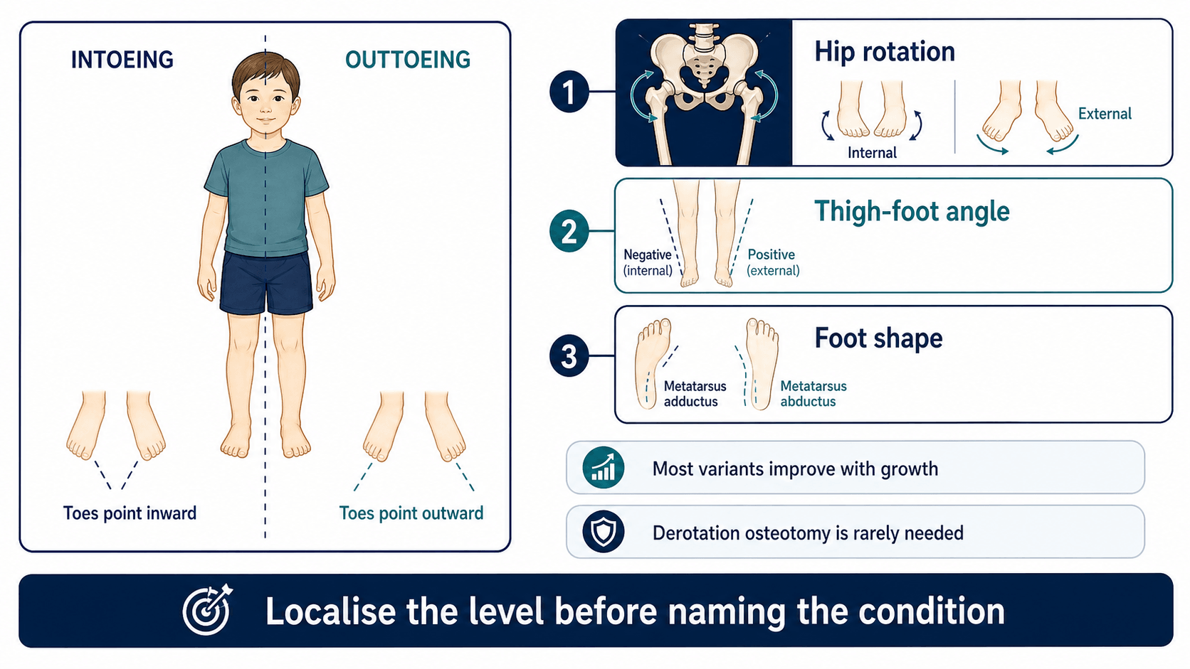
Localise the rotational level before reassuring or escalating
- The three classic in-toeing causes are metatarsus adductus, internal tibial torsion and femoral anteversion.
- Most painless symmetric developmental in-toeing improves with growth and does not need braces or special shoes.
- Patella direction during gait helps separate femoral from tibial contribution.
- Painful adolescent out-toeing requires hip assessment and imaging when SUFE or other hip pathology is possible.
- Derotation osteotomy is reserved for selected older children with severe persistent functional deformity.
- “Watch the child walk before putting them on the couch.
- “Name the level: foot, tibia, femur, hip or neuromuscular.
- “W-sitting is associated with femoral anteversion but is not itself an indication for surgery.
- “Reassurance is only safe after pain, asymmetry, progression and neurological signs have been considered.
Typical developmental in-toeing is painless, often symmetric and often improving. Pain, limp, asymmetry, neurological signs, progression or adolescent out-toeing should change the diagnostic pathway.
Images and Diagrams
| Finding | Likely level | Clinical meaning |
|---|---|---|
| Curved lateral foot border | Foot | Metatarsus adductus; assess flexibility. |
| Patella forward, feet inward | Tibia | Internal tibial torsion. |
| Patellae inward, high hip internal rotation | Femur | Femoral anteversion. |
| Painful adolescent out-toeing | Hip until proven otherwise | Assess for SUFE or other hip pathology. |
WALKClinic Flow
Hook:WALK keeps the clinical assessment sequence practical.
FITCommon In-toeing Levels
Hook:FIT helps localise painless developmental in-toeing.
PAINEscalation Clues
Hook:PAIN prevents unsafe reassurance.
Overview/Epidemiology
Rotational concerns are one of the commonest reasons children are referred to orthopaedics. Parents may describe in-toeing, out-toeing, tripping, unusual running, shoe wear, W-sitting or cosmetic concern. The clinician's job is to identify whether this is a normal developmental variant, a structural rotational deformity causing function problems, a painful hip problem, or a neuromuscular gait issue.
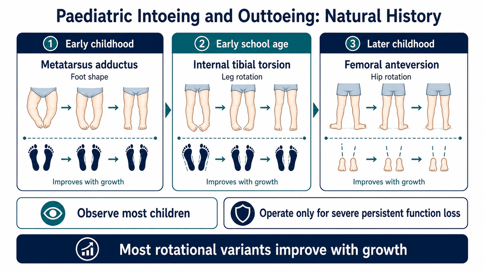
Most typical in-toeing is painless and improves with growth. The common developmental sequence is metatarsus adductus in infancy, internal tibial torsion in toddlers and femoral anteversion in preschool or early school-age children. This timeline is useful, but it is not a substitute for examination.
Reassurance should be specific. Families should hear which level is responsible, what natural history is expected, why braces or special shoes are not useful for typical torsion, and what symptoms should prompt reassessment.
Pathophysiology
The foot progression angle is the visible result of multiple segments. A foot can point inward because the forefoot is adducted, the tibia is internally rotated, the femur is anteverted, or the child has neuromuscular tone and motor-control issues. The patella is a key clue because it reflects femoral orientation more than foot orientation.
Metatarsus adductus is a foot-shape problem, often related to intrauterine packaging. Flexible forms often improve. Rigid forms require more careful foot assessment.
Internal tibial torsion is common in toddlers. During gait, the patella may face forward while the feet point inward. Many children improve as tibial rotation changes with growth.
Femoral anteversion produces increased hip internal rotation and reduced external rotation. During gait, the patellae point inward, and W-sitting is common. Most cases improve gradually, but severe persistent anteversion can cause tripping, cosmetic concern and functional limitations.
External tibial torsion and femoral retroversion can cause out-toeing. External tibial torsion may become more apparent with growth and may contribute to patellofemoral symptoms. Painful adolescent out-toeing is a different problem: the hip must be considered first, especially SUFE.
Classification
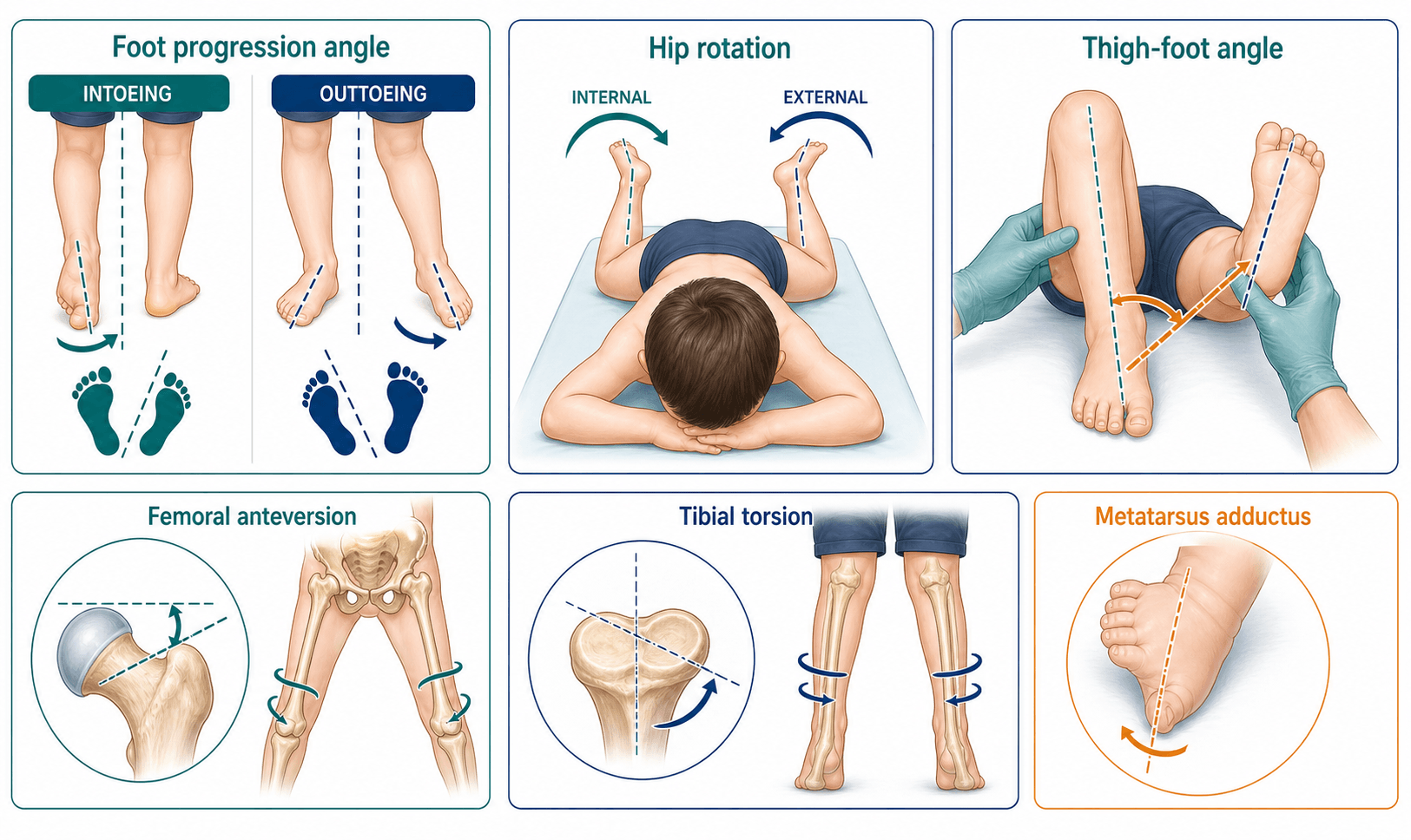
- Metatarsus adductus: curved lateral border of the foot, forefoot adduction and abnormal heel bisector.
- Internal tibial torsion: patella forward but feet point inward; measured with thigh-foot angle.
- Femoral anteversion: patellae point inward, hip internal rotation is increased and external rotation is reduced.
- Neuromuscular in-toeing: tone, weakness, asymmetry or lever-arm dysfunction changes the pattern.
Clinical Presentation
History
Ask what the family sees during real activity. A child may walk acceptably in clinic but trip when tired, run awkwardly or struggle with sport. Establish age of onset, whether the pattern is improving, symmetry, pain, functional limitation and developmental context.
Ask about:
- In-toeing or out-toeing during walking, running or fatigue.
- Tripping, falling, shoe wear, sport limitation or cosmetic concern.
- Pain location: hip, knee, shin, ankle or foot.
- Asymmetry, worsening or sudden change.
- W-sitting, sitting preferences and family history.
- Pregnancy, birth history, milestones and prematurity.
- Neurological symptoms, known cerebral palsy or developmental delay.
Examination
Observe from the front, side and behind. Estimate foot progression angle during natural walking, not just when the child is concentrating. Watch patella direction: inward-facing patellae suggest femoral anteversion, while forward-facing patellae with inward feet suggest tibial torsion.
Measure:
- Hip internal and external rotation, usually prone or supine.
- Thigh-foot angle prone with knees flexed to 90 degrees.
- Foot shape, lateral border, heel bisector and flexibility.
- Coronal alignment, limb length and knee tracking.
- Tone, reflexes, selective motor control, strength and balance when atypical.
- Hip range and obligatory external rotation in painful out-toeing adolescents.
The useful answer is not "the child in-toes". The useful answer is "this is painless symmetric internal tibial torsion" or "this is femoral anteversion with no red flags".
Investigations
Typical painless symmetric in-toeing is a clinical diagnosis and usually does not require imaging. Imaging is for red flags, painful presentations, atypical asymmetry, neuromuscular assessment, suspected hip pathology or surgical planning.
| Question | Investigation | Decision it informs |
|---|---|---|
| Typical painless in-toeing? | No routine imaging | Clinical diagnosis, reassurance and review triggers. |
| Painful adolescent out-toeing? | AP pelvis and lateral hip radiographs | Excludes SUFE or other hip pathology. |
| Severe torsion considered for surgery? | CT or MRI rotational profile | Quantifies femoral and tibial version for planning. |
| Neuromuscular gait? | Video, gait analysis and relevant imaging in selected cases | Links torsion to lever-arm dysfunction and treatment planning. |
| Coronal deformity also present? | Standing alignment radiographs | Separates rotational and coronal contributors. |
Rotational CT should not be ordered just because a toddler in-toes. It should answer a specific treatment question, usually in an older child with severe persistent functional torsion or complex deformity.
Differential Diagnosis
The differential is best worked through by level (foot, tibia, femur, hip, neuromuscular control), then by whether the picture is benign developmental variation or a pathological signal. The table below distils the discriminating features that separate look-alikes at each level.
| Diagnosis | Discriminating feature | Typical age | What separates it from look-alikes |
|---|---|---|---|
| Metatarsus adductus | Curved (convex) lateral foot border, forefoot adduction, abnormal heel bisector; hindfoot normal | Infant | Deformity is in the foot only; hip rotation and thigh-foot angle are normal. |
| Internal tibial torsion | Patella faces forward but feet point inward; internal (negative) thigh-foot angle | Toddler (1 to 3 years) | Foot and hip are normal; the rotation is in the tibial segment. |
| Femoral anteversion | Patellae point inward; hip internal rotation increased and external rotation reduced; W-sitting | Child 3 to 10 years | Patella direction (inward) distinguishes it from tibial torsion where patella faces forward. |
| Neuromuscular in-toeing (cerebral palsy) | Asymmetry, tone, brisk reflexes, toe-walking, lever-arm dysfunction | Any age | Abnormal neurology and gait quality; not a smooth symmetric developmental pattern. |
| Skewfoot or residual clubfoot | Forefoot adduction with hindfoot valgus (skewfoot) or cavus/equinus residua | Infant to child | Mid- and hindfoot deformity, often rigid; needs dedicated foot assessment and weight-bearing radiographs. |
| Diagnosis | Discriminating feature | Pain or red flag | Action |
|---|---|---|---|
| Physiological external rotation hip contracture | Bilateral out-toeing in the new walker that resolves with growth | None | Reassure and review. |
| External tibial torsion | Feet point outward with patella forward; external (positive) thigh-foot angle | May cause patellofemoral pain | Observe; consider osteotomy only if severe and symptomatic. |
| Femoral retroversion | Increased hip external rotation, reduced internal rotation | Linked to SCFE risk | Examine hip; image if painful. |
| Slipped capital femoral epiphysis (SCFE) | Obligate external rotation on hip flexion (Drehmann sign), limp, hip/thigh/knee pain | Major red flag | Non-weight bearing; urgent AP and frog-lateral pelvis radiographs. |
| Post-traumatic or post-surgical rotational malunion | Unilateral change after fracture or osteotomy | Asymmetric | Compare sides; rotational imaging if surgery considered. |
Management
| Diagnosis | Typical management | When to escalate |
|---|---|---|
| Flexible metatarsus adductus | Observation, parental stretching or casting depending flexibility and severity. | Rigid severe deformity, skin crease, failed improvement or associated foot disorder. |
| Internal tibial torsion | Observation, normal activity and explanation that braces or special shoes do not untwist the tibia. | Older child with severe persistent functional limitation after expected improvement window. |
| Femoral anteversion | Observation and reassurance when painless and symmetric. | Severe persistent in-toeing with tripping, functional limitation, marked cosmetic concern in an older child, or neuromuscular lever-arm dysfunction. |
| External tibial torsion | Assess knee symptoms, patellar tracking and neuromuscular context. | Patellofemoral pain or instability, brace intolerance, progressive external foot progression or major lever-arm dysfunction. |
| Painful adolescent out-toeing | Treat as hip pathology until proven otherwise. | Immediate pelvis and lateral hip imaging if SUFE is possible; keep non-weight bearing when suspicious. |
Typical painless in-toeing is managed with explanation, normal activity and observation. The explanation should be anatomical and practical: identify the level, explain expected improvement and list red flags. Braces, twister cables and special shoes do not reliably remodel typical femoral or tibial torsion and should not be presented as corrective treatment.
| Step | Femoral derotation | Tibial derotation |
|---|---|---|
| Indication | Severe persistent femoral anteversion or retroversion causing functional gait limitation after natural improvement is unlikely. | Severe tibial torsion causing functional limitation, patellofemoral symptoms, brace difficulty or neuromuscular lever-arm dysfunction. |
| Measurement | Clinical hip rotation plus CT or MRI version when surgery is being planned. | Thigh-foot angle, transmalleolar axis and CT/MRI torsion when surgical planning requires precision. |
| Level | Usually proximal or subtrochanteric femoral osteotomy depending surgeon preference and fixation strategy. | Usually distal tibial osteotomy for isolated tibial torsion, with fibular osteotomy when required. |
| Fixation | Plate or intramedullary fixation depending age, bone size and osteotomy level. | Plate, pins, screws or external fixation depending technique and soft-tissue risk. |
| Major risks | Malrotation, non-union, hardware irritation, hip or knee symptoms and missed combined torsion. | Compartment syndrome, peroneal nerve injury, malrotation, non-union and hardware symptoms. |
Complications
Early
- Over-investigation of normal variants.
- Anxiety from vague reassurance.
- Braces or special shoes prescribed as if they correct torsion.
- Missed SUFE in painful adolescent out-toeing.
- Missed neurological diagnosis in asymmetric or progressive gait.
Late
- Persistent severe torsion with tripping or sport limitation.
- Patellofemoral symptoms, especially with external tibial torsion.
- Lever-arm dysfunction in neuromuscular disease.
- Surgical complications after derotation osteotomy.
- Residual cosmetic concern despite normal function.
Good reassurance is not "they will grow out of it" in isolation. Good reassurance names the level, explains the expected change and gives clear review triggers.
Decision-Making in Practice
Rotational assessment is mainly a clinical skill. Most children with intoeing or out-toeing have physiological variation that improves or becomes asymptomatic, but the clinician must identify asymmetry, progression, pain, neurological disease, slipped epiphysis, patellar instability, severe functional limitation or torsion that will not remodel.
| Finding | Interpretation | Management consequence |
|---|---|---|
| Metatarsus adductus | Forefoot-driven intoeing in infancy | Observe, stretch or cast depending flexibility and severity |
| Internal tibial torsion | Toddler intoeing with inward thigh-foot angle | Usually observation; surgery only for severe persistent functional deformity |
| Femoral anteversion | Older child intoeing, high hip internal rotation and W-sitting | Usually observation; derotation osteotomy only for severe persistent disability |
| External tibial torsion | Out-toeing, patellofemoral symptoms or lever-arm dysfunction | May worsen with growth and can require osteotomy when severe |
| Asymmetry or pain | Unilateral change, limp, hip pain, knee pain or neurological signs | Investigate for pathology rather than reassuring |
A complete rotational profile records foot progression angle, hip rotation, thigh-foot angle, transmalleolar axis, heel bisector, foot shape and gait. The examination should be performed prone and walking, with comparison between sides. Radiographs are not needed for every intoeing child; they are used when there is pain, asymmetry, deformity outside the expected pattern, suspected hip pathology, neuromuscular disease or surgical planning.
Derotation osteotomy is a functional operation, not a cosmetic operation. Indications include severe persistent deformity causing tripping, pain, brace difficulty, patellofemoral instability, lever-arm dysfunction or major gait limitation after physiological improvement is no longer expected.
Guidelines, Registries & Global Practice
Rotational concerns are among the most common reasons children are referred to orthopaedics worldwide, yet they generate little registry data because most are treated non-operatively. The clinically important point for any global exam is that named societies broadly agree on a conservative, profile-based approach, with surgery reserved for the rare severe symptomatic older child.
Global epidemiology
- In-toeing peaks in toddlers and preschoolers and is one of the commonest paediatric musculoskeletal referrals across all health systems.
- Internal tibial torsion is the commonest cause of in-toeing in toddlers; femoral anteversion predominates in children aged roughly 3 to 10 years; metatarsus adductus presents in infancy.
- Out-toeing is less common than in-toeing and tends to occur in older children; painful adolescent out-toeing carries a disproportionate risk burden because of SCFE.
- Femoral version decreases from around 30 to 40 degrees at birth to roughly 15 degrees by skeletal maturity, which underpins the natural-history counselling given everywhere.
Side-by-side guidance
| Source | Imaging stance | Treatment emphasis |
|---|---|---|
| AAOS / OrthoInfo (US) | No routine imaging for typical in-toeing; image for atypical or painful cases. | Reassurance and observation; braces, casts and special shoes not recommended for typical torsion. |
| BOA / BSCOS (UK) | Clinical diagnosis for benign torsion; radiographs reserved for red flags and suspected hip pathology. | Conservative management with clear safety-netting; specialist referral for pain, asymmetry or progression. |
| AO Foundation / paediatric trauma & deformity | Cross-sectional (CT or low-dose) version measurement when surgery is planned. | Derotation osteotomy at the correct level for severe symptomatic deformity; precise correction and fixation. |
| EFORT / European paediatric consensus | Selective imaging; emphasises rotational profile and gait assessment. | Observation for physiological variants; multilevel planning and gait analysis for neuromuscular lever-arm dysfunction. |
| SICOT (global) literature | CT confirmation of version before osteotomy in symptomatic anteversion. | Derotation osteotomy effective in carefully selected symptomatic children (Naqvi et al.). |
Registry and practice notes
- There is no dedicated international registry for paediatric rotational deformity because the overwhelming majority resolve without surgery; high-quality outcome data come from single-centre series and gait-analysis cohorts rather than arthroplasty-style registries.
- Where derotation osteotomy is performed, most published evidence is Level IV (case series), so practice variation in surgical thresholds is wide and driven by symptom severity and surgeon judgement.
High- versus limited-resource practice variation
- In well-resourced settings, instrumented 3D gait analysis and CT/MRI version measurement support surgical planning, especially in cerebral palsy single-event multilevel surgery.
- In limited-resource settings, the clinical rotational profile (foot progression angle, hip rotation, thigh-foot angle, heel bisector) remains the backbone; it is accurate, free and exam-relevant everywhere. Plain radiographs are universally available for the critical task of excluding SCFE in painful out-toeing.
- The one universal, resource-independent rule: painful adolescent out-toeing is a hip problem until pelvic radiographs prove otherwise.
Controversies and Areas of Uncertainty
- Surgical threshold for idiopathic anteversion. There is no agreed degree of version or foot progression angle that mandates surgery. Decisions rest on function and symptoms in an older child, and the evidence base is largely uncontrolled case series.
- Value of routine version imaging. CT quantifies version but adds radiation; low-dose CT, biplanar (EOS-type) imaging and MRI are increasingly preferred, but no single modality is universally mandated.
- Effectiveness of conservative devices. Twister cables, derotation braces and orthopaedic shoes are not supported for typical torsion; their continued use in some settings reflects parental expectation rather than evidence.
- Predictability of femoral derotation osteotomy. Outcomes are reported as more predictable in adults than children for neuromuscular internal-rotation gait, and recurrence or under-correction can occur; this informs timing and counselling.
- Miserable malalignment. Combined femoral anteversion and external tibial torsion can co-exist and partly cancel out clinically while still causing patellofemoral symptoms; isolated single-level correction may be insufficient and combined torsion must be actively excluded before surgery.
Evidence Signals
Most rotational variations are benign but must be classified clinically
- In-toeing is usually metatarsus adductus in the infant, internal tibial torsion in the toddler and femoral anteversion in the child younger than 10 years.
- Out-toeing patterns largely result from external rotation hip contracture, external tibial torsion and external femoral torsion.
- A deliberate rotational profile assessment is needed to separate benign variation from true structural abnormality.
Normative rotational profile values guide management
- 1,000 normal lower extremities were studied to establish age-related normal values for the rotational profile.
- Out-toeing in infants, medial tibial torsion in toddlers and medial femoral torsion in young children are extremes of a normal developmental pattern.
- In the vast majority, these rotational variations fall within the broad range of normal and require no treatment.
Derotation osteotomy resolves in-toeing in selected symptomatic children
- 21 children (35 limbs, mean age 13.3 years) with symptomatic idiopathic femoral anteversion (mean version 40.8 degrees on CT) underwent proximal femoral derotation osteotomy.
- Foot progression angle improved from 15.2 degrees internal to 7.7 degrees external and tripping, falling and hip pain resolved in all patients.
- Knee pain failed to improve in 3 patients, underlining careful patient selection.
Clinical Reasoning Notes
Structured clinical approach
Start with the gait and localise:
- "The child has inward foot progression during gait."
- "The patellae face forward, so the main contribution appears tibial."
- "The thigh-foot angle confirms internal tibial torsion."
- "There is no pain, asymmetry, progression or neurological sign."
- "This is a developmental variant; I would reassure and avoid braces."
For a different child:
- "The adolescent has painful out-toeing and a limp."
- "I would not call this external tibial torsion without assessing the hip."
- "I would obtain AP pelvis and lateral hip imaging to exclude SUFE."
Common pitfalls
- Calling every in-toeing femoral anteversion.
- Measuring foot progression but not hip rotation or thigh-foot angle.
- Ignoring patella direction during gait.
- Ordering CT for a typical toddler.
- Missing adolescent SUFE.
- Prescribing braces as if they change bone version.
- Operating too young for cosmetic concern.
- Forgetting neurological screening in asymmetric cases.
Evidence Base
Primary-care framework for lower extremity rotational and angular problems
- In-toeing is caused by metatarsus adductus, internal tibial torsion and femoral anteversion; out-toeing by external tibial torsion and femoral retroversion.
- Most children have rotational and angular findings within two standard deviations of the mean and resolve spontaneously with growth.
- Orthotics are not beneficial, radiographs are not routinely required, and surgery is rarely needed (typically over age 8 with severe functional deformity).
In-toeing is commonly over-treated with braces and special footwear
- The usual causes of in-toeing are excessive femoral anteversion, internal tibial torsion and metatarsus adductus.
- Because of poor understanding of natural history, in-toeing is commonly over-treated with braces or special footwear.
- Early diagnosis of pathological causes such as cerebral palsy and developmental dysplasia of the hip is essential.
Out-toeing and obligate external rotation should be presumed SCFE until excluded
- Any child with hip, thigh or knee pain, or with out-toeing, decreased internal rotation or obligate external rotation on flexion, should be presumed to have SCFE until proven otherwise.
- There is a high rate of diagnostic delay, allowing deformity progression and conversion of stable to unstable slips with higher avascular necrosis risk.
- Diagnosis is confirmed with AP and frog-lateral pelvis radiographs and treatment is urgent and surgical.
Drehmann sign reflects proximal femoral deformity and impingement in SCFE
- In 92 SCFE hips, a positive Drehmann sign (obligatory external rotation with hip flexion) correlated with higher alpha-angle and radiologic cam-type femoroacetabular impingement.
- Drehmann sign was present in 25 percent, 75 percent and 100 percent of mildly, moderately and poorly remodelled hips respectively.
- The sign is a valuable clinical marker of the anatomic change underlying out-toeing after SCFE.
AAOS family education on in-toeing
- Common causes of in-toeing include metatarsus adductus, tibial torsion and femoral anteversion.
- Most children improve without braces, casts or surgery.
- Persistent severe functional problems require specialist review.
Clinical Decision Scenarios
Practise clinical reasoning and management decisions out loud
“A three-year-old has painless symmetric in-toeing and trips occasionally. How do you assess and counsel?”
“An adolescent presents with painful out-toeing and a limp. What must you exclude?”
“A 10-year-old has marked in-toeing with inward-facing patellae, trips frequently when running and is teased at school. Hip internal rotation is 80 degrees and external rotation is 10 degrees. Parents ask for surgery. How do you decide?”
In-toeing
- Metatarsus adductus
- Internal tibial torsion
- Femoral anteversion
- Neuromuscular
Measure
- Foot progression
- Patella direction
- Hip rotation
- Thigh-foot angle
- Foot shape
Red Flags
- Pain
- Asymmetry
- Progression
- Neurology
- Adolescent out-toeing
Treat
- Reassure typical
- No special shoes
- Image red flags
- Gait analysis selected
- Derotation selected older child