Post-traumatic calcification at the MCL femoral attachment | AP knee radiograph is diagnostic | Usually self-limiting | Surgery only for refractory cases
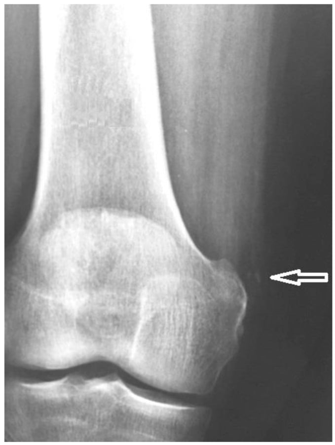
- What it is: post-traumatic calcification or ossification at or near the femoral attachment of the medial collateral ligament of the knee, classically seen as a curvilinear density on AP radiograph medial to the femoral condyle
- Mechanism: typically follows an MCL sprain or direct blow to the medial knee; the calcification appears 3 to 6 weeks after injury and may represent heterotopic ossification within the damaged ligament
- Clinical picture: medial knee pain, stiffness, and restricted flexion after a recent knee injury; local tenderness over the medial femoral epicondyle; the patient may have forgotten the original injury
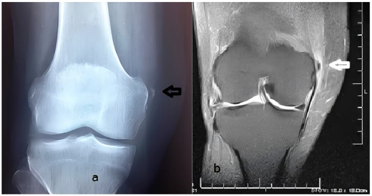
- Imaging: AP radiograph of the knee is usually diagnostic - look for calcification or ossification medial to the femoral condyle at the MCL origin; MRI shows low-signal deposit and assesses MCL integrity
- Management: predominantly conservative (rest, protected weight-bearing, physiotherapy, NSAIDs); surgical excision is rarely needed and only for refractory cases after at least 6 to 12 months of failed conservative treatment
- “The Pellegrini-Stieda lesion is one of the few orthopaedic diagnoses you can make from a single AP knee radiograph - look for the characteristic curvilinear calcification medial to the femoral condyle
- “Named after Pellegrini (1905) who described the radiographic finding and Stieda (1908) who described the associated medial knee injury - examiners love a quick historical note
- “Do NOT confuse with a medial femoral condyle avulsion fracture: the Pellegrini-Stieda calcification is separate from the cortex and lies within the MCL substance, whereas an avulsion fragment is a detached piece of bone
Overview
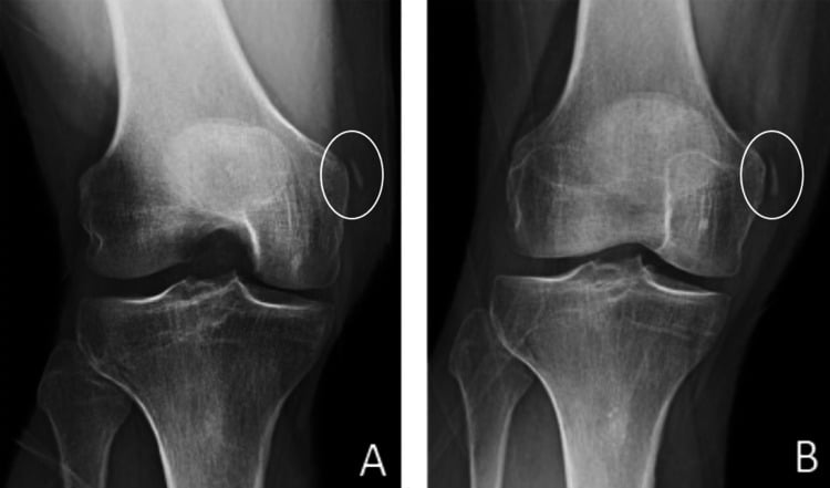
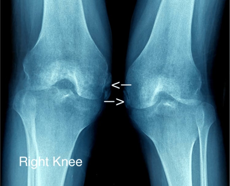
The Pellegrini-Stieda lesion is post-traumatic calcification or ossification at or near the femoral attachment of the medial collateral ligament (MCL) of the knee. On the AP radiograph it is a curvilinear or comma-shaped density lying medial to the femoral condyle, and it is one of the classic spot diagnoses in orthopaedic radiology.
What it is. Neither a true fracture nor a ligament tear, it is a form of heterotopic ossification: new bone formed within the substance of the injured MCL. "Pellegrini-Stieda disease" and "Pellegrini-Stieda syndrome" are used interchangeably with "lesion" and name the same process, not a separate disease entity.
The name. Pellegrini (1905) first described the radiographic appearance of medial knee calcification, and Stieda (1908) reported on the associated medial knee injury.
The story. It typically develops weeks after an MCL sprain or a direct blow to the medial knee. The calcification is not visible on the initial radiograph and typically appears 3 to 6 weeks after the injury, which is itself a diagnostic clue.
Where it comes up. The lesion joins anatomy (the MCL origin), pathology (heterotopic ossification), radiology (reading the AP knee) and management (conservative patience) in a single topic, and it can surface in several parts of the exam:
- Radiology viva - a spot diagnosis on the AP knee, to be told apart from avulsion fractures and other calcifications
- Sports medicine - a complication of MCL injuries in athletes, and one reason some medial knee sprains take longer than expected to settle
- Trauma - it follows knee injuries including fractures and ligament disruptions, and its delayed appearance teaches the timeline of heterotopic ossification
- Basic science - a way into the biology of heterotopic ossification: mesenchymal stem cell differentiation, BMP signalling and mineralisation
- Operative surgery - a rare indication for excision, but the principles (protect the MCL, excise the mass, rehabilitate) are examinable
Anatomy
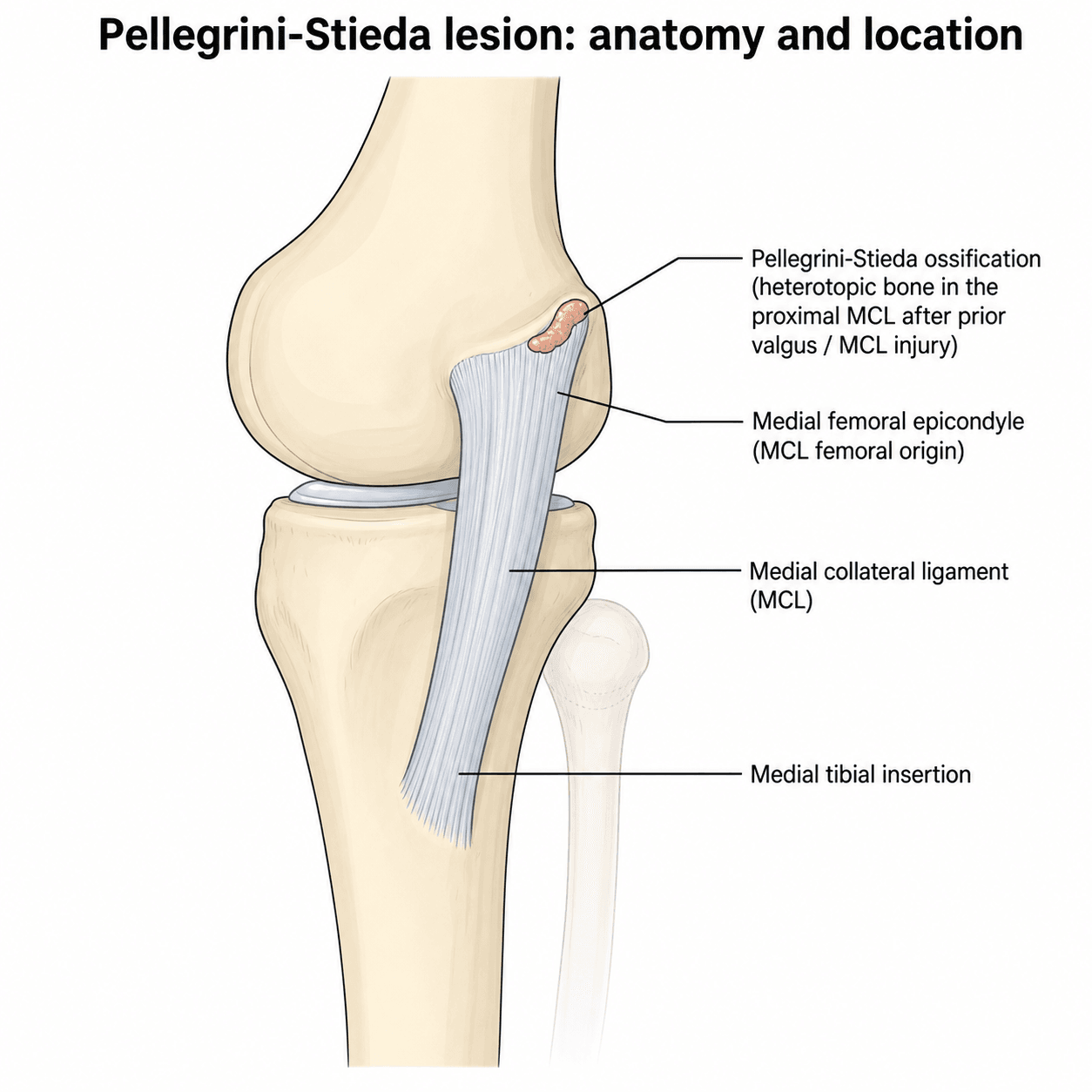
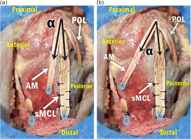
The ligament. The MCL is a broad, flat ligament on the medial side of the knee. It originates from the medial femoral epicondyle and inserts onto the medial tibial metaphysis, about 5 to 7 cm distal to the joint line.
Two layers. The superficial MCL (tibial collateral ligament) is the larger, more superficial portion, running from the medial epicondyle to the proximal tibia. The deep MCL blends with the joint capsule and attaches firmly to the medial meniscus.
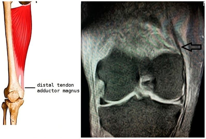
Where the lesion forms. At or near the femoral attachment, where the ligament is tightly bound to the medial epicondyle. The adductor magnus tendon and the medial head of gastrocnemius lie close by and may contribute to the pattern of calcification in some cases.
Pathophysiology
The prevailing theory. The exact mechanism remains debated, but the lesion is thought to be heterotopic ossification within the damaged MCL, forming in a sequence:
- Trauma - an MCL sprain (often a valgus injury) or a direct blow to the medial knee damages the ligament fibres at or near the femoral attachment
- Inflammatory response - the damaged tissue releases inflammatory mediators and growth factors that stimulate mesenchymal stem cell differentiation into osteoblasts; BMP signalling is the key molecular driver
- Mineralisation - over the following weeks calcium is deposited in the damaged tissue, forming an immature calcific deposit that progressively matures
- Maturation - the deposit may mature into true lamellar bone with organised trabeculae, or it may remain as amorphous calcification
Familiar biology. The process parallels the heterotopic ossification seen after hip arthroplasty and spinal cord injury.
The time course. The delay between injury and a visible deposit is a hallmark of the lesion, and it is what distinguishes it from an acute avulsion fracture:
- 0 to 2 weeks - injury; the initial radiograph is normal; inflammatory phase with local swelling and pain
- 2 to 6 weeks - the calcification becomes visible on the radiograph; the patient may notice persistent medial knee pain and stiffness
- 6 weeks to 6 months - the calcification may enlarge or mature; symptoms typically plateau
- 6 to 12 months - gradual resorption in most cases; the calcification may partially or fully resolve
Classification
There is no single universally accepted classification. The most practical approach is to describe the lesion by its radiographic maturity, which correlates with clinical behaviour and management.
- Radiograph
- Thin, faint, curvilinear calcification adjacent to the medial femoral epicondyle
- Timing after injury
- 2 to 6 weeks
- Clinical
- Often asymptomatic, or mild local pain and stiffness
- Management and prognosis
- Usually resolves with conservative treatment over weeks to months; excellent prognosis, most settle without intervention
- Radiograph
- Denser, more prominent calcification or ossification; may be rounded, oval or comma-shaped
- Timing after injury
- 6 weeks to 6 months
- Clinical
- May cause persistent medial knee pain and restricted flexion
- Management and prognosis
- Conservative treatment (physiotherapy, NSAIDs) often effective but takes longer; good with patience, but recovery is measured in months
- Radiograph
- Well-formed bony spur or beak projecting from the medial femoral epicondyle; may show cortical continuity and a trabecular pattern
- Timing after injury
- More than 6 months
- Clinical
- May remain symptomatic with pain and a mechanical block to flexion
- Management and prognosis
- Surgical excision considered if refractory to prolonged conservative treatment (at least 6 to 12 months); surgical outcomes generally good, but recurrence is possible
If asked to classify, describe what you see on the radiograph (thin and curvilinear vs dense and beak-like) and relate it to the stage of maturation. The practical implication is that early lesions almost always settle, while mature beak-type lesions are the ones that occasionally need surgery.
Clinical Presentation
History. A preceding MCL injury (valgus stress, a direct blow or a sporting injury) 3 to 6 weeks earlier, which the patient may have forgotten or considered minor. Medial knee pain comes on gradually and worsens with activity, and may be worse at night or after exercise. Stiffness makes bending difficult, especially beyond 90 degrees of flexion: pain and loss of flexion from the calcific mass are the main clinical problem.
Examination. Look for:
- Localised tenderness over the medial femoral epicondyle, the MCL origin
- Restricted knee flexion compared with the contralateral side
- Mild medial joint-line swelling or fullness
- Pain on valgus stress testing, with the MCL usually stable rather than lax (see the next section)
- A limp from pain and stiffness
Not every lesion hurts. Many are discovered incidentally on radiographs taken for other reasons and cause no symptoms. Others present late: because the calcification appears weeks after the injury, the patient returns with medial knee pain that was expected to have resolved by now.
Bilateral lesions are rare but possible, typically in patients with bilateral knee injuries or systemic conditions predisposing to heterotopic ossification.
Assessing the Underlying MCL: Valgus Stress Testing
Interpreting the valgus stress test is the key examination skill for the underlying MCL injury that produced the lesion. Test at 30 degrees of flexion and in full extension, then grade the injury:
- Finding
- Medial joint-line opening with a firm endpoint
- Interpretation
- ISOLATES the superficial MCL (cruciates and posterior capsule relaxed) - the standard test for MCL injury
- Finding
- Medial opening in full extension
- Interpretation
- A RED FLAG - implies posteromedial-corner and/or cruciate involvement, i.e. a more severe combined injury, not an isolated MCL sprain
- Finding
- Tenderness, no laxity, firm endpoint
- Interpretation
- Partial fibre injury; heals quickly
- Finding
- Increased laxity at 30 degrees but a firm endpoint
- Interpretation
- Partial tear
- Finding
- Laxity with a soft or absent endpoint
- Interpretation
- Complete tear (most isolated grade III MCL injuries still heal non-operatively in a hinged brace)
In the Pellegrini-Stieda knee. Valgus stress at 30 degrees typically reproduces pain, but the ligament is stable: the calcification sits within a healed MCL. Frank laxity instead signals ongoing MCL insufficiency.
Always test in FULL EXTENSION too. Medial opening there points to a posteromedial-corner or cruciate injury and changes the whole management, so it must not be missed.
Investigations
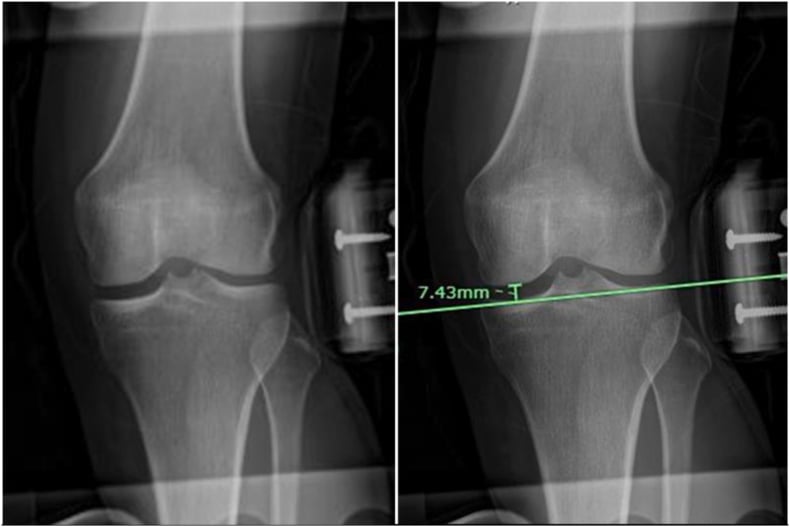
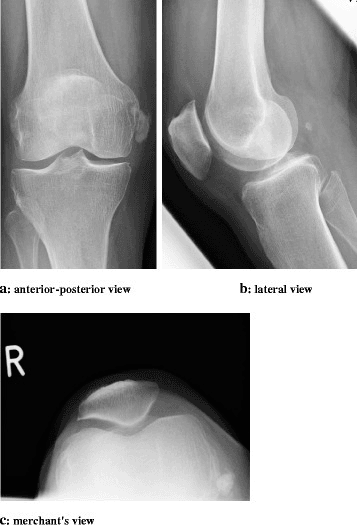
Plain radiographs. The AP knee is first-line in any patient with medial knee pain after injury, and often the only investigation needed. The lesion is a curvilinear or comma-shaped area of calcification or ossification just medial to the femoral condyle, at the expected site of the MCL femoral attachment. It lies within the MCL substance, separate from the femoral cortex (unlike an osteophyte or an avulsion fragment) and not extending into the joint (unlike a loose body), although a mature beak-type lesion may show cortical continuity.
The lateral view is always obtained alongside the AP as part of the standard knee series, but it is less helpful. The calcification is superimposed on the femur, though a large deposit may be projected anteriorly.
MRI is not routinely required but is useful when:
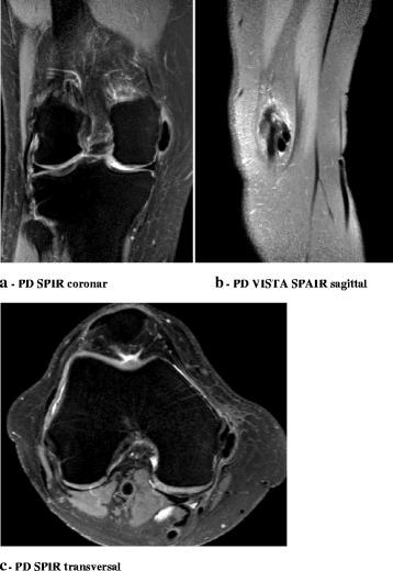
- the diagnosis is uncertain, to confirm the calcification lies within the MCL and exclude other pathology
- there is concern about associated ligament or meniscal injury
- surgical excision is being considered, to plan the approach and assess MCL integrity
The lesion returns low signal on all sequences (T1, T2 and PD) at the MCL femoral attachment, consistent with calcification. There may be surrounding soft-tissue oedema in the acute phase.
CT is occasionally used for pre-operative planning. It defines the exact size, shape and location of a mature ossified lesion and its relationship to the femoral cortex, which helps plan the approach and the extent of excision.
Differential diagnosis. The most common mistake is confusing the lesion with a medial femoral condyle avulsion fracture or a medial meniscus calcification. The alternatives, and what separates them:
- Avulsion fracture of the medial epicondyle - a detached piece of cortical bone rather than calcification within the MCL; if a density is seen on a film taken immediately after trauma, think avulsion instead
- Chondrocalcinosis (CPPD) - calcification within the meniscus or articular cartilage, inside the joint rather than at the MCL
- Loose body in the medial gutter - a mobile calcified body within the joint, not fixed at the MCL attachment
- Femoral cortex irregularity - a cortical ridge or groove may mimic the lesion; look for continuity with the cortex
- Calcific tendinitis of the medial gastrocnemius - can cause medial knee pain, but the calcification is more posterior and distal
- Infection or osteomyelitis - periosteal reaction at the medial condyle, with systemic signs and a different clinical picture
- Other soft-tissue calcification - from renal failure, tumoral calcinosis or previous surgery
Correlate the radiographic finding with the clinical history and examine the patient. If the calcification is not at the MCL femoral attachment, or the story does not fit, consider the alternative diagnoses.
Management
Conservative management is the mainstay. The vast majority of lesions resolve with non-operative treatment, most settling over 3 to 12 months, and patience is key before considering surgery. Treatment follows a progressive approach.
Acute phase (0-6 weeks). Rest from provocative activities, with protected weight-bearing on crutches if needed, to avoid stressing the healing MCL; a hinged knee brace protects it from valgus stress. Simple analgesia or NSAIDs control pain and inflammation, and cryotherapy (ice) reduces local swelling. Avoid passive stretching or forced flexion.
Subacute phase (6 weeks to 3 months). Gentle active-assisted range-of-motion exercises regain flexion without aggravating the calcification. Progress weight-bearing as tolerated, strengthen the quadriceps and hamstrings, and continue NSAIDs if needed. The calcification may now be visible on the radiograph, and is expected at this stage.
Remodelling phase (3-12 months). Progressive strengthening and stretching, then a gradual return to sport and full activity. Serial radiographs may show a gradual reduction in the size of the calcification, and most patients achieve full or near-full recovery by 6 to 12 months. Document range of motion and pain scores throughout, because that record is what justifies the timing of any intervention.
When to operate. The indications for excision are:
- persistent, functionally limiting pain despite at least 6 to 12 months of conservative treatment
- significant restriction of knee flexion (a mechanical block) from a large, mature lesion
- a beak-type (Type III) lesion causing impingement or a mechanical block
Surgical technique. Excision of the calcified or ossified mass, with careful preservation of the MCL:
- Medial incision over the area of calcification
- Identify and protect the MCL fibres
- Carefully excise the calcific or ossific mass from within the ligament substance
- Preserve or repair the MCL if its integrity is compromised
- Immobilise in a hinged brace after surgery, followed by gradual rehabilitation
Outcomes. Reported results of excision are generally good, but the evidence base is limited to small case series and case reports. The key principle is to exhaust conservative treatment first.
In a viva, always start with conservative management and explain why you would persevere with it. Only mention surgery as a last resort, after documenting failure of at least 6 to 12 months of non-operative treatment. The examiner wants to hear that you understand the natural history and would not rush to operate.
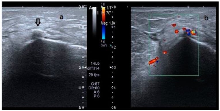
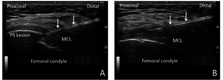
Percutaneous treatment. Ultrasound-guided percutaneous treatment of the deposit has been reported in individual cases (see the Evidence Base).
Complications and Prognosis
- Frequency
- Common in the first 3-6 months; most settle with time
- Management
- Continue conservative treatment; NSAIDs; reassure patient about expected time course
- Frequency
- Common, especially with larger or beak-type lesions
- Management
- Gentle physiotherapy; surgical excision if mechanical block persists beyond 6-12 months
- Frequency
- Rare - the underlying ligament is usually intact
- Management
- Assess MCL stability clinically; brace if mild laxity; consider MCL reconstruction if unstable
- Frequency
- Reported in some case reports
- Management
- Careful surgical technique with preservation of MCL fibres; post-operative bracing; consider NSAID prophylaxis
- Frequency
- Very common - many lesions leave a small residual calcific density
- Management
- No treatment needed if the patient is pain-free and functional
Prognosis. The overall prognosis is very good. Most lesions resolve or become asymptomatic within 6 to 12 months of conservative management.
Heterotopic Ossification: Prophylaxis and Context
- How it works
- Inhibit prostaglandin-mediated osteoblast differentiation from mesenchymal precursors
- Practical use
- A short course (commonly about 2 to 6 weeks) after a high-risk injury or after excision; gastrointestinal and renal caution
- How it works
- Same anti-prostaglandin effect with less gastrointestinal risk
- Practical use
- Alternative where indomethacin is poorly tolerated
- How it works
- Blocks proliferation of pluripotent mesenchymal cells
- Practical use
- Reserved for very high-risk cases (mainly hip surgery), given within roughly 24 hours before to 72 hours after surgery
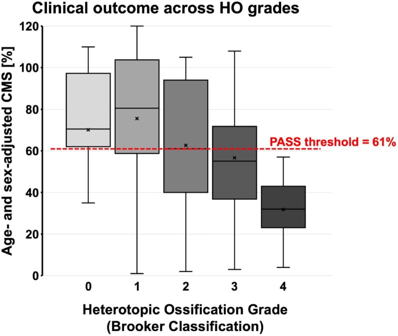
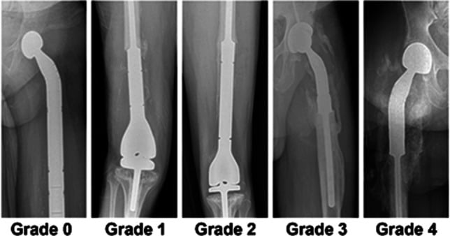
Grading. The reference grading for heterotopic ossification, around the hip, is the Brooker classification:
- I - islands of bone in soft tissue
- II - spurs leaving over 1 cm between opposing surfaces
- III - spurs leaving under 1 cm
- IV - apparent ankylosis
Risk factors include male sex, prior heterotopic ossification, ankylosing spondylitis or DISH, and extensive surgical dissection.
For the Pellegrini-Stieda lesion specifically there is NO high-level evidence for prophylaxis. A short post-injury or post-excision NSAID course is used by extrapolation to reduce formation or recurrence.
Guidelines, Registries and Global Practice
- No specific guidelines exist for the Pellegrini-Stieda lesion from any major orthopaedic society (AAOS, BOA, EFORT). Management is based on expert opinion, case series, and general principles of heterotopic ossification treatment.
- Conservative management is the universal standard across all regions: rest, protected mobilisation, physiotherapy, and NSAIDs. The time course and expected recovery are the same regardless of geography.
- Surgical excision is reported in case series from multiple countries (Japan, India, Europe, North America) with broadly similar indications: persistent symptoms after at least 6 to 12 months of failed conservative treatment, with a mechanical block from a mature lesion.
- Prevention: some surgeons recommend a short course of NSAIDs after MCL injury to reduce the risk of heterotopic ossification, extrapolating from the hip arthroplasty literature, but there is no high-quality evidence specific to the Pellegrini-Stieda lesion.
- Global epidemiology: the lesion is reported worldwide with no clear geographic or ethnic predilection. It is more common in athletes and active individuals who sustain MCL injuries, and may be under-diagnosed in regions where knee injuries are managed non-operatively without follow-up radiography.
- Minimally invasive techniques such as ultrasound-guided percutaneous lavage and extracorporeal shockwave therapy have been reported in individual cases, but remain experimental without Level 1 or 2 evidence.
Exam Viva Scenarios
Practise clinical reasoning and management decisions out loud
“The examiner shows you an AP knee radiograph with a curvilinear calcification medial to the femoral condyle. They ask: 'What is this and what is its significance?'”
“A 28-year-old footballer presents with persistent medial knee pain and stiffness 8 weeks after a valgus injury to the knee. Initial X-rays at the time of injury were normal. Examination shows tenderness over the medial femoral epicondyle and flexion limited to 90 degrees. How do you investigate and manage this?”
“A 45-year-old woman has had medial knee pain and progressive loss of flexion for 14 months after a fall onto the medial aspect of her knee. AP radiograph shows a well-formed bony spur projecting from the medial femoral epicondyle. Flexion is limited to 80 degrees. She has had physiotherapy and NSAIDs for 10 months without improvement. What is your management?”
Definition and Pathology
- Post-traumatic calcification or ossification at the MCL femoral attachment
- A form of heterotopic ossification within the damaged ligament
- Appears 3-6 weeks after injury on radiograph
- Named after Pellegrini (1905) and Stieda (1908)
Radiographic Appearance
- AP knee: curvilinear or comma-shaped calcification medial to femoral condyle
- Separate from the femoral cortex (not an avulsion fracture)
- Three stages: thin/curvilinear (early) to dense (established) to bony spur/beak (mature)
- MRI: low signal on all sequences at MCL origin
Clinical Features
- Medial knee pain 3-6 weeks after MCL sprain or direct blow
- Tenderness over the medial femoral epicondyle
- Restricted knee flexion (may be a mechanical block in mature lesions)
- MCL is usually stable on valgus testing
Management
- Conservative first: rest, brace, physiotherapy, NSAIDs
- Most resolve in 3-12 months
- Surgery only after 6-12 months of failed conservative treatment
- Surgical excision: medial approach, protect MCL, remove calcific mass
Differential Diagnosis
- Avulsion fracture of the medial epicondyle (cortical fragment, present on initial X-ray)
- Chondrocalcinosis / CPPD (calcification in meniscus or cartilage)
- Medial loose body (mobile, within the joint)
- Soft tissue calcification from renal failure or tumoral calcinosis
Evidence Base
The Pellegrini-Stieda Lesion of the Knee: An Anatomical and Radiological Review
- Cadaveric study of 6 fresh-frozen knees: the superficial MCL origin (n=3) or the gastrocnemius muscle origin (n=3) was marked with radio-opaque fluid and imaged to recreate the conditions under which Pellegrini and Stieda studied the lesion
- On conventional radiographs the markings showed a random distribution; the Stieda/gastrocnemius group showed no overlap of markings at all
- No position comparable to the original Pellegrini and Stieda images could be reproduced from the cadaveric markings
- Conventional X-ray of the knee could NOT reproduce a distinction between the superficial MCL and the gastrocnemius as the origin of the lesion
Ultrasound-guided Percutaneous Lavage as Treatment for Pellegrini-Stieda Syndrome
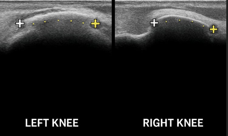
- Case report of a 52-year-old woman with one week of severe right medial knee pain and stiffness from a calcific Pellegrini-Stieda lesion within the MCL, treated with ultrasound-guided percutaneous lavage (UGPL / barbotage)
- The technique involves needle aspiration and saline lavage of the calcific deposit under ultrasound guidance, similar to the treatment of calcific tendinitis of the shoulder
- Pain-free walking and bending at follow-up; Lower Extremity Functional Scale improved to 64/80 at 1 week and 72/80 at 1 month, with no complications
- The authors proposed that the pathomechanism may be similar to calcific tendinitis of the rotator cuff (reactive calcification with pre-calcific, formative, resorptive and healing phases)
Unicompartmental Knee Arthroplasty for Knee Osteoarthritis With the Pellegrini-Stieda Lesion: A Case Report
- Case report of end-stage medial-compartment knee osteoarthritis with a co-existing Pellegrini-Stieda lesion (ossification at the MCL origin) treated by unicompartmental knee arthroplasty (UKA)
- During postoperative follow-up the ossification lesion gradually DISAPPEARED and did not relapse
- The authors attributed the resolution to UKA correcting the abnormal biomechanics of the varus osteoarthritic knee
- A co-existing Pellegrini-Stieda lesion did not preclude a successful UKA
The Pellegrini-Stieda Lesion Dissected Historically
- Historical and systematic review (4,997 PubMed/MEDLINE articles screened, 37 analysed) tracing the original descriptions by Pellegrini (1905) and Stieda (1908) and the evolving views on the lesion's origin
- Proposed origins of the medial knee calcification include the MCL, medial gastrocnemius, adductor magnus, vastus medialis, and the deep/superficial MCL
- The MCL was most often cited as the origin (54% overall; 25% on MRI, 57% at surgery), but many cases remained undecided (e.g. 50% on MRI)
- The eponym fits because Pellegrini and Stieda held two different views on the origin; MRI is a noninvasive and relatively accurate method to determine it