Complete separation of the superior from the inferior hemipelvis through the acetabulum — the most demanding acetabular revision scenario
- Definition: complete separation of the superior hemipelvis (ilium) from the inferior hemipelvis (ischium and pubis) through the acetabulum — a fracture or non-union through both anterior and posterior columns
- Risk factors: female sex, rheumatoid arthritis, pelvic irradiation, massive osteolysis, prior acetabular fracture, multiple revisions
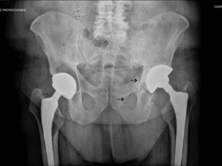
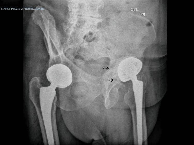
- Radiographic triad on AP pelvis: visible fracture line crossing anterior AND posterior columns, break in Kohler (ilioischial) line, asymmetry or rotation of the obturator ring; confirm with Judet views and CT with metal artefact reduction
- Intraoperative confirmation: after implant removal, apply a stress to the inferior hemipelvis (lever on the ischium or push on superior dome) and look for independent motion between superior and inferior segments
- Treatment philosophy splits into COMPRESSION (aim for union: plating plus cup, distraction) versus BRIDGING/BYPASS (accept non-union: cup-cage, custom triflange)
- Structural allograft plus conventional cage is a historic solution with high mechanical failure because the cage has no biologic fixation and fatigues
- “A hemispherical cup alone — however large — cannot reliably treat chronic discontinuity: micromotion at the non-union causes early loosening
- “The acetabular distraction technique deliberately springs the discontinuity open by 2 to 3 mm so that elastic recoil of the pelvis compresses the oversized porous cup
- “In the cup-cage, the cage protects the porous shell from load while ingrowth occurs; the polyethylene liner is CEMENTED into the cage so version can be adjusted independently of cage position
- “The ischial limb of a cup-cage may be slotted ONTO or INTO the ischium; intraosseous placement risks less sciatic nerve irritation from a prominent flange
Discontinuity missed on planning films leads to intraoperative disaster: a jumbo cup impacted into an unrecognised discontinuity displaces the inferior hemipelvis and cannot obtain stability. Scrutinise Kohler line and obturator ring on every revision AP pelvis; obtain CT with metal artefact reduction for any Paprosky IIIA or IIIB defect.
Biology dictates strategy. Acute fracture in healthy bone: compress and heal (plate plus cup). Chronic discontinuity with dead, irradiated or rheumatoid bone: union will not occur — bridge it with a cup-cage or custom triflange. Applying a compression-only strategy to non-healing bone is a classic failure pathway.
The sciatic nerve is at risk during ischial dissection for cage or triflange limbs and during distraction/lengthening of a chronically shortened limb. Intrapelvic structures (external iliac vessels, obturator neurovascular bundle) are at risk from medial screws, retained cement and quadrant violations — remember the Wasielewski quadrant system.
Massive osteolysis causing discontinuity may be septic. Full infection workup — inflammatory markers, aspiration with cell count and culture — before any single-stage reconstruction. Reconstructing a discontinuity onto infected bone guarantees failure.
Definition, Pathoanatomy and Aetiology
Pelvic discontinuity is a complete separation of the superior hemipelvis (ilium and acetabular dome) from the inferior hemipelvis (ischium and pubis), through the acetabulum, involving both the anterior and posterior columns. It represents either an acute fracture, an established non-union, or gradual dissolution of the columns by osteolysis.
Distinguish two entities:
- Acute intraoperative discontinuity — created during component removal or cup impaction, usually in reasonable-quality bone. Behaves like an acute transverse-pattern acetabular fracture: high healing potential if compressed and stabilised.
- Chronic discontinuity — established at presentation, usually with severe associated bone loss (classically Paprosky IIIB, occasionally IIIA). The fracture surfaces are sclerotic, atrophic or osteolytic; union potential is low, particularly in irradiated bone.
Risk factors
- Female sex (thinner columns, osteoporosis) — the large majority of reported cases are women
- Rheumatoid and other inflammatory arthropathies
- Prior pelvic irradiation (osteonecrotic, avascular bed — essentially zero healing potential)
- Massive particle-induced osteolysis
- Prior acetabular fracture, prior multiple revisions, developmental dysplasia with deficient columns
The two hemipelves move independently under load — the inferior segment typically flexes and internally rotates relative to the ilium. Any implant fixed to only one segment, or spanning the gap without rigid fixation to both, experiences cyclical micromotion at the interface, preventing ingrowth and causing early loosening or implant fracture.
Classification and Decision Framework
- Best indication
- Acute intraoperative discontinuity; chronic discontinuity with good bone quality and healing potential
- Principle
- Compression osteosynthesis of the discontinuity — treat it as fracture surgery: compress and heal
- Fixation
- Plate across the posterior column; porous cup with screws into both superior and inferior segments
- Key weakness
- Fails if bone cannot heal — chronic atrophic non-union, poor vascularity, irradiated pelvis
- Best indication
- Chronic discontinuity — the workhorse; moderate residual ilium and ischium available for shell contact
- Principle
- Porous tantalum shell for biologic ingrowth, protected by an overlying ilioischial cage while ingrowth occurs
- Fixation
- Cage limbs to ilium and ischium; liner cemented into the cage, converting the construct to a single unit
- Key weakness
- Requires enough host bone for shell contact; sciatic nerve risk with the ischial limb
- Best indication
- Chronic discontinuity with adequate columns to grip an oversized cup; elastic rather than rigid non-union
- Principle
- Under-ream, then impact a jumbo porous cup 2 to 3 mm larger, springing the discontinuity open; elastic recoil compresses host bone onto the cup
- Fixation
- Press-fit by distraction plus multiple ilium and ischium screws; augments as needed
- Key weakness
- Technique-sensitive; risk of iatrogenic column fracture; unsuitable for a rigid, stiff non-union
- Best indication
- Massive bone loss, failed cage, destroyed landmarks, irradiated pelvis with no healing potential
- Principle
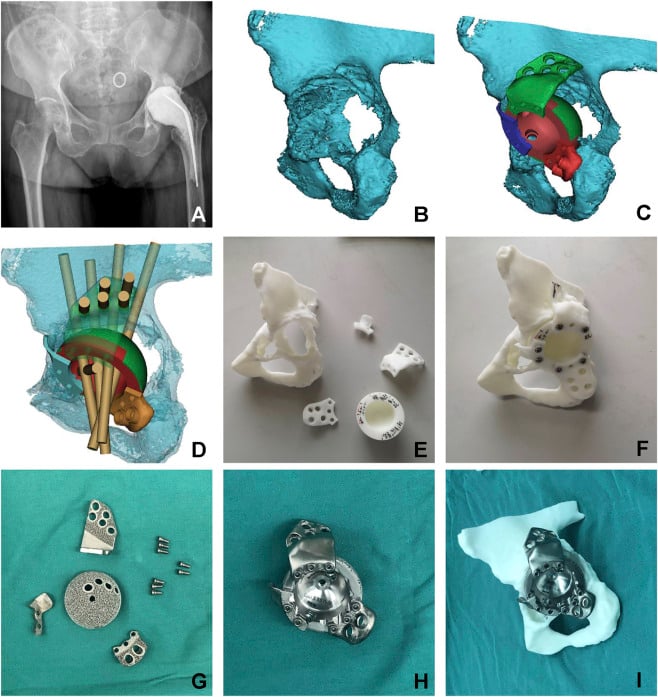
- CT-designed monoblock component spanning and bridging the discontinuity rather than healing it
- Fixation
- Locked screws into all three flanges — ilium, ischium and pubis
- Key weakness
- Cost, lead time of weeks, no intraoperative adjustability, wound and nerve complications
- Best indication
- Historic; occasionally a young patient in whom bone stock restoration is the goal
- Principle
- Allograft fills the defect; the cage bridges and protects it during incorporation
- Fixation
- Cage screws into ilium with an ischial flange — no porous surface, no ingrowth
- Key weakness
- No biologic fixation — fatigue failure and high mid-term loosening rates; largely superseded
- 1Rung 1 — Posterior column plate plus porous cupAcute intraoperative discontinuity, or chronic with viable bone. Compress and heal, then support with a hemispherical cup screwed into both segments.
- 2Rung 2 — Acetabular distractionChronic discontinuity with intact enough columns. Jumbo porous cup 2 to 3 mm oversized springs the discontinuity open; elastic recoil delivers compression.
- 3Rung 3 — Cup-cage constructChronic workhorse when moderate ilium and ischium remain. Porous tantalum shell for ingrowth, protected by an ilioischial cage; liner cemented into the cage.
- 4Rung 4 — Custom triflangeUnreconstructable pelvis: absent landmarks, failed cage, irradiation. CT-designed monoblock locked into ilium, ischium and pubis. Accept weeks of lead time.
- 5Off-ladder — Structural allograft plus conventional cageHistoric option with no biologic fixation. Mention only to dismiss, except in the young patient where bone stock restoration is the aim.
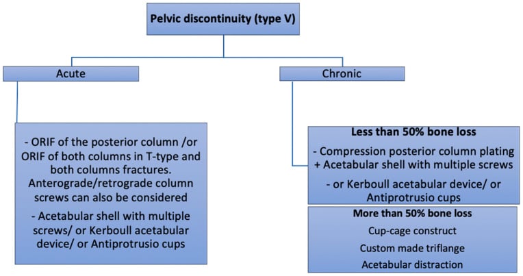
- 1Rung 1 — Posterior column plate plus porous hemispherical cupAcute intraoperative discontinuity, or chronic discontinuity with good bone quality and genuine healing potential. Compression osteosynthesis across the discontinuity, then a hemispherical porous cup with screws into both the superior (iliac) and inferior (ischiopubic) segments. Fails when the bone cannot heal — the chronic atrophic non-union.
- 2Rung 2 — Acetabular distraction (Sporer and Paprosky)Chronic discontinuity where both columns remain adequate to grip an oversized cup. Under-ream, then impact a jumbo porous cup 2 to 3 mm larger than the reamed diameter, springing the discontinuity open; elastic recoil compresses host bone onto the porous surface. Technique-sensitive: requires an elastic rather than rigid non-union and risks iatrogenic column fracture.
- 3Rung 3 — Cup-cage constructThe chronic workhorse when moderate residual ilium and ischium remain for shell contact. A porous tantalum shell provides the biologic ingrowth surface, protected from load by an overlying ilioischial cage while ingrowth occurs, with the liner cemented into the cage. Beware the sciatic nerve when seating the ischial limb.
- 4Rung 4 — Custom triflange (patient-specific)Massive bone loss, failed previous cage, destroyed landmarks or an irradiated pelvis. CT-designed monoblock spanning ilium, ischium and pubis with locked screws into all three segments. Costs and lead time of several weeks, no intraoperative adjustability, and appreciable wound and nerve complications.
- 5Off the ladder — structural allograft plus conventional cageHistoric; occasionally considered in the young patient to restore bone stock. Allograft fills the defect and the cage bridges and protects it, but there is no biologic fixation of the cage — fatigue failure and high mid-term loosening rates mean it is largely superseded.
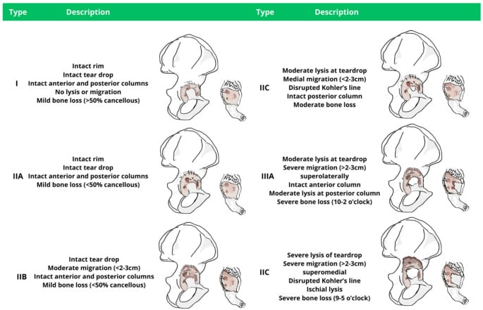
The Paprosky acetabular classification frames the bone loss; discontinuity is a modifier most commonly attached to type IIIB (superior migration greater than 3 cm, Kohler line broken, severe ischial lysis, unsupportive rim). Berry and colleagues (Mayo) additionally stratified discontinuity by associated bone loss and prior irradiation — the two variables that still drive treatment.
Decision framework — three questions:
- Is it acute or chronic? Acute equals fracture surgery: compress and heal.
- Can it heal? Healing potential depends on bone quality, vascularity (irradiation), inflammatory disease and defect size.
- Can host bone support a porous implant? If contact with viable ilium and ischium is achievable, cup-cage or distraction works; if landmarks are destroyed, custom triflange.
- Best indication
- Acute intraoperative discontinuity; chronic with good bone and healing potential
- Principle
- Compression osteosynthesis of the discontinuity; cup with screws into both segments
- Key weakness
- Fails if bone cannot heal — chronic atrophic non-union
- Best indication
- Chronic discontinuity, workhorse; moderate residual ilium and ischium
- Principle
- Porous tantalum shell for biologic ingrowth, protected by an overlying ilioischial cage while ingrowth occurs; liner cemented into cage
- Key weakness
- Requires enough host bone for shell contact; ischial limb sciatic risk
- Best indication
- Chronic discontinuity with adequate columns to grip an oversized cup
- Principle
- Under-ream, then impact a jumbo porous cup 2 to 3 mm larger, springing the discontinuity open; elastic recoil compresses bone onto the cup
- Key weakness
- Technique-sensitive; risk of iatrogenic column fracture; needs elastic, not rigid, non-union
- Best indication
- Massive bone loss, failed cage, absent landmarks, irradiated pelvis
- Principle
- CT-designed monoblock spanning ilium, ischium and pubis with locked screws into all three
- Key weakness
- Cost, lead time (weeks), no intraoperative adjustability, wound/nerve complications
- Best indication
- Historic; occasionally young patients to restore bone stock
- Principle
- Allograft fills defect; cage bridges and protects it
- Key weakness
- No biologic fixation of the cage — fatigue failure and high mid-term loosening rates; largely superseded
Diagnosis: Imaging and Intraoperative Confirmation
- 1Step 1 — Well-centred revision AP pelvis
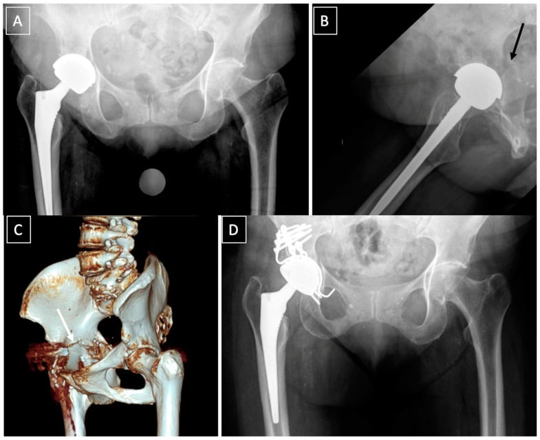
Systematically seek the three signs in every acetabular revision: a lucency crossing both columns (anterior column at the superior pubic ramus to posterior column at the ilioischial region), a break, step or offset in Kohler (ilioischial) line indicating medial displacement of the inferior segment, and obturator ring asymmetry or rotation from the inferior hemipelvis flexing and internally rotating.
Any sign present, or any Paprosky III pattern, mandates further imaging — no sign present does not exclude discontinuity.
- 2Step 2 — Judet oblique views
The iliac oblique profiles the posterior column and anterior wall; the obturator oblique profiles the anterior column and posterior wall.
A true discontinuity requires demonstrable disruption of BOTH columns.
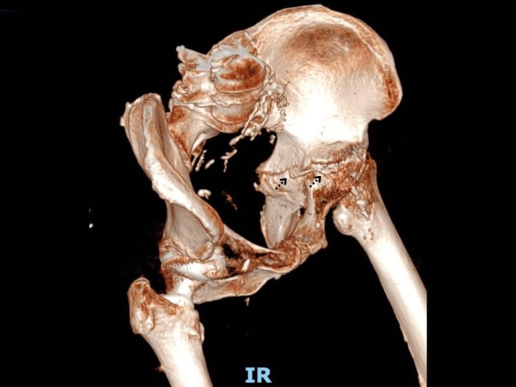
- 3Step 3 — CT with metal artefact reduction (MARS)
The definitive investigation: confirms discontinuity, quantifies remaining bone stock in the ilium and ischium, and distinguishes established non-union from a partially healed fracture.
Reconstruction strategy (cup-cage, distraction, triflange) is set preoperatively.
- 4Step 4 — Mandatory intraoperative stress test
Preoperative imaging is falsely negative in a substantial minority, so discontinuity must be confirmed or excluded in theatre in EVERY Paprosky III defect.
Direct visual confirmation, not inference from radiographs.
- 5Step 5 — Probe the acetabular floor
Any transverse membrane-filled cleft in the floor is probed directly.
Fibrous tissue within a mobile cleft indicates non-union — a discontinuity.
- 6Step 6A — Discontinuity confirmed
Independent segment motion or a mobile fibrous cleft is demonstrated.
Failure to stress test is the classic viva trap: a 'IIIB defect' that is really a discontinuity will defeat a jumbo cup alone.
- 7Step 6B — Discontinuity excluded
No independent motion between superior and inferior segments on firm stress after full debridement.
Document the negative stress test explicitly.
Surgical Techniques
chronic pelvic discontinuity with Paprosky IIIA/IIIB bone loss where sufficient host ilium and ischium remain for shell contact. Why over alternatives: combines the biologic fixation of a highly porous shell with the immediate mechanical span of a cage — the cage stress-shields the discontinuity and protects the shell-bone interface from micromotion during ingrowth.
- Position: lateral decubitus, secure pelvic supports (accurate cup version depends on stable pelvic position); radiolucent table.
- Imaging/equipment: image intensifier available; highly porous (tantalum or equivalent) revision shells including jumbo sizes, matched ilioischial cage system, augments, long screws, cement; explant tools; neuromonitoring optional.
- Preparation: full infection workup completed; cell salvage; cross-match — these are high blood-loss cases.
- Approach: posterior approach with extensile capability; identify and protect the sciatic nerve throughout, especially during ischial exposure. Trochanteric slide or extended trochanteric osteotomy if femoral component removal needed.
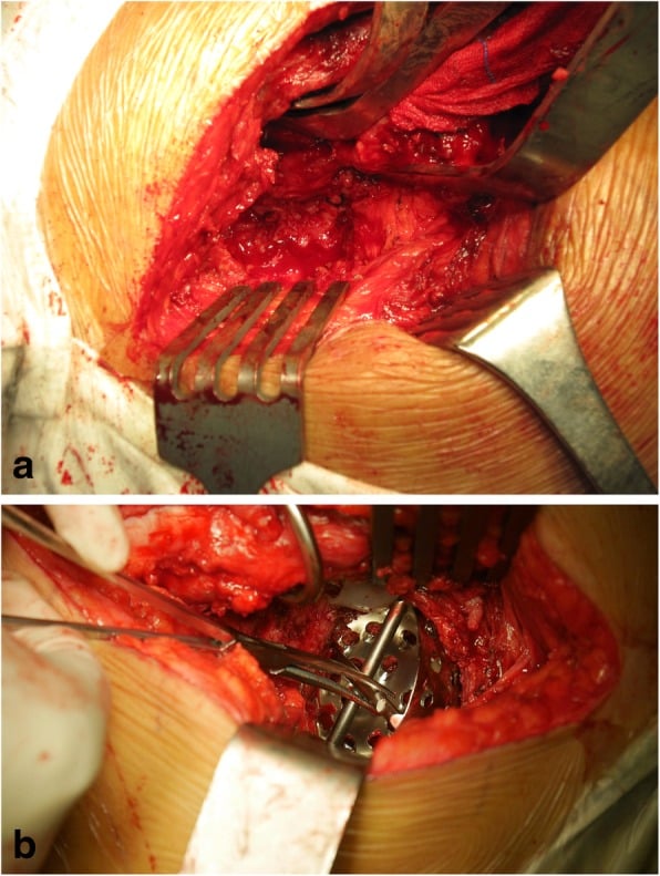
- Dissection: remove components and all membrane; send tissue for culture and frozen section. Expose the ischium distally and the outer ilium proximally for cage limbs. Confirm the discontinuity by stress testing.
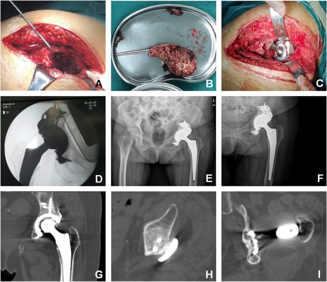
- Reconstruction: ream cautiously for maximal host contact (both segments). Impact the porous shell — augments fill superolateral or posterior defects and are unitised to the shell with cement. Fix the shell with multiple screws into ilium AND ischium/pubis where possible.
- Cage: contour the cage inside the shell. The iliac flange is fixed to the outer ilium with multiple screws; the ischial limb is either slotted into the ischium (intraosseous — lower sciatic irritation) or screwed onto its surface.
- At-risk structures: sciatic nerve (ischial limb), superior gluteal bundle (iliac screws/flange), intrapelvic vessels (anterosuperior/anteroinferior quadrant screws — Wasielewski), obturator bundle.
- Liner: cement a polyethylene liner into the cage in appropriate anteversion and inclination — decoupling liner orientation from cage position is a core advantage. Consider a constrained or dual-mobility bearing given high instability risk from abductor deficiency.
- Closure/aftercare: meticulous closure; protected weight bearing (toe-touch to partial) for 8 to 12 weeks; abduction precautions.
- Pitfalls/salvage: inadequate shell-host contact (add augments, do not rely on cage alone); vertical cemented liner (dislocation); cage limb fracture indicates failed shell ingrowth — salvage is usually custom triflange.
Sciatic nerve: at direct risk during ischial exposure, ischial cage/triflange limb placement and from limb lengthening when correcting a chronically migrated hip centre — limit acute lengthening and check the nerve is lax with the knee flexed. Intrapelvic vessels: screws in the anterosuperior and anteroinferior quadrants threaten the external iliac and obturator vessels; a migrated intrapelvic cup may require preoperative CT angiography and a retroperitoneal-capable surgical plan for extraction.
SPANCup-cage construct logic
Hook:The cage SPANs the gap while the shell grows in.
Outcomes, Failure Modes and Complications
- Where
- Posterior column, behind ischium and greater sciatic notch
- Mechanism
- Retractor pressure, traction during large exposure or triflange placement, screws exiting the notch
- Avoid by
- Hip extended and knee flexed, no blind notch retraction, direct palpation of the notch before column plating
- If it happens
- Release traction, remove offending screw, document deficit, foot-drop splint and expectant management; nerve injury is a recognised triflange complication
- Where
- Anterior column and quadrant, medial wall
- Mechanism
- Medial cup or graft perforation, anterior column screws, protrusion component extraction
- Avoid by
- Keep screws in the posterosuperior safe quadrants, CT angiography when the component is intrapelvic, vascular surgeon on standby
- If it happens
- Pack, do not chase the bleeding; intrapelvic approach for proximal control
- Where
- Obturator foramen, inferomedial to the acetabulum
- Mechanism
- Inferior flange or ischial limb fixation, medial graft placement, corona mortis anastomosis
- Avoid by
- Subperiosteal dissection of the inferior ramus, know the corona mortis lies on the superior pubic ramus
- If it happens
- Ligate the bleeding vessel under direct vision
- Where
- Across the anterior and posterior columns
- Mechanism
- Failure to compress, chronic sclerotic non-viable edges, no biologic surface across the defect
- Avoid by
- Compress acute discontinuity with a posterior column plate plus cup; treat chronic discontinuity by distraction or cup-cage with graft and porous ingrowth surface
- If it happens
- Acute plate plus cup is the best-prognosis group; chronic recurrent motion needs escalation to cup-cage or custom triflange
- Where
- Ischial limb or iliac flange
- Mechanism
- Load never transferred to ingrown bone, so the metal cycles to failure — the hallmark of failed biologic fixation of the underlying shell or graft
- Avoid by
- Achieve ingrowth on a porous shell first, cage only as a bridge; avoid allograft plus cage as definitive treatment given historic high mechanical failure
- If it happens
- Do not just replace the cage: CT for bone stock, exclude infection, escalate — usually to a custom triflange
- Where
- Acetabular shell and columns
- Mechanism
- Inadequate initial host bone contact, or occult infection
- Avoid by
- Aim for three-point fixation from ilium to ischium, maximise viable host contact
- If it happens
- Reaspirate and screen for infection, revise to cup-cage or triflange
- Where
- Bearing
- Mechanism
- Abductor deficiency and extensive exposure — the leading complication after custom triflange
- Avoid by
- Cement a dual-mobility or constrained liner into the cage or triflange, restore version and offset
- If it happens
- Bracing for a first event; recurrent instability needs a constrained or dual-mobility revision
- Where
- Whole construct
- Mechanism
- Long operative time, high blood loss, multiply revised soft tissues — rates exceed routine revision
- Avoid by
- Optimise host, minimise dead space, meticulous haemostasis and appropriate prophylaxis
- If it happens
- Full workup; debridement with liner exchange only if fixation is sound, otherwise staged revision
- Acute discontinuity, plate plus cup: the best-prognosis group; most heal when compressed in healthy bone.
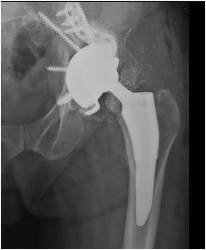
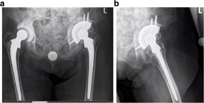
- Chronic discontinuity, cup-cage: modern series report survivorship for aseptic failure commonly in the 80 to 90 percent range at 5 to 10 years — the benchmark against which alternatives are judged.
- Distraction: encouraging mid-term results from the originating centres, with radiographic stability in the large majority; longer-term multicentre data still accruing.
- Custom triflange: good fixation survivorship in most series but a high overall complication rate — dislocation is the leading complication (deficient abductors, large exposure), followed by infection and nerve injury.
- Allograft plus cage: historic mid-term mechanical failure and loosening rates were high; the construct is now rarely used as definitive treatment for discontinuity.
Failure modes to recognise:
- Cage or flange fatigue fracture — hallmark of failed biologic fixation of the underlying shell or graft
- Shell migration with recurrent discontinuity motion — inadequate initial contact or infection
- Dislocation — abductor deficiency; mitigate with dual-mobility or constrained liners cemented into the cage
- Periprosthetic joint infection — these are long, high-blood-loss, multiply revised cases; infection rates exceed routine revision
A broken ischial cage limb on follow-up radiographs means the load never transferred to ingrown bone — the shell is loose. Do not simply replace the cage: reassess bone stock with CT, exclude infection, and plan escalation, usually to a custom triflange.
Early Migration Is Not Automatically Failure
Two independent series, two different constructs, the same observation — and it changes what you do at the six-week film.
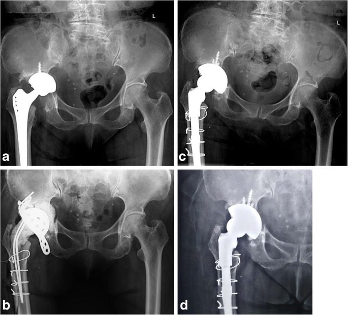
- Cup-cage (Abolghasemian, 26 discontinuities): three hips showed early radiographic migration of the components, then stabilised and finished with a successful outcome.
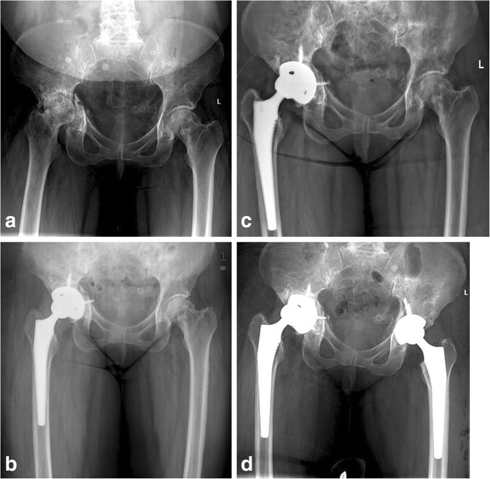
- Distraction with porous tantalum (Sporer, 20 analysed): four patients migrated early, then remained clinically asymptomatic and radiographically stable.
Why it happens. Both constructs are designed to settle. A cup-cage relies on biological ingrowth into a shell that the cage protects while union occurs; a distraction construct relies on the elastic recoil of the columns gripping an oversized cup. In each case a small early positional change can represent the construct seating rather than failing.
The condition attached to it. Abolghasemian's authors are specific: early migration does not necessarily mean failure provided the continuity of the bone graft at the discontinuity site is not disrupted. That is the discriminator — graft intact and the patient comfortable means serial radiographs; graft disrupted, progressive migration or pain means re-revision.
What to do. Do not re-revise on a single early film. Repeat the radiograph at a defined interval, compare the same landmarks, and treat progression rather than position as the failure signal. Equally, do not use this as reassurance for a construct that is migrating and symptomatic.
Guidelines, Registries & Global Practice
- Global epidemiology: published series put discontinuity at roughly 1 to 5 percent of acetabular revisions, but quote the anchor rather than the range where you can: Berry's Mayo series found it in 31 of 3505 acetabular revisions — 0.9 percent — which sits below the commonly repeated range and is the largest denominator any of these figures rests on. The spread across series reflects how hard the diagnosis is to make rather than genuine geographic variation, since a discontinuity missed preoperatively and not stress-tested at operation is simply recorded as a type IIIB defect. As revision burden rises worldwide with ageing arthroplasty cohorts, absolute case numbers are increasing in every registry region.
- Registry perspective: national registries (NJR, AJRR, AOANJRR, SHAR, Norwegian, NZJR) capture revision for acetabular loosening and bone loss but do not code discontinuity as a discrete entity, so evidence rests on institutional series and multicentre cohorts rather than registry survivorship curves — an important limitation to state in an examination answer.
- Society guidance: AAOS and NICE address hip revision pathways (infection exclusion, centralisation of complex revision) rather than discontinuity-specific technique; the AO principles of column fixation underpin the plate-plus-cup strategy for acute discontinuity. In several health systems (for example UK revision networks and similar European models), complex acetabular revision including discontinuity is increasingly regionalised to high-volume revision centres.
- Resource-setting variation: custom triflanges and porous tantalum systems are costly and require CT-to-implant infrastructure; in resource-limited settings, plate-plus-cup, ring/cage constructs with impaction or structural graft, and staged strategies remain in use, accepting higher mechanical failure risk. Distraction with a standard jumbo porous cup is comparatively resource-efficient where porous shells are available.
Controversies & Areas of Uncertainty
- Cup-cage versus distraction versus custom triflange for chronic discontinuity: no comparative trials exist; selection is driven by bone stock, surgeon experience and implant availability. Distraction advocates cite single-component simplicity; triflange advocates cite fixation into all three residual bones.
- Should the discontinuity be made to heal at all? Bridging constructs succeed without union; whether adding compression plating or grafting to a cup-cage improves durability is unproven.
- Bearing choice: dual-mobility versus constrained liners cemented into cages — constrained liners reduce dislocation but transmit torque to a fragile fixation interface; many revision surgeons now favour dual-mobility.
- 3D-printed custom implants: expanding indications and enthusiastic early series, but cost-effectiveness and long-term survivorship compared with off-the-shelf cup-cage remain undefined.
- Weight-bearing protocols: protected weight bearing for 8 to 12 weeks is conventional but evidence-light; earlier loading may be tolerated by well-fixed distraction or triflange constructs.
MCQ Practice Points
Q: What anatomically defines pelvic discontinuity? A: Complete separation of the superior hemipelvis from the inferior hemipelvis through the acetabulum, requiring disruption of BOTH the anterior and posterior columns — not an isolated wall or single-column defect.
Q: Which AP pelvis findings suggest discontinuity? A: A fracture line crossing both columns, a break or offset in the Kohler ilioischial line, and asymmetry or rotation of the obturator ring; confirm with Judet views and metal artefact reduction CT.
Q: Which Paprosky defect is most associated with discontinuity? A: Type IIIB — superior migration greater than 3 cm, broken Kohler line, severe ischial lysis and an unsupportive rim; discontinuity should be actively sought and stress-tested in all IIIB revisions.
Q: In the acetabular distraction technique, how much is the discontinuity distracted? A: 2 to 3 mm, achieved by impacting a porous cup 2 to 3 mm larger than the final reamer; pelvic elastic recoil then compresses both segments onto the cup.
Q: Why is the liner cemented into the cage rather than locked into the shell? A: Cementing decouples bearing orientation from cage and shell position, allowing independent correction of anteversion and inclination — cages rarely sit in ideal bearing orientation.
Q: Why did structural allograft plus conventional cage fail for chronic discontinuity? A: The cage has no ingrowth surface; as allograft resorbs, the unsupported cage undergoes cyclical loading and fatigue fracture, giving high mid-term mechanical failure — the rationale for the cup-cage.
Exam Viva Scenarios
Practise clinical reasoning and management decisions out loud
“A 72-year-old woman with rheumatoid arthritis presents 18 years after total hip arthroplasty with progressive groin pain and shortening. The AP pelvis shows a superiorly migrated, medialised cup. How do you assess for pelvic discontinuity and how does it change your plan?”
“During cup impaction in a first-time acetabular revision in a fit 60-year-old man, you feel a crack and the trial cup will no longer achieve a stable press-fit. On stressing the hemipelvis the ischial segment moves independently of the ilium. What now?”
“You have elected a cup-cage for a chronic Paprosky IIIB discontinuity. Talk me through the construct: how each element works, how you place the cage limbs, and how you control version.”
“A 68-year-old woman has a chronic pelvic discontinuity. In one scenario she has a mobile fibrous non-union with reasonable column bone; in another she had pelvic irradiation for cervical cancer and has grossly atrophic columns with a previously failed cage. Justify your reconstruction in each.”
Definition and risks
- Complete separation of superior from inferior hemipelvis through the acetabulum — both columns disrupted
- Acute intraoperative (heals) versus chronic (usually will not heal)
- Risks: female, rheumatoid, pelvic irradiation, massive osteolysis, prior acetabular fracture, multiple revisions
- Most associated with Paprosky IIIB defects
Diagnosis
- AP pelvis triad: both-column fracture line, broken Kohler line, obturator ring asymmetry/rotation
- Judet views profile each column; MARS CT is definitive and plans custom implants
- Intraoperative confirmation mandatory: stress the ischium and look for independent segment motion
- Always exclude infection before reconstruction
Treatment selection
- Acute or high healing potential: posterior column plate plus multi-hole porous cup (compression)
- Chronic workhorse: cup-cage — porous shell for ingrowth, cage spans and protects, liner cemented in corrected version
- Distraction (Sporer/Paprosky): oversized jumbo porous cup, 2 to 3 mm distraction, recoil compression; needs elastic columns
- Massive loss/failed cage/irradiated: custom triflange into ilium, ischium and pubis
- Allograft plus cage: historic, fatigue failure — avoid as definitive treatment
Hazards and outcomes
- Sciatic nerve: ischial limb dissection and acute lengthening; superior gluteal bundle at iliac flange
- Dangerous screw quadrants: anterosuperior and anteroinferior (external iliac and obturator vessels)
- Cup-cage survivorship commonly 80 to 90 percent mid-term; dislocation leading complication of triflanges
- Broken cage limb equals failed biologic fixation — CT, exclude infection, escalate reconstruction
- Protected weight bearing 8 to 12 weeks; dual-mobility or constrained bearing for abductor deficiency
Evidence Base
Pelvic Discontinuity in Revision Total Hip Arthroplasty
- Discontinuity was found in 31 of 3505 acetabular revisions - 0.9 PER CENT, which is the origin of the incidence figure
- 28 of the 31 hips were in WOMEN; female sex (p less than 0.001) and rheumatoid arthritis (p = 0.003) were both significant risk factors
- The radiographic triad: a visible fracture line through both columns, MEDIAL TRANSLATION of the inferior hemipelvis (a break in Kohler's line) and ROTATION of the inferior hemipelvis (asymmetry of the obturator rings) on a true AP
- Results stratified by bone loss and irradiation: satisfactory in 3 of 3 type IVa, 10 of 19 type IVb, and 3 of 5 irradiated type IVc
- NINE of 27 hips needed a further operation - four for aseptic loosening, FOUR FOR RECURRENT DISLOCATION and one for infection
- A mechanically stable construct was achieved in 17 of 24 hips
The Challenge of Pelvic Discontinuity: Cup-Cage Reconstruction Does Better Than Conventional Cages in Mid-Term
- 26 pelvic discontinuities in 24 patients treated by cup-cage (mean follow-up 82 months) compared with 19 discontinuities in 19 patients treated by ilioischial cage (mean 69 months)
- Failure from septic or aseptic loosening: 4 of 26 (15 per cent) cup-cage against 13 of 19 (68 PER CENT) cage alone
- Seven-year survivorship 87.2 per cent for cup-cage against 49.9 per cent for cage alone (p = 0.009)
- Radiological union of the discontinuity occurred in ALL successful cup-cage cases but in only three of the successful cage cases
- THREE cup-cage hips migrated early on radiographs, then STABILISED and went on to a successful outcome
Acetabular Distraction: An Alternative for Severe Defects with Chronic Pelvic Discontinuity?
- 28 patients with chronic pelvic discontinuity reconstructed by acetabular distraction with a porous tantalum elliptical component, alone or with a modular tantalum augment
- Only 20 were analysed: three died and five were lost before two years - a 29 per cent attrition before the minimum follow-up point
- One of 20 required re-revision for aseptic loosening; 15 remained radiographically stable
- FOUR patients migrated early and then remained clinically asymptomatic and radiographically stable - the same behaviour Abolghasemian reports for the cup-cage
- Mean improvement in the modified Merle d'Aubigne-Postel score was 6.6 in those not re-revised, with NO postoperative dislocations
- Complications included one infection, one VASCULAR injury and one BOWEL injury
Acetabular Defect Classification and Surgical Reconstruction in Revision Arthroplasty. A 6-Year Follow-Up Evaluation
- 147 cemented acetabular components revised to cementless hemispheric press-fit components, average follow-up 5.7 years (range 3-9)
- Defects typed 1 to 3 by the pattern of bone loss and matched to particulate graft, bulk femoral head graft, cup superiorisation or structural distal femur/proximal tibia allograft
- Six of 147 components (4.0 per cent) were radiographically and clinically unstable and warranted revision - ALL of them type 3B
- Moderate lateral allograft resorption was seen radiographically, but host-graft union was confirmed at revision
- The paper's own conclusion is that adequate remaining HOST bone must be present for ingrowth, whatever the graft does