Growth Plate Anatomy & Physiology
- The physis (growth plate) is the cartilaginous structure responsible for LONGITUDINAL bone growth via endochondral ossification, located between epiphysis and metaphysis.
- It is organised into zones from epiphyseal to metaphyseal side: RESTING (reserve) -> PROLIFERATIVE -> HYPERTROPHIC (maturation/degeneration/provisional calcification) -> the chondro-osseous junction where bone is formed.
- Longitudinal growth occurs principally in the PROLIFERATIVE zone (chondrocyte division and column formation); the hypertrophic zone adds height by cell ENLARGEMENT.
- The HYPERTROPHIC zone (specifically the zone of provisional calcification / degeneration) is the WEAKEST and is the plane through which physeal (Salter-Harris) fractures usually propagate.
- The physis has a DUAL blood supply: EPIPHYSEAL vessels nourish the resting/proliferative zones (their disruption arrests growth), and METAPHYSEAL vessels supply the ossification front; the perichondral RING OF LaCROIX gives mechanical support and the GROOVE OF RANVIER provides appositional (latitudinal) growth.
- Chondrocyte proliferation and differentiation are governed by a PTHrP-Indian hedgehog (Ihh) NEGATIVE-FEEDBACK LOOP and modulated by growth hormone/IGF-1, thyroid and sex hormones, BMP/TGF-beta, Wnt/beta-catenin and vitamin D.
- “Order the zones from the epiphysis down: Reserve, Proliferative, Hypertrophic, (provisional) Calcification - 'Real Patients Have Cartilage'.
- “Physeal fractures go through the HYPERTROPHIC zone (weak, mineralising, low matrix) - but the germinal reserve/proliferative zones on the epiphyseal side are usually spared, which is why most heal without growth arrest.
- “Disruption of the EPIPHYSEAL (not metaphyseal) blood supply causes growth arrest, because it feeds the dividing proliferative cells.
The hypertrophic zone has the largest cells, the least extracellular matrix per unit volume, and a partially mineralised, transitional structure - mechanically the weakest layer. Shear/avulsion forces therefore propagate through it, which is why Salter-Harris physeal fractures run through the hypertrophic zone.
The germinal reserve and proliferative zones lie on the epiphyseal side of the fracture plane and keep their epiphyseal blood supply, so most physeal fractures heal without growth disturbance. Growth arrest follows when the germinal layers or their blood supply are damaged (crush/SH-V, or a bar across the plate).
Overview & Development
The physis develops from cartilage-committed mesenchymal cells expressing the SOX family of genes and is the site of endochondral ossification - the orderly conversion of a cartilage template into bone that drives longitudinal growth. Its multilayered structure is built by chondrocyte proliferation and hypertrophy, with synthesis of an extracellular matrix rich in collagens (mainly types II, IX, X and XI) and proteoglycans (aggrecan, decorin). Disturbances of physeal development and physiology produce the skeletal dysplasias.
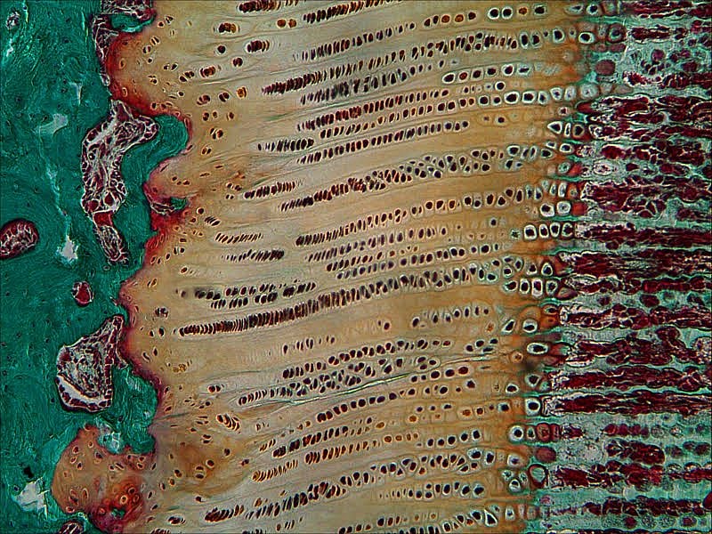
Histology & Zones
The physis is read from the epiphyseal side to the metaphyseal side:
| 0 | 1 | 2 | 3 |
|---|---|---|---|
| Reserve (resting) | Stem-like chondrocytes; matrix synthesis/storage | Low oxygen, scattered cells; the germinal source | Source layer; damage here harms future growth |
| Proliferative | Rapidly dividing flattened cells in columns | Highest oxygen/glycogen; site of LONGITUDINAL growth | Fed by epiphyseal vessels; arrest if supply lost |
| Hypertrophic | Enlarging cells -> apoptosis (maturation/degeneration/provisional calcification) | Most cell volume, least matrix; mineralisation begins | WEAKEST zone - Salter-Harris fracture plane |
| Provisional calcification / chondro-osseous junction | Matrix mineralises; vessels invade; osteoblasts form bone | Primary spongiosa; metaphyseal ingrowth vessels | Site of new bone; rickets widens this region |
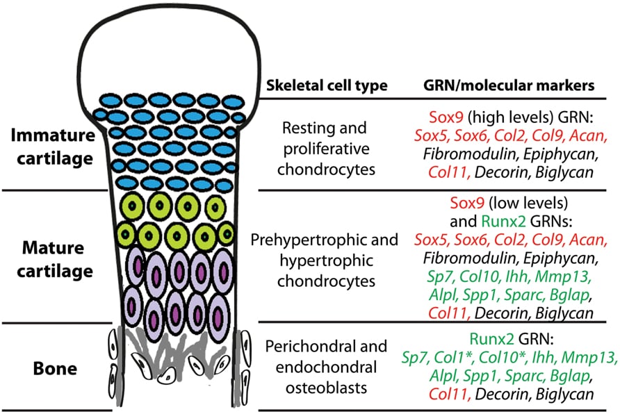
The resting and proliferative zones lay down a type II / IX / XI collagen and aggrecan-rich matrix. In the hypertrophic zone, terminally differentiated chondrocytes switch to type X collagen, release alkaline phosphatase, calcium and matrix vesicles, and the matrix mineralises (provisional calcification) before the cells undergo apoptosis and metaphyseal vessels bring in osteoblasts to form the primary spongiosa.
Blood Supply & Surrounding Structures
- Epiphyseal vessels supply the reserve and proliferative zones. Their disruption is catastrophic for growth because it starves the dividing cells -> growth arrest.
- Metaphyseal vessels supply the zone of provisional calcification / ossification front and remove debris; loss impairs ossification but the germinal cells survive.
- Perichondral ring of LaCroix - a fibrous collar around the physis providing mechanical support and limiting shear.
- Groove of Ranvier - a wedge of cells at the periphery that adds chondrocytes to the periphery, driving latitudinal (appositional / width) growth of the physis.
Longitudinal growth comes from the proliferative-zone column formation plus hypertrophic cell enlargement; latitudinal (width) growth of the physis comes from the groove of Ranvier at the periphery. Examiners like the pairing: proliferative zone = length, groove of Ranvier = width, ring of LaCroix = support.
Molecular & Endocrine Regulation
The pace of chondrocyte maturation is set by a negative-feedback loop. As proliferating chondrocytes begin to mature, they secrete Indian hedgehog (Ihh), which (directly and via the perichondrium) stimulates production of parathyroid hormone-related protein (PTHrP) near the ends of the bone. PTHrP acts on its receptor to keep chondrocytes proliferating and delay their hypertrophic differentiation. As cells move away from the PTHrP source they escape its influence, hypertrophy, and release more Ihh - closing the loop. This PTHrP-Ihh axis tightly controls the proportion of proliferating versus hypertrophic cells and therefore the rate of growth.
| 0 | 1 |
|---|---|
| PTHrP | Keeps chondrocytes proliferating; delays hypertrophy (with Ihh, the master local loop) |
| Indian hedgehog (Ihh) | From prehypertrophic cells; stimulates PTHrP and proliferation; couples to osteoblast formation |
| Growth hormone / IGF-1 | Stimulates proliferative-zone chondrocytes (GH acts partly directly, partly via IGF-1) |
| Thyroid hormone | Drives chondrocyte hypertrophy and ossification; deficiency delays bone age |
| Sex steroids (oestrogen) | Accelerate maturation and ultimately cause physeal CLOSURE (oestrogen is the key closer in both sexes) |
| BMP / TGF-beta, Wnt/beta-catenin | Regulate chondrocyte differentiation and the cartilage-to-bone transition |
| Vitamin D / glucocorticoids | Vitamin D needed for mineralisation; glucocorticoids suppress growth |
Because these pathways converge on the physis, their disturbance produces recognisable disease: rickets (defective mineralisation) widens the hypertrophic zone and frays the metaphysis; achondroplasia results from an activating FGFR3 mutation that inhibits proliferation; oestrogen ultimately drives physeal closure (precocious puberty -> early closure/short stature; deficiency -> tall stature); and growth-hormone excess/deficiency alters longitudinal growth.
Clinical Significance
Physeal (Salter-Harris) fractures run through the hypertrophic zone; most heal without sequelae, but injury to the germinal zones or a physeal bar causes growth arrest, angular deformity or limb-length discrepancy.
The physis is the seat of skeletal dysplasias, the route of haematogenous osteomyelitis (metaphyseal sluggish flow), and the target of guided-growth surgery (e.g. tension-band plating / epiphysiodesis) that deliberately modulates the plate to correct deformity or length.
Evidence & Key Studies
Morphology and physiology of the epiphyseal growth plate
- Describes the multilayer growth plate: resting (protein synthesis/germinal), proliferative (rapid duplication), transformation/hypertrophic and degenerative zones with intensive mineralisation and chondrocyte apoptosis.
- Matrix is composed of collagens (mainly II, IX, X, XI) and proteoglycans (aggrecan, decorin); regulation involves growth/thyroid/sex hormones, beta-catenin, BMPs, IGF, TGF-beta and vitamin D.
- Identifies PTHrP (from the perichondrium) as the most significant factor, stimulating proliferation and delaying hypertrophy, with an Indian hedgehog (Ihh) feedback loop; disturbances cause skeletal dysplasia.
Imaging findings of metabolic bone disease
- Impaired chondrocyte development and failure to mineralise growth-plate cartilage in rickets lead to WIDENED growth plates and frayed metaphyses at sites of greatest growth.
- Thyroid hormone regulates endochondral bone formation; untreated congenital hypothyroidism delays bone age and produces irregular/fragmented epiphyses.
- Illustrates how systemic metabolic derangements manifest specifically at the physis and metaphysis.
According to PubMed, the zonal structure, matrix composition and the PTHrP-Ihh regulatory loop described here are drawn from the cited reviews. The classic descriptions of the perichondral ring of LaCroix, the groove of Ranvier and the dual epiphyseal/metaphyseal blood supply are standard physeal anatomy used in these and related works.
Clinical Decision Scenarios
Practise clinical reasoning and management decisions out loud
“Describe the histological zones of the growth plate from the epiphysis to the metaphysis, and tell me which zone fails in a physeal fracture and why.”
“How is chondrocyte proliferation and maturation regulated at the growth plate, and how does the periphery of the physis grow?”
Mnemonics & Memory Aids
RPHC
Hook:'Real Patients Have Cartilage' - Reserve, Proliferative, Hypertrophic, Calcification, top (epiphysis) to bottom (metaphysis).
RANVIER
Hook:Groove of RANVIER grows the physis wider; ring of LaCroix holds it together.
Zones (epiphysis -> metaphysis)
- Reserve (resting): germinal, matrix storage, low O2
- Proliferative: dividing columns = LONGITUDINAL growth; high O2/glycogen
- Hypertrophic: cells enlarge/mineralise/apoptose; WEAKEST = Salter-Harris plane
- Provisional calcification / chondro-osseous junction: vessel invasion, primary spongiosa
Blood supply & periphery
- Epiphyseal vessels -> reserve/proliferative (loss = growth arrest)
- Metaphyseal vessels -> ossification front
- Groove of Ranvier = width (latitudinal) growth; Ring of LaCroix = mechanical support
Regulation
- PTHrP-Ihh feedback loop = master local control (PTHrP delays hypertrophy)
- GH/IGF-1 (proliferation), thyroid (hypertrophy), oestrogen (closes the plate in both sexes)
- BMP/TGF-beta, Wnt/beta-catenin, vitamin D modulate differentiation/mineralisation
Clinical links
- Physeal fractures through hypertrophic zone; growth arrest if germinal zones/bar
- Rickets widens the hypertrophic/calcification zone; achondroplasia = FGFR3
- Guided growth (tension-band plate / epiphysiodesis) modulates the plate