Reverse Bankart | McLaughlin | Capsulorrhaphy | Bone Loss
POSTERIOR INSTABILITY TYPES
Critical Must-Knows
- 2-5% of dislocations but frequently MISSED
- FAIR position of risk (flexion, adduction, internal rotation)
- Reverse Bankart = posterior labral tear
- McLaughlin lesion = reverse Hill-Sachs on anterior humeral head
- Posterior bone loss rare but important
Clinical Pearls
- "Jerk test and Kim test specific for posterior labral lesions
- "Lightbulb sign on AP X-ray = locked posterior dislocation
- "Seizure patients have high rate of bilateral posterior dislocations
- "Avoid surgery for voluntary dislocators
Clinical Imaging
Posterior Shoulder Instability MRI Findings


Critical Exam Concepts
Often Missed
Posterior dislocations frequently missed (up to 60% initially). Always get axillary or scapular Y view. Suspect with seizure/electrocution.
FAIR Position
Flexion, Adduction, Internal Rotation is position of risk. Mechanism is posterior force with arm in this position.
Reverse Bankart
Posterior labral tear = reverse Bankart. Key pathology for surgical stabilization. Analogous to anterior Bankart.
McLaughlin Lesion
Reverse Hill-Sachs = impaction fracture anterior humeral head. McLaughlin procedure transfers subscapularis into defect.
Posterior vs Anterior Instability
| Feature | Posterior | Anterior |
|---|---|---|
| Frequency | 2-5% | 95%+ |
| Position of risk | FAIR (flex, add, IR) | ABER (abd, ext rot) |
| Labral lesion | Reverse Bankart (posterior) | Bankart (anterior) |
| Humeral lesion | McLaughlin (anterior) | Hill-Sachs (posterior) |
| Bone loss | Posterior glenoid (reverse bony Bankart) | Anterior glenoid |
SEEPPosterior Dislocation Causes
| S | Seizure Grand mal seizures (bilateral common) |
| E | Electrocution Massive muscle contraction |
| E | Ethanol Alcohol withdrawal seizures |
| P | Posterior trauma Direct blow to anterior shoulder |
| S | Seizure Grand mal seizures (bilateral common) | E | Ethanol Alcohol withdrawal seizures |
| E | Electrocution Massive muscle contraction | P | Posterior trauma Direct blow to anterior shoulder |
Hook:SEEP causes arm to SEEP backward!
FAIRPosition of Risk
| F | Flexion Shoulder flexed forward |
| A | Adduction Arm across body |
| I | Internal rotation Arm internally rotated |
| R | Risk position This is position of posterior instability |
| F | Flexion Shoulder flexed forward | I | Internal rotation Arm internally rotated |
| A | Adduction Arm across body | R | Risk position This is position of posterior instability |
Hook:FAIR position is NOT fair to posterior shoulder!
JKPosterior Instability Tests
| J | Jerk test Patient supine, arm flexed 90, adduct and axial load |
| K | Kim test Patient seated, arm 90 abduction, posterior force with elevation |
| J | Jerk test Patient supine, arm flexed 90, adduct and axial load |
| K | Kim test Patient seated, arm 90 abduction, posterior force with elevation |
Hook:JK tests for posterior labral tears!
Overview and Epidemiology
Frequently Missed
Posterior dislocations missed in up to 60% of initial presentations. Always suspect with seizure, electrocution, or failure to externally rotate. Get axillary view.
Epidemiology
- 2-5% of shoulder dislocations
- Higher in epileptic patients
- Overhead athletes (backhanders, linemen)
- Males greater than females
- Often bilateral with seizure
Mechanism
- Seizure/electrocution: Massive muscle contraction
- Trauma: Posterior force in FAIR position
- Sports: Repetitive microtrauma (overhead)
- Voluntary: Psychological component
- Internal rotators overpower external (seizure)
Pathophysiology and Mechanisms
Posterior Stabilizers
Bone: Posterior glenoid rim provides bony stability.
Labrum: Posterior fibrocartilaginous rim deepens socket.
Capsule: Posterior capsule and posterior band IGHL.
Muscles: Infraspinatus, teres minor (external rotators).
The posterior capsule is thinner than anterior - less robust stabilization.
Locked Posterior Dislocation
Locked posterior dislocation = humeral head trapped behind glenoid with McLaughlin engaging posteriorly. Requires specific reduction technique or open reduction.
Classification Systems
Posterior Instability Types
| Type | Mechanism | Features | Treatment Approach |
|---|---|---|---|
| Traumatic acute | Seizure, electrocution | Often locked, bilateral | Closed reduction if possible |
| Recurrent traumatic | Previous dislocation | Apprehension, recurrence | Surgical stabilization |
| Atraumatic recurrent | Microtrauma, laxity | Athletes, overhead sports | Rehab first, surgery if fails |
| Voluntary | Patient induced | Psychological aspect | Avoid surgery |
Clinical Assessment
History
- Mechanism: Seizure, electrocution, FAIR trauma
- Pain: Posterior shoulder
- Restricted motion: Cannot externally rotate
- Recurrence: Prior events
- Voluntary: Patient can demonstrate
Examination
- Position: Arm held in IR, adducted
- ROM: Blocked external rotation (key finding)
- Jerk test: Positive for posterior labral tear
- Kim test: Posterior subluxation
- Posterior apprehension: FAIR position loading
Jerk Test Technique
Patient supine. Arm at 90 degrees flexion and internal rotation. Examiner applies axial load and adducts across body. Positive: Posterior subluxation followed by clunk as it reduces when arm is returned to neutral.
Key Clinical Pearls
Blocked external rotation: Inability to externally rotate should raise suspicion for locked posterior dislocation.
Lightbulb sign: On AP view, humeral head appears like a lightbulb (internally rotated appearance) - suggests locked posterior dislocation.
Differential Diagnosis
Distinguishing Posterior Instability from Mimics
| Diagnosis | Key discriminator | Best test |
|---|---|---|
| Posterior instability | Pain/apprehension in FAIR; positive jerk and Kim tests | MR arthrogram + dynamic exam |
| Anterior instability | Apprehension in ABER (abduction-external rotation) | Anterior apprehension/relocation |
| Multidirectional instability (MDI) | Global laxity, sulcus sign, often bilateral, atraumatic | Sulcus sign, generalised laxity (Beighton) |
| Locked posterior dislocation | Fixed internal rotation, blocked external rotation | Axillary radiograph (humeral head behind glenoid) |
| SLAP / biceps pathology | Pain on overhead throwing, positive O'Brien | MR arthrogram, dynamic labral tests |
| Posterior cuff/scapular dyskinesis | Weakness, scapular winging, no true subluxation | Scapular assistance test, cuff strength |
Investigations
X-ray Assessment
Views: AP, axillary (essential), scapular Y.
Lightbulb sign: Humeral head internally rotated on AP view.
Rim sign: Increased space between humeral head and glenoid.
Through sign: Overlap of head and glenoid (Y view).
Axillary view is essential and diagnostic - posterior position of head.
Lightbulb Sign
Lightbulb sign = internally rotated humerus on AP view looks like a lightbulb. Indicates locked posterior dislocation due to inability to externally rotate. DO NOT MISS.
Management Algorithm
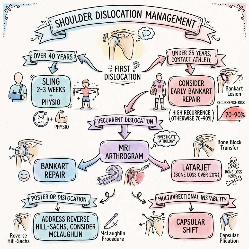
Acute Posterior Dislocation
Acute Management
High suspicion with seizure/electrocution. Get axillary view. Check for locked dislocation.
CT to assess humeral head defect size. Less than 20% good prognosis.
Traction, gentle external rotation. If less than 6 weeks old, may succeed.
Brace in neutral or external rotation. Avoid internal rotation.
Surgical Technique
Posterior Labral Repair (Arthroscopic)
Setup: Beach chair or lateral decubitus.
Portals: Standard posterior (viewing), anterior, and posterolateral (working).
Technique:
- Mobilize labrum from glenoid
- Prepare glenoid rim bleeding bone bed
- Place suture anchors (2-4) on glenoid face
- Shuttle sutures through labrum
- Tie knots to reduce labrum to glenoid
Consider capsular plication for laxity.
Avoid Overtightening
Avoid overtightening posterior capsule which leads to loss of internal rotation and anterior subluxation. Balance is important.
Complications
| Complication | Cause | Prevention | Management |
|---|---|---|---|
| Recurrence | Missed pathology, undertightening | Address all lesions | Revision surgery |
| Stiffness | Overtightening, immobilization | Balanced repair | Physical therapy |
| Anterior instability | Overtightening posterior | Avoid overtightening | Rare, may need revision |
| Axillary nerve | Portal placement | Safe portal placement | Observation usually |
Recurrence Rate
Recurrence after posterior stabilization: 5-15% in experienced hands. Worse with voluntary instability (avoid surgery), bone loss, and unrecognized pathology.
Postoperative Care
Posterior Stabilization Rehabilitation
Sling in neutral rotation (not internal rotation). Pendulums, elbow and wrist ROM.
Progressive ROM. Avoid combined flexion, adduction, internal rotation. Start external rotation.
Progressive strengthening. Rotator cuff and scapular stabilizers. Pool exercises.
Sport-specific training. Full return at 6-9 months if strength and stability adequate.
Avoid FAIR Position
Avoid FAIR position (flexion, adduction, internal rotation) during early rehabilitation - this stresses the posterior repair. Progress to full ROM gradually.
Outcomes and Prognosis
Prognosis by Type
Traumatic recurrent: Good outcomes with surgery. 85-90% stability.
Atraumatic: May respond to physiotherapy. Surgery if fails.
Voluntary: Poor surgical outcomes. Avoid surgery. Psychological counseling.
Large McLaughlin: Worse prognosis. May need arthroplasty if engaging.
Controversies and Areas of Uncertainty
Unresolved Questions
- Bone-loss threshold: the exact posterior glenoid loss at which soft-tissue repair fails (often quoted 20-25%) is not well defined and remains debated.
- Open vs arthroscopic bone block: posterior bone-block techniques are less standardised than the anterior Latarjet, with no high-level comparative data.
- Optimal immobilisation: neutral vs external-rotation bracing after reduction or repair lacks robust evidence.
Evolving Practice
- Classification: the Stanmore polar (structural / muscular-patterning / atraumatic) framework increasingly guides whether to operate, replacing rigid traumatic vs voluntary dichotomy.
- Throwing athletes: consistently return to pre-injury level less often, so counselling and graded return-to-throw protocols are emphasised.
- Thermal capsulorrhaphy: abandoned due to chondrolysis and capsular necrosis - mentioned only to be rejected in vivas.
Evidence Base and Key Studies
Arthroscopic Posterior Capsulolabral Reconstruction (100 shoulders)
- 91 athletes (100 shoulders) with unidirectional recurrent posterior instability, mean 27-month follow-up
- Mean ASES score improved from 50.4 to 85.7 (p less than 0.001)
- 89% returned to sport; 67% returned at the same level
- Posterior-instability shoulders had significantly greater chondrolabral and osseous retroversion than controls
Arthroscopic Posterior Capsulolabral Reconstruction (200 shoulders)
- 183 athletes (200 shoulders), mean 36-month follow-up - the largest single-series cohort
- Mean ASES score improved from 45.9 to 85.1 (p less than 0.001)
- 90% returned to sport; suture-anchor plication gave significantly higher ASES and return-to-play rates than anchorless repair
- Contact athletes did as well as the overall cohort
Exam Viva Scenarios
Use these scenarios to practise clinical reasoning and management decisions
Scenario 1: Post-Seizure Shoulder
"A 35-year-old epileptic presents after a grand mal seizure with bilateral shoulder pain. He cannot externally rotate either shoulder. AP X-rays appear normal. How would you manage this?"
Scenario 2: Recurrent Posterior Subluxation
"A 25-year-old tennis player has recurrent posterior shoulder subluxation episodes during serving. He has failed 6 months of physiotherapy. MRI shows a posterior labral tear. What would you recommend?"
Scenario 3: Voluntary Dislocator
"A 20-year-old can voluntarily dislocate her shoulders by positioning them. She has some pain and asks for surgical stabilization to stop the dislocations. What is your approach?"
MCQ Practice Points
Position of Risk
Q: What position causes posterior shoulder instability? A: FAIR - Flexion, Adduction, Internal Rotation. This is opposite to ABER for anterior instability.
Most Common Cause
Q: Most common cause of acute posterior dislocation? A: Seizure (grand mal). Also electrocution. Internal rotators overpower external rotators during convulsion.
Lightbulb Sign
Q: What is the lightbulb sign? A: Internally rotated humeral head on AP X-ray with locked posterior dislocation. Looks like a lightbulb.
McLaughlin Lesion
Q: What is a McLaughlin lesion? A: Reverse Hill-Sachs. Impaction fracture of anteromedial humeral head. May engage on posterior glenoid.
Diagnostic View
Q: What X-ray view is diagnostic for posterior dislocation? A: Axillary view. Shows posterior position of humeral head relative to glenoid. AP can miss 60%.
Voluntary Instability
Q: Should you operate on voluntary dislocators? A: NO. Poor surgical outcomes, high recurrence. Physiotherapy and psychological assessment indicated.
Guidelines, Registries & Global Practice
Global Epidemiology
- Posterior instability is around 2-5% of all glenohumeral instability
- Acute locked posterior fracture-dislocation incidence around 0.6 per 100,000 per year (Robinson, Edinburgh population)
- Peak in middle-aged men (seizure, electrocution, high-energy trauma)
- A second peak in young overhead/collision athletes (atraumatic, repetitive microtrauma)
- Up to 50-79% of acute posterior dislocations are missed at first presentation
Society Positions (Consensus)
- No single dedicated international guideline exists - practice is consensus and registry-informed
- AAOS / ASES (US), BESS-BOA (UK), AO Foundation, and EFORT/SECEC (Europe) converge on the same principles
- Axillary or modified axial radiograph is mandatory in every suspected dislocation
- Atraumatic instability: structured physiotherapy first; surgery only after a failed dedicated rehab trial
- Voluntary/positional (muscular-pattern) instability: non-operative, with psychological input where relevant
Side-by-Side Guidance: Diagnosis & Treatment of Posterior Instability
| Theme | AAOS / ASES (US) | BESS-BOA (UK) | AO Foundation / EFORT (Europe) |
|---|---|---|---|
| Imaging for suspected dislocation | Axillary lateral mandatory; CT for bone loss | Trauma series + axial/Velpeau view; CT if locked | AO: axial view essential; CT to template head defect |
| First-line for atraumatic instability | Supervised rehab, scapular/cuff control | Physiotherapy-led muscle-patterning programme first | Conservative rehab; surgery reserved for structural lesions |
| Surgery for recurrent structural instability | Arthroscopic capsulolabral repair with anchors | Arthroscopic labral repair / capsular plication | Arthroscopic repair; posterior bone block (autograft) for bone loss |
| Reverse Hill-Sachs / McLaughlin | Subscap or lesser-tuberosity transfer (20-40%) | Modified McLaughlin; arthroplasty if greater than 40-50% | AO: ORIF + grafting acutely; transfer/arthroplasty if neglected |
| Voluntary / muscular-pattern | Avoid surgery; psychological assessment | Non-operative; biofeedback rehab (Stanmore type 3) | Conservative; surgery only if proven structural lesion |
Registry & Resource-Setting Notes
- Registry data for posterior instability are limited compared with arthroplasty (no large dedicated instability registry). Most evidence is single-centre cohorts and the DeLong/Bradley meta-analysis. National arthroplasty registries (NJR, AJRR, AOANJRR) capture only the small subset progressing to reverse shoulder arthroplasty for chronic locked fracture-dislocation.
- High-resource settings: MR arthrography and CT bone-loss templating are routine; arthroscopic suture-anchor repair is the default.
- Limited-resource settings: diagnosis rests on a careful axillary/Velpeau radiograph and clinical exam; open posterior repair, open McLaughlin/lesser-tuberosity transfer, and ORIF remain valid where arthroscopy or advanced imaging is unavailable. The priority everywhere is recognising the missed dislocation early.
Orthopaedic Exam Relevance
Posterior shoulder instability is high-yield because it is commonly missed. Know the FAIR position of risk, the lightbulb sign and mandatory axillary view, why voluntary/muscular-pattern dislocators should not have surgery, and the size-based treatment of the reverse Hill-Sachs (McLaughlin) lesion. Examiners worldwide expect the same principles regardless of country.
POSTERIOR SHOULDER INSTABILITY
Clinical summary
Key Differences from Anterior
- •2-5% vs 95% of dislocations
- •FAIR position (flex, add, IR) vs ABER
- •Reverse Bankart (posterior labrum)
- •McLaughlin lesion (anterior head)
Causes (SEEP)
- •Seizure (most common)
- •Electrocution
- •Ethanol (withdrawal seizures)
- •Posterior trauma
Imaging
- •Lightbulb sign on AP = locked posterior
- •Axillary view is DIAGNOSTIC
- •CT for McLaughlin size
- •MRI for labral tear
Key Clinical Tests
- •Jerk test: axial load and adduction
- •Kim test: posterior force with elevation
- •Blocked external rotation = locked
- •Posterior apprehension in FAIR
McLaughlin Lesion Treatment
- •Less than 20%: conservative/soft tissue
- •20-40%: McLaughlin procedure
- •Greater than 40%: allograft or arthroplasty
- •Fill the engaging defect
Critical Pearls
- •Often missed - 60% initially
- •Always get axillary view
- •Avoid surgery for voluntary
- •Immobilize in neutral (not IR)