Ischial Tuberosity Origin | 2- vs 3-Tendon Tear | Sciatic Proximity
- Biceps femoris (long head) and semitendinosus share a conjoint tendon off the ischial tuberosity
- Semimembranosus has a separate origin (more anterolateral, deeper on the tuberosity)
- Mechanism = forced hip flexion with knee extension (waterskiing, sprinting, hurdling)
- MRI is the gold standard: shows tendon stump location, retraction distance, and sciatic relationship
- Complete 3-tendon avulsion or any avulsion with greater than 2 cm retraction is a surgical indication
- “Surgical repair within 4 weeks gives the best functional outcome
- “Sciatic nerve lies posterior to the hamstring origin - identify first when operating
- “Prone position, knee flexed, transverse or oblique gluteal crease incision
- “Suture anchors (3-5) into the ischial tuberosity, knee flexed to reduce tension
- “Neglected avulsions scar to the sciatic nerve and cause hamstring syndrome
Overview and Epidemiology
Proximal hamstring avulsion is frequently misdiagnosed as a simple hamstring strain, and the window for an excellent operative result is narrow. Acute repair within 4 weeks reliably returns high-demand athletes to their prior sport. Conservative care or delayed repair of a complete 3-tendon avulsion is associated with persistent weakness, sitting pain and sciatic symptoms.
A high index of suspicion and a timely MRI are the two exam-favoured actions.
Who. The peak is in 30-50 year-old recreational and competitive athletes, with a slight male predominance in the waterskiing and sprint populations. Sprinting, hurdling, waterskiing, rugby, football, dance and martial arts are the sports that produce it. Bilateral avulsion is rare, reported in sprinters and waterskiing cohorts.
The cost of a missed tear. If untreated, knee flexion and hip extension strength drop by 20-50%. The patient is also left with discomfort sitting on hard chairs, from ischial compression of the stump, and with sciatic symptoms from scar tethering: paraesthesia and dysaesthesia.
Anatomy and Pathophysiology
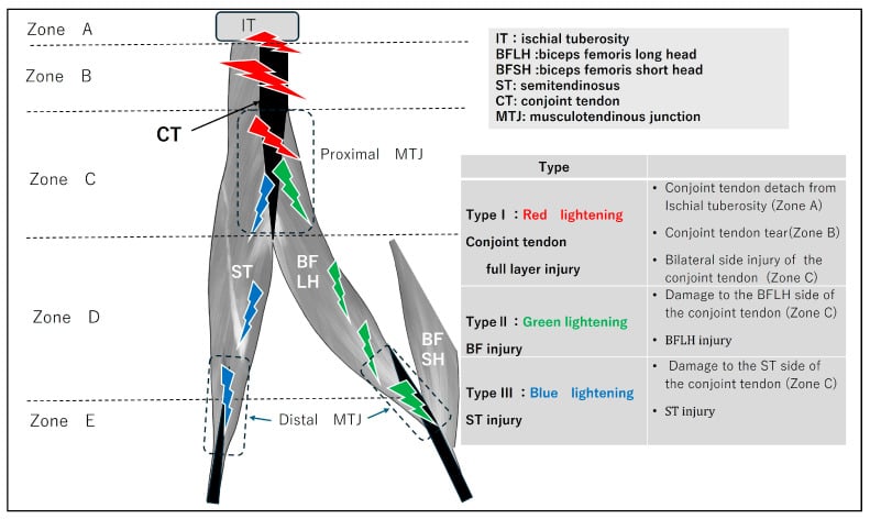
Two footprints. The hamstrings arise from two separate footprints on the ischial tuberosity. Biceps femoris long head and semitendinosus share a conjoint tendon with a single, rounded footprint on the posteromedial tuberosity. Semimembranosus arises separately, from a C-shaped or linear facet that is more anterolateral and deeper.
Why it matters. The anatomy explains the injury patterns: a 2-tendon avulsion is usually the conjoint, which is most commonly avulsed as a unit, and a 3-tendon avulsion is the complete origin. Semimembranosus may avulse independently or with the conjoint.
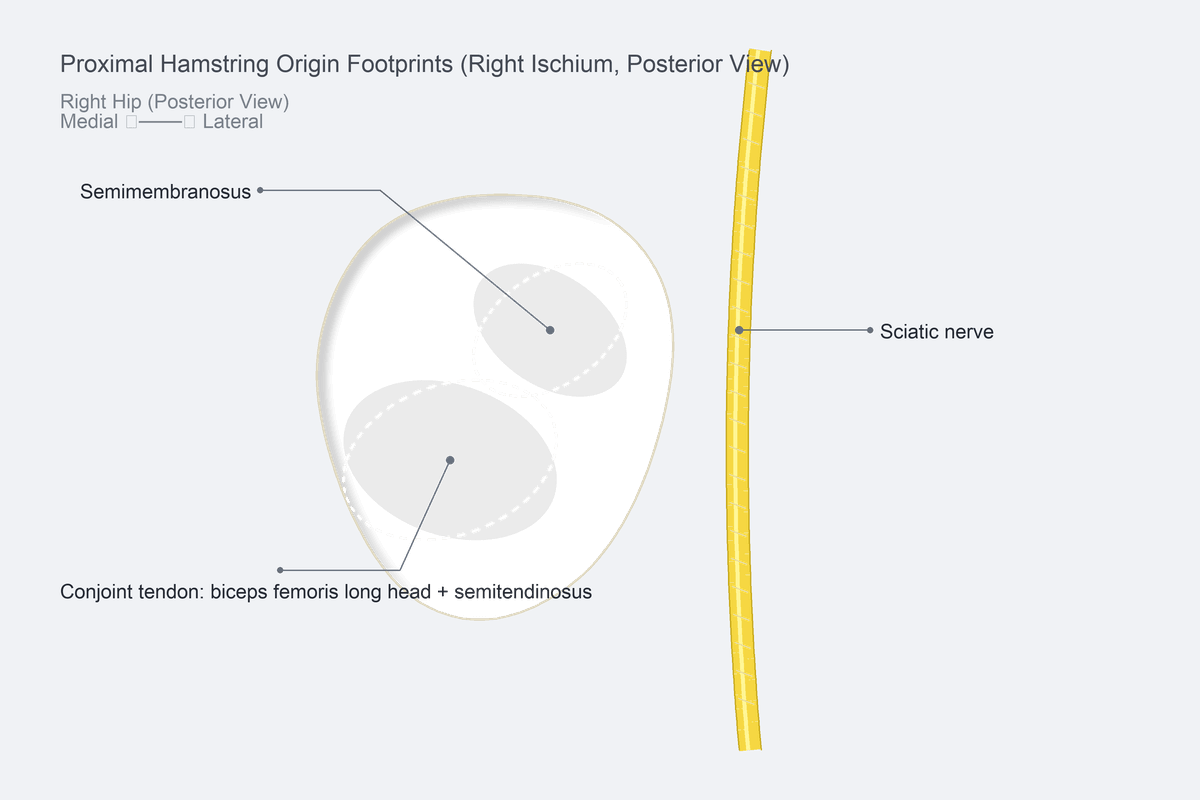
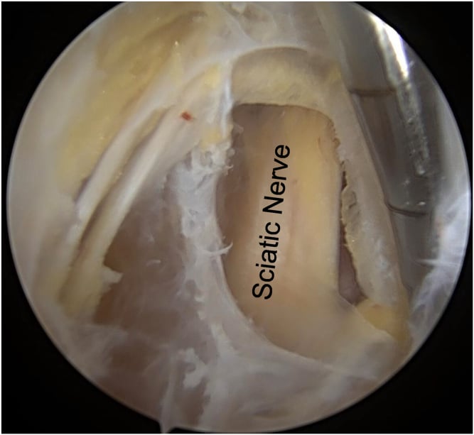
The nerves. The sciatic nerve lies posterior to the hamstring origin, descending just lateral to the tuberosity and on down the thigh. In many cases it is within 1 cm of the stump, and it is at risk both during surgery and from chronic scar. The inferior gluteal nerve and artery exit below piriformis, superior to the hamstring origin, and are protected during gluteal retraction.
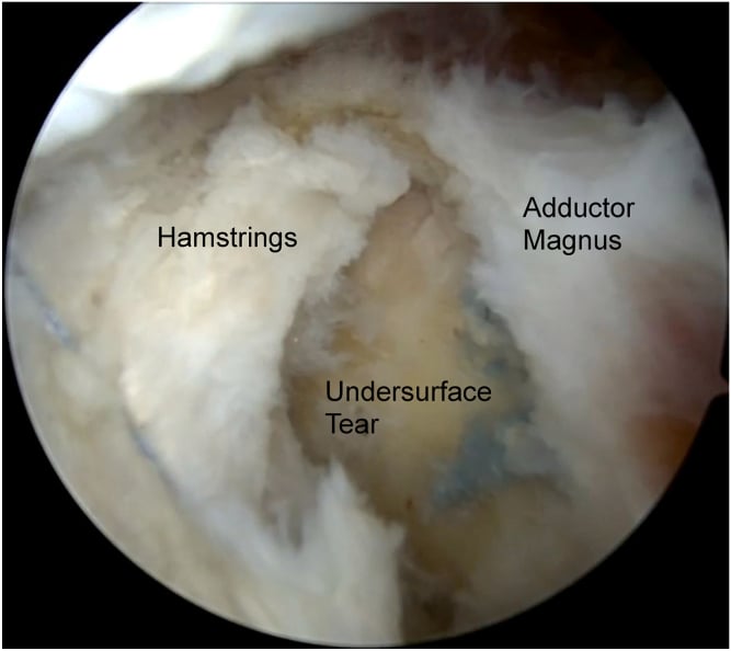
Mechanism. Forced hip flexion with the knee extended lengthens the hamstring while it contracts under load, an eccentric overload. The tendon avulses from bone or fails at the musculotendinous junction. The classic scenarios:
- A waterskiing fall, knee extended and hip flexing
- A sprint start or acceleration
- Hurdling, splits and kicking sports
- A dashboard injury in a road traffic collision
Warm-up deficit, fatigue, hamstring tightness and a prior hamstring strain amplify the risk.
After the tear. With time the stump retracts, adheres to the sciatic nerve and scars, and the repair becomes harder at each stage; the stages are set out under acuity below.
Classification and Types
The Wood grading, made on MRI or at operation, is the most widely used system and drives operative decision-making. The number of tendons off and the time since injury are described alongside it.
- Description
- Osseous avulsion chip, minimal displacement
- Typical management
- Conservative (often heal with rest and rehab)
- Description
- Musculotendinous junction, less than 2 cm retraction
- Typical management
- Controversial - trial conservative or repair in athletes
- Description
- Complete tendon avulsion, 2-5 cm retraction
- Typical management
- Operative repair (strong indication)
- Description
- Complete avulsion, greater than 5 cm retraction
- Typical management
- Operative repair (urgent referral, can be technically demanding)
Type I and low Type II injuries usually respond to structured rehab. Type III and IV are the surgical core.
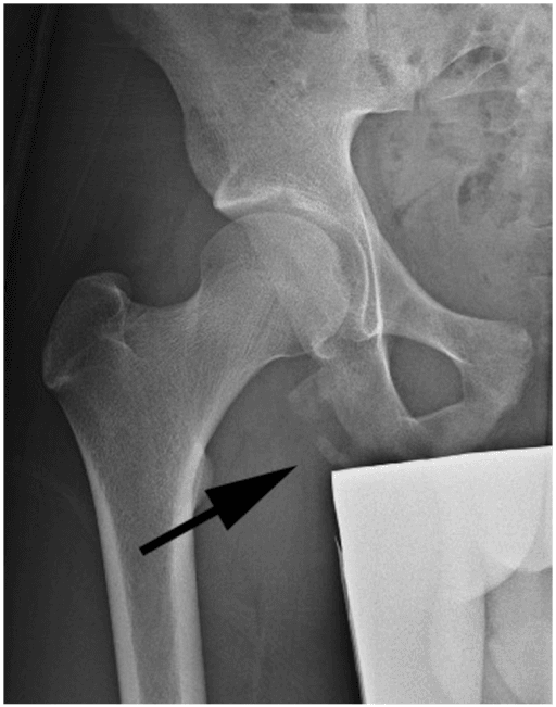
The adolescent variant. In the skeletally immature athlete the same sprint, hurdle or split mechanism avulses the bony apophysis instead of tearing the tendon: an ischial tuberosity apophyseal avulsion fracture. The ischial apophysis is the last pelvic apophysis to fuse, around 20-25 years, and the injury is typically seen in sprinters, gymnasts and footballers of 14-18. It presents with a pop and lower-buttock pain and is diagnosed on a pelvic radiograph, which shows a displaced bony fragment at the ischium, where the adult tendinous avulsion would need MRI.
Treating it. X-ray first, and treat by fragment displacement, not by the adult tendon-retraction rules. A minimally displaced fragment, commonly under about 1.5-2 cm, is treated conservatively with rest, protected weight-bearing and graded rehab. A widely displaced fragment (greater than about 2 cm), a symptomatic non-union or painful exuberant callus is treated with ORIF when acute, or fragment excision with tendon reattachment when chronic or non-united.
The classic trap is mistaking the bony fragment, or its hypertrophic healing callus, for a tumour or chronic osteomyelitis.
Clinical Assessment
History. The story is forced hip flexion with the knee extended, then an audible or felt pop in the buttock or posterior thigh and immediate posterior thigh and ischial pain. The patient cannot sprint or accelerate, sit comfortably or climb stairs with power, and loses push-off and knee flexion power; sitting on a hard chair reproduces the pain. Bruising tracks down the posterior thigh over 24-48 hours.
Examination. Look for swelling and ecchymosis at the gluteal crease and tenderness over the ischial tuberosity, then examine prone and compare with the other side. The gap lies at or just distal to the gluteal crease and is best felt with the hip slightly extended. Knee flexion power is reduced, especially at 15-30 degrees.
- Technique
- Prone, hip extended, knee flexed to 15-30 degrees, resisted flexion
- Positive finding
- Pain at ischium + weakness
- Sensitivity / Note
- Useful screening test
- Technique
- Prone, knee flexed 90 degrees, palpate gluteal crease
- Positive finding
- Soft tissue gap between ischium and stump
- Sensitivity / Note
- Specific when present
- Technique
- Prone knee flexion against graded resistance
- Positive finding
- Reduced compared with other side
- Sensitivity / Note
- Suggests 2- or 3-tendon tear
- Technique
- Tinel sign over ischial region, foot dysaesthesia
- Positive finding
- Paraesthesia radiating to foot
- Sensitivity / Note
- Suggests scar tethering or acute displacement
Always document the tibial and peroneal divisions. Tingling or dysaesthesia in the foot calls for an urgent MRI.
Differential diagnosis. At the ischium the alternatives are:
- Lumbar disc sciatica - pain radiates below the knee, the straight-leg raise is positive and there is no palpable gap
- Ischiogluteal bursitis - chronic pain on sitting, with no acute event and no weakness
- Mid-belly hamstring strain - tender in the posterior thigh rather than at the ischium, with no gap
- Apophysitis or apophyseal avulsion - similar ischial pain in skeletally immature athletes (14-25 years); the pelvic X-ray shows a widened apophysis
- Piriformis syndrome - buttock pain in a sciatic distribution, but no ischial defect and no post-traumatic onset
The chronic cousin: proximal hamstring tendinopathy. Not every ischial complaint is an avulsion. Proximal (high) hamstring tendinopathy is a chronic insertional or overuse tendinopathy of the conjoint origin, classically in distance runners and sprinters. The pain is deep in the buttock and ischium, worst on sitting and during the acceleration or lengthening phase of running, and it comes on insidiously with no pop, no palpable gap and preserved strength.
Separating it. It overlaps with, and must be separated from, ischiogluteal bursitis and deep gluteal (sciatic entrapment) syndrome. MRI or ultrasound shows tendon thickening, intratendinous signal and peritendinous oedema without a retracted stump. This is the answer to "how does it differ from hamstring syndrome and from an avulsion?".
Treating the tendinopathy. Management is non-operative and load-based: relative rest and sitting modification, then a progressive eccentric or heavy-slow-resistance hamstring programme, which is the mainstay. Extracorporeal shockwave therapy and a carefully placed imaging-guided peritendinous injection, avoiding intratendinous corticosteroid and the sciatic nerve, are adjuncts. Surgical debridement or tenotomy is reserved for the refractory minority.
Investigations
Radiographs first. An AP pelvis, a frog-leg lateral and a dedicated ischial tuberosity view look for an osseous avulsion fragment, ischial apophyseal irregularity in adolescents, and pre-existing enthesopathy. A bony chip near the ischium in a sprinting athlete is diagnostic of Wood Type I.
MRI is the gold standard. A dedicated hamstring protocol uses axial and coronal T2 fat-saturated, sagittal T2 and axial T1 sequences, and all three planes are mandatory. It shows the fluid-filled gap at the ischial tuberosity, the retracted stump, sciatic nerve oedema or tethering, and muscle oedema when acute or atrophy when chronic.
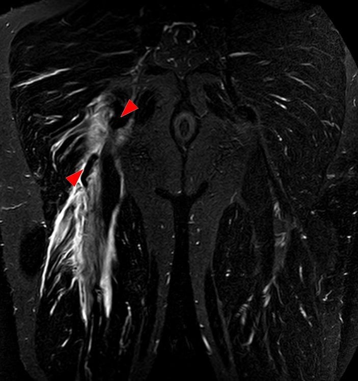
Three things to write in the MRI report for a proximal hamstring avulsion:
- Which tendons are off - conjoint only, conjoint + semimembranosus, semimembranosus only.
- Retraction distance - measure in cm from the ischial footprint to the most distal retracted tendon end on coronal T2 fat-sat.
- Sciatic nerve relationship - displaced, tethered, oedematous, or clear.
These three lines drive the surgical decision more than any other finding.
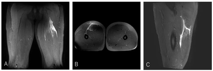
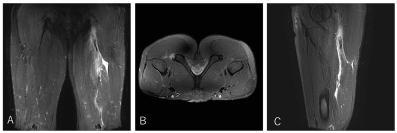
Ultrasound when MRI is indeterminate. It is used for an equivocal MRI, a claustrophobic patient, or to assess tendon continuity dynamically, and shows discontinuity of the tendon and dynamic gapping with knee flexion. It is operator dependent, and the deeper sciatic anatomy is less well seen.
CT in selected cases. Before surgery for a bony avulsion with intra-articular extension or a large osseous fragment, CT defines fragment size, displacement and articular involvement, and so the choice between ORIF and anchor repair.
Management Algorithm
When to operate. The indications for repair:
- A complete 3-tendon avulsion
- A 2-tendon avulsion with greater than 2 cm retraction, measured on coronal or sagittal MRI
- An elite or young active patient, or a manual labourer
- Sciatic nerve symptoms or tethering on MRI
- Failure of conservative care
Timing. Repair within 4 weeks is easier and stronger, and acute repair consistently outperforms delayed repair and conservative care. The case for early repair comes from retrospective series in which outcome tracked the delay to surgery (see the Evidence Base).
- Diagnosis
- MRI: all three tendons off tuberosity
- Treatment
- Operative repair within 4 weeks
- Key Pearl
- Best results
- Diagnosis
- MRI retraction 2-5 cm, no scar
- Treatment
- Operative repair (acute) or delayed
- Key Pearl
- Younger, active patients benefit most
- Diagnosis
- MRI shows tendon near tuberosity
- Treatment
- Trial of conservative care
- Key Pearl
- Re-image at 6-12 weeks if no progress
- Diagnosis
- MRI: fluid at footprint, intact conjoint
- Treatment
- Conservative, structured rehab
- Key Pearl
- Most do well without surgery
Goal. For a Type III or IV injury, a 3-tendon avulsion or a symptomatic 2-tendon avulsion: reattach the avulsed tendons to the ischial tuberosity, free the sciatic nerve, and restore hamstring strength and length.
Positioning. Prone on a standard table with bolsters under the chest and pelvis and both arms abducted. Prep widely from the lower lumbar spine to the popliteal fossa and drape the entire leg free so the knee can be flexed. A tourniquet is not required, as the dissection is in the buttock.
Approach. A transverse or oblique incision in the gluteal crease, centred over the ischial tuberosity, typically 6-10 cm. Identify the sciatic nerve first, lateral to the tuberosity and lying on the short external rotators, and tag it with a vessel loop. The tendon end is usually retracted 2-5 cm distally, and gentle blunt dissection frees it from the surrounding fascia.
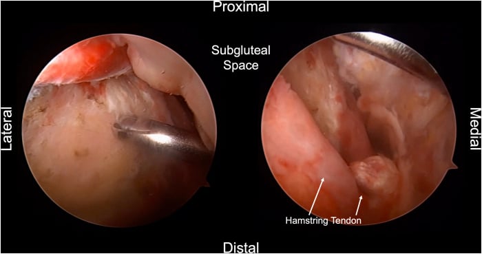
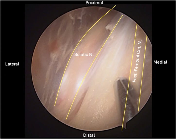
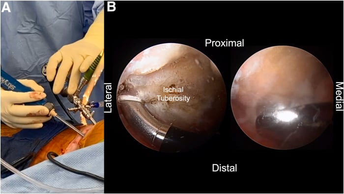
The footprint. Remove residual tendon and decorticate the ischial tuberosity to bleeding cancellous bone. The conjoint and semimembranosus footprints are separate, so inspect both even if the MRI suggested a 2-tendon tear: failing to inspect the semimembranosus footprint is a recognised cause of missed injury.
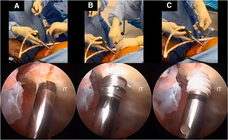
Fixation. Place three to five double-loaded suture anchors along the footprint (5.5 mm titanium or PEEK, with high-strength suture), pass tendon-grasping sutures through the stump, and tie with the knee in 60-90 degrees of flexion. The flexed knee brings the hamstring muscle closer to the footprint and dramatically reduces the tensile load on the repair; many early re-ruptures are technical failures from tying in extension under too much tension. Confirm solid reattachment without strangulating the tendon.
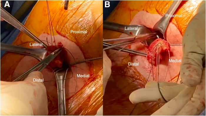
Closure and aftercare. A closed-suction drain stays in the buttock for 24-48 hours. The patient goes into a hip-knee brace locked at 30-60 degrees of knee flexion for 4-6 weeks and is non-weight-bearing on crutches for 6 weeks, with progressive loading thereafter.
SCAROperative Repair Sequence
Hook:The repair leaves a SCAR - sciatic first, clear footprint, anchors in, reattach with the knee flexed.
- Indication
- Acute avulsion with mobile stump
- Outcome
- Best functional scores; return to sport in 6-9 months
- Complications
- Re-rupture (less than 5 percent), sciatic dysaesthesia
- Indication
- Subacute injury with sciatic scar
- Outcome
- Good sciatic symptom relief; strength gains
- Complications
- Persistent nerve symptoms, scar recurrence
- Indication
- Large, displaced osseous fragment
- Outcome
- Anatomic bony healing
- Complications
- Hardware irritation, non-union
Complications
- Incidence
- 5-15 percent dysaesthesia; less than 1 percent motor
- Risk Factors
- Chronic scar, delayed surgery, aggressive retraction
- Management
- Neurolysis at index procedure; late neurolysis for scar
- Incidence
- 3-8 percent in modern series
- Risk Factors
- Tying under tension, premature rehab, anchor pullout
- Management
- Revision repair; consider augmentation
- Incidence
- 1-2 percent
- Risk Factors
- Buttock incision, drain, haematoma
- Management
- Irrigation and debridement; antibiotics
- Incidence
- 1-3 percent
- Risk Factors
- Prone position, hamstring immobility, obesity
- Management
- Mechanical prophylaxis; chemical prophylaxis per local protocol
- Incidence
- 10-20 percent even after repair
- Risk Factors
- Tuberosity prominence, scar tethering
- Management
- Cushion, padding, occasional bursectomy
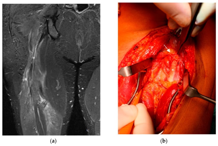
Chronic, untreated avulsions scar the sciatic nerve to the retracted stump, and hamstring syndrome is common after missed or conservatively managed complete tears. Patients develop sitting pain (ischial compression), posterior thigh tightness, persistent weakness and sciatic-type dysaesthesia in the foot. Treatment is late sciatic neurolysis with or without repair, but outcomes are less reliable than acute surgery. This is the single most important reason to MRI early and operate on complete avulsions in active patients.
Outcomes and Prognosis
- Patient
- Complete tear, high demand
- Return to sport
- 80-90 percent
- Strength restoration
- 85-95 percent of contralateral
- Notes
- Best functional scores across series
- Patient
- Subacute, sciatic symptoms
- Return to sport
- 60-75 percent
- Strength restoration
- 75-85 percent of contralateral
- Notes
- Sciatic relief in most; strength gains later
- Patient
- Late presentation, hamstring syndrome
- Return to sport
- 40-60 percent
- Strength restoration
- 60-75 percent of contralateral
- Notes
- Improved but not normal; graft may be required
- Patient
- Partial tear, low retraction
- Return to sport
- 70-85 percent
- Strength restoration
- 80-90 percent of contralateral
- Notes
- Most do well if not complete 3-tendon
- Patient
- Missed or declined surgery
- Return to sport
- 30-50 percent
- Strength restoration
- 50-70 percent of contralateral
- Notes
- Sitting pain, sciatic symptoms common
Prognostic factors. The favourable picture is acute repair within 4 weeks of a complete tendon off with a mobile stump, in a young active patient with no sciatic involvement, done in a single operation. The unfavourable one is chronicity beyond 12 weeks, sciatic scar, a retracted stump that will not reach, re-rupture, multiple prior repairs, age greater than 60, and low demand with significant pain.
4 weeks - outcomes decline measurably beyond this window in most series.
Guidelines, Registries & Global Practice
- Incidence: Proximal hamstring avulsions are uncommon but increasing as participation in sprinting, water sports, and high-intensity training grows worldwide
- Populations at risk: Sprinters (track and field), rugby, football (soccer and American), water skiers, dancers, martial artists
- Mechanism distribution: Eccentric lengthening in sport dominates; dashboard and falls are second-tier causes
- Bilateral avulsions: Reported in sprint and water-ski cohorts; rare but reported
- High-resource: dedicated MRI within 48 hours, sports orthopaedic referral, suture anchor repair, structured sport-specific rehab
- Limited-resource: MRI may be delayed; ultrasound used for screening; conservative care is the default for partial tears; surgical repair is concentrated in tertiary centres
- Universal principle: outcome is determined more by timing of surgery (less than 4 weeks) and accurate MRI grading than by implant choice
- Surgery: most high-volume centres use 3-5 suture anchors to the ischial tuberosity with the knee flexed
- Diagnosis emphasis
- MRI with retraction measurement; classify by Wood
- Operative threshold
- Complete 3-tendon or retraction greater than 2 cm in active patients
- Rehab / return to sport
- Brace at 30-60 degrees flexion, return at 6-9 months
- Diagnosis emphasis
- MRI in elite athletes with suspected avulsion
- Operative threshold
- Operative for complete 2- or 3-tendon in athletes
- Rehab / return to sport
- Eccentric rehab, sport-specific progression, RTS testing
- Diagnosis emphasis
- Clinical exam plus MRI; sciatic assessment
- Operative threshold
- Operative for 3-tendon, 2-tendon with retraction, or sciatic symptoms
- Rehab / return to sport
- MDT-led rehab; phased return to running
- Diagnosis emphasis
- Standardised Wood classification; MRI and operative correlation
- Operative threshold
- Acute operative repair less than 4 weeks when indicated
- Rehab / return to sport
- Eccentric loading, Nordic hamstrings in return phase
There is no dedicated registry for proximal hamstring avulsions, unlike ACL or hip arthroplasty registries. The evidence base is dominated by retrospective case series and small prospective cohorts comparing acute vs delayed repair. Randomised trials are ethically and logistically difficult because the operative indication (complete 3-tendon tear in an active patient) is widely accepted. National joint registries and sports injury databases are gradually including hamstring data, but the field remains principle-based: MRI grading, sciatic assessment, and acute surgery when indicated.
Record in every suspected proximal hamstring avulsion:
- Mechanism (eccentric lengthening during hip flexion + knee extension)
- Examination findings (palpable gap, strength deficit, sciatic Tinel)
- MRI report including which tendons off and retraction in cm
- Sciatic nerve symptoms (subjective and objective)
- Patient demand (elite, recreational, low demand)
- Operative plan and timing (within 4 weeks window if indicated)
Delayed diagnosis and missed operative windows are the most common medicolegal issues in this injury worldwide. Document the assessment and the discussion with the patient explicitly.
Controversies & Areas of Uncertainty
Some centres report acceptable outcomes from structured rehab for 2-tendon avulsions with less than 2 cm retraction in non-elite patients, but most sports orthopaedic groups now favour repair in any active patient, citing faster return to sport and lower chronic pain. The exact threshold is debated.
Three to five anchors along the footprint is conventional, but there is no head-to-head trial of 2-anchor vs 5-anchor constructs. Biomechanical cadaveric studies favour wider anchor spread, but clinical correlation is limited.
Bracing at 30-60 degrees of knee flexion is widely accepted, but the optimum angle, duration, and weight-bearing status vary between centres. Early protected weight-bearing in some protocols appears safe but is not yet supported by high-level evidence.
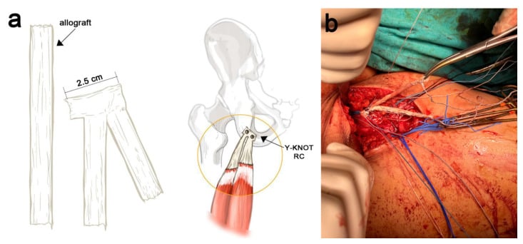
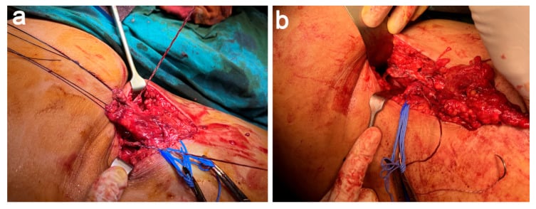
In chronic avulsions with gap greater than 5 cm, allograft (Achilles or hamstring) interposition is the standard off-the-shelf option, but synthetic tapes and other biologics are emerging. Long-term outcome data are lacking.
MCQ Practice Points
Q: Which two tendons share the conjoint origin at the ischial tuberosity? A: Biceps femoris (long head) and semitendinosus. They share a common footprint on the posteromedial ischial tuberosity. The semimembranosus has a separate, more anterolateral origin. This anatomy explains why a 2-tendon avulsion is usually the conjoint and a 3-tendon avulsion is the complete origin.
Q: What is the classic mechanism of a proximal hamstring avulsion? A: Forced hip flexion with the knee in extension - eccentric overload of a contracting hamstring. Common in water skiing (fall with knee extended), sprint starts, hurdling, and dashboard injuries. A pop is typically felt in the buttock, followed by immediate pain and weakness.
Q: In the Wood classification, what retraction distance indicates a surgical repair? A: Greater than 2 cm of retraction (Type III or Type IV). Type I (osseous avulsion, minimal displacement) and Type II (less than 2 cm retraction) may be treated conservatively depending on patient demand. Type III (2-5 cm) and Type IV (greater than 5 cm) are operative indications.
Q: What is the ideal timing for surgical repair of an acute proximal hamstring avulsion? A: Within 4 weeks of injury (acute window). Acute repair produces better strength recovery, higher return-to-sport rates, and lower complication rates than delayed repair. Beyond 12 weeks, the stump retracts and may not reach the ischium, sometimes requiring allograft.
Q: What is the relationship of the sciatic nerve to the proximal hamstring origin, and why does it matter? A: The sciatic nerve lies posterior to the ischial tuberosity and the hamstring origin, passing distally into the thigh. It is at risk during surgical repair (must be identified first) and can become tethered by scar tissue in chronic avulsions (causing hamstring syndrome with sitting pain and sciatic dysaesthesia).
Q: What is the post-operative bracing protocol after proximal hamstring avulsion repair? A: Hip-knee brace locked at 30-60 degrees of knee flexion for 4-6 weeks, non-weight-bearing with crutches for 6 weeks, then progressive loading. The knee is flexed at brace application to offload the repair, since tying was performed with the knee flexed. Eccentric strengthening starts at 6-8 weeks; return to sport typically at 6-9 months.
Exam Viva Scenarios
Practise clinical reasoning and management decisions out loud
“A 24-year-old professional rugby player describes a pop in his left buttock while sprinting to catch a kick 6 days ago. He has bruising tracking down the posterior thigh, weakness with knee flexion, and pain on sitting. MRI shows a complete avulsion of the biceps femoris long head, semitendinosus, and semimembranosus with 3 cm of retraction. The sciatic nerve is displaced but not oedematous. What is your management plan?”
“A 47-year-old recreational marathon runner presents 9 months after a hamstring injury misdiagnosed as a strain. He has persistent buttock pain, sitting discomfort, and intermittent tingling on the lateral border of his foot. Examination reveals a palpable gap at the ischium, reduced hamstring strength, and a positive Tinel sign posteriorly. MRI confirms a complete 3-tendon avulsion with 4 cm of retraction and sciatic nerve tethering. He wants to return to running. How would you manage this?”
Key Anatomy
- Conjoint tendon (biceps long head + semitendinosus) and semimembranosus arise separately from the ischial tuberosity
- Sciatic nerve lies posterior to the hamstring origin - at risk in chronic scar and during surgery
- Mechanism = forced hip flexion with the knee in extension (eccentric lengthening)
Diagnosis
- Mechanism, pop, buttock pain, ecchymosis tracking down the posterior thigh
- Palpable gap at the ischium with the patient prone and the knee flexed
- Reduced knee flexion strength; Puranen test positive
- MRI is gold standard - measure retraction in cm, identify which tendons are off, check sciatic nerve
Classification (Wood)
- Type I = osseous avulsion, minimal displacement (often conservative)
- Type II = musculotendinous, retraction less than 2 cm (contested)
- Type III = complete tendon avulsion, retraction 2-5 cm (operative)
- Type IV = complete avulsion, retraction greater than 5 cm (operative, urgent)
Operative Indications
- Complete 3-tendon avulsion
- 2-tendon avulsion with retraction greater than 2 cm in active patients
- Sciatic nerve symptoms or MRI tethering
- Failed conservative care of partial avulsions
Surgical Pearls
- Prone position, transverse or oblique gluteal crease incision
- Identify and protect the sciatic nerve first
- 3-5 suture anchors to the ischial tuberosity; reattach conjoint and semimembranosus separately
- Tie sutures with the knee flexed to 60-90 degrees to offload the repair
- Acute repair (less than 4 weeks) is best; chronic cases may need allograft
Complications
- Sciatic nerve dysaesthesia (5-15 percent) - identify nerve first
- Re-rupture (3-8 percent) - tied under tension is the usual cause
- Hamstring syndrome in chronic missed injuries - scar tethering of sciatic
- DVT, infection, persistent sitting pain from tuberosity prominence
Evidence Base and Key Trials
Functional outcome after repair of proximal hamstring avulsions
- Thirty-four complete proximal hamstring ruptures identified on bowstring sign and MRI; 23 attended for subjective, functional and isokinetic Cybex testing
- Nine acute and fourteen chronic repairs, mean follow-up 43.3 months
- Twenty-one of 23 returned to an average of 95 per cent of pre-injury activity level at a mean of 9.8 months; 18 excellent, four good, one fair
- Hamstring strength averaged 93 per cent and 90 per cent of the uninvolved limb at 240 and 180 degrees per second
Complete proximal hamstring avulsions: a series of 41 patients with operative treatment
- Forty-one patients (21 men, 20 women) with complete proximal hamstring avulsion, mean follow-up 37 months (range 12 to 72)
- Nineteen excellent and 10 good results; five moderate and seven poor
- Delay from injury to surgery averaged 2.4 months in the excellent or good group versus 11.7 months in the moderate or poor group (p less than 0.001)
- Considerable symptomatic improvement was still achieved in chronic cases, but early surgery gave the better result
Hamstring injuries in professional football players: magnetic resonance imaging correlation with return to play
- Thirty-eight professional American football players (43 cases) with acute hamstring strains - not avulsions - graded on MRI and correlated with games missed
- Players missed an average of 2.6 plus or minus 3.1 games; the ten players who had retraction on MRI missed 5.5
- Radiological grade tracked absence: grade I 1.1 games, grade II 1.7, grade III 6.4
- Involvement of less than 50 per cent of the muscle cross-section meant 1.8 games missed, against 3.2 games when more than 75 per cent was involved
Outcomes following adolescent athlete proximal hamstring apophyseal avulsion bone fragment excision and direct tendon-ischial tuberosity reattachment
- Eighteen consecutive athletes (7 elite, 11 recreational) treated by one surgeon over 13 years, mean age at injury 14.6 years
- Avulsed fragment excised and the tendon reattached directly to the ischial tuberosity with bone anchors, assessed on the validated hamstring-specific SHORE score
- Mean scores improved from 11.89 to 33.31, finishing within 0.3 to 6.1 per cent of pre-injury values
- Every case returned to pre-injury level of sport by one year with no significant complications; acute surgery within 3 months improved scores by 247.7 per cent at one year against 59.0 per cent for chronic nonunion surgery