Joint Infection | Cartilage Destruction | Surgical Emergency
- Septic arthritis is a surgical emergency - glycosaminoglycan loss begins within hours, and the irreversible collagen loss follows later
- Synovial fluid WCC over 50,000 with over 90% PMNs strongly suggests septic arthritis
- Native joint sepsis = washout (repeated if not improving) + antibiotics. Prosthetic joint = DAIR or two-stage revision
- Kocher criteria (a HISTORY of fever, non-weight bearing, ESR of at least 40, WCC over 12) predict paediatric septic hip
- Australian eTG recommends flucloxacillin plus ceftriaxone for empiric therapy
- “Gonococcal arthritis is the most common in sexually active young adults
- “Lyme arthritis is monoarticular large joint - serology positive
- “Prosthetic joint infection under 3 months = acute, debride and retain (DAIR)
- “Kingella kingae is common in children under 4 years - difficult to culture
Overview
Septic arthritis is a bacterial infection of a joint and a true orthopaedic emergency. Bacterial toxins and the host's own inflammatory mediators act together to destroy articular cartilage rapidly, and understanding how they do it is what makes the urgency of treatment obvious.
The clock. Glycosaminoglycan loss begins within hours of bacterial invasion; collagen loss, the irreversible step, comes later. Surgical drainage is required within 24 hours to prevent permanent joint damage.
Routes of Infection
Haematogenous spread. The most common route, 80% of cases, and the usual one in native joints. A transient bacteraemia seeds the synovium, which is highly vascular and has no basement membrane, so bacteria penetrate easily into the joint space. Intravenous drug use, immunosuppression and diabetes are risk factors.
Direct inoculation. The cause is iatrogenic (joint injection, arthroscopy, arthroplasty) or traumatic (penetrating injury, open fracture). The initial bacterial load is typically lower, and symptoms may be delayed.
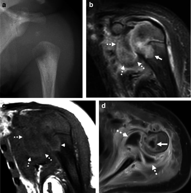
Contiguous spread. Infection spreads from adjacent osteomyelitis or soft tissue infection, osteomyelitis especially in children. The hip, shoulder and elbow have an intra-articular metaphysis, so metaphyseal infection ruptures into the joint, and neonatal osteomyelitis commonly causes septic arthritis. In adults, vertebral osteomyelitis can seed the disc.
How the Cartilage Is Destroyed
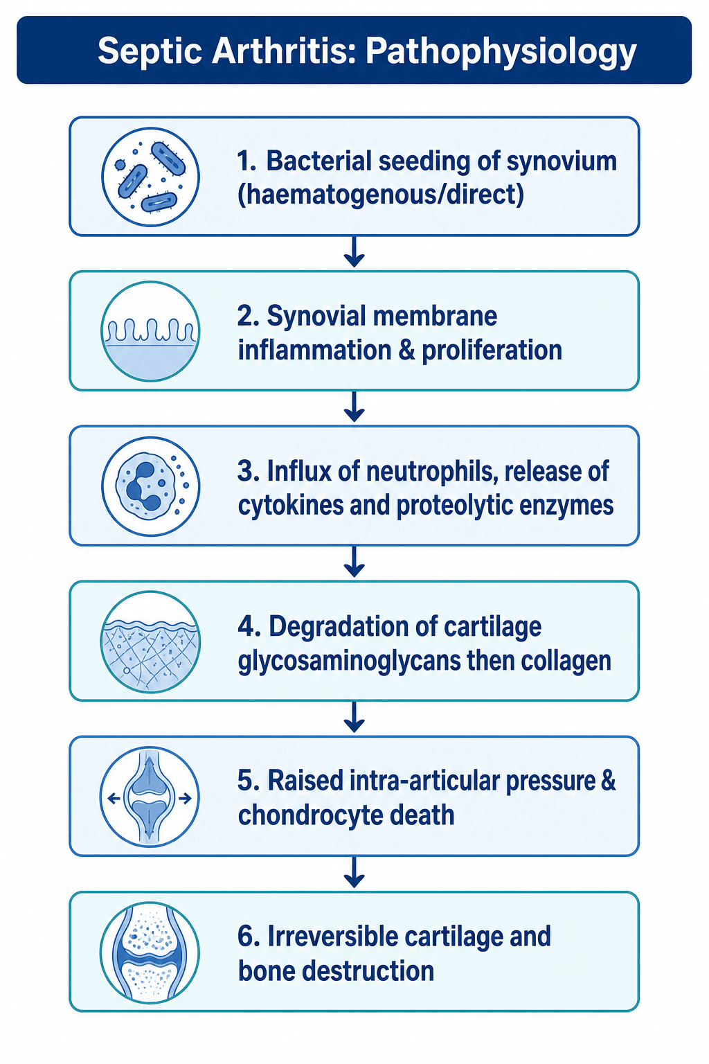
The host response. The host paradoxically contributes to the destruction. Infiltrating neutrophils release proteolytic enzymes, elastase, cathepsin G, collagenase and matrix metalloproteinases, that degrade the cartilage matrix, and a cytokine cascade of IL-1 and TNF-alpha drives continued inflammation. Bacterial toxins damage chondrocytes directly.
Pressure. Synovial inflammation raises intra-articular pressure, which compromises cartilage nutrition.
Biofilm. Bacteria can form biofilms on cartilage and synovial surfaces. The glycocalyx matrix protects them from antibiotics and host immune cells, and quorum sensing lets them communicate and coordinate toxin production. Persistent infection develops if a biofilm establishes on damaged cartilage.
The sequence. In the experimental model proteoglycan is lost first and collagen weeks later.
Progressive Joint Destruction
Bacteria bind to the synovium and proliferate. They multiply rapidly in nutrient-rich synovial fluid and trigger the inflammatory response.
Massive neutrophil infiltration and enzyme release. Glycosaminoglycan loss is already under way: starting antibiotics as early as 4 to 12 hours after inoculation still left an average 18% GAG loss.
Glycosaminoglycan is stripped from the matrix first. By 48 hours experimental cartilage had lost 20% (E. coli) to 42% (S. aureus) of its GAG, and chondrocytes are dying as rising pressure compromises the epiphyseal blood supply. The collagen framework is still largely intact, which is why urgent decompression and antibiotics can still preserve the cartilage architecture.
The collagen scaffold fails late, and that is the irreversible step. Collagen loss was not significant before the third week, by which point about 42% of collagen and up to 73% of GAG had gone. Once the collagen framework is destroyed the cartilage cannot be reconstituted.
Pannus formation and fibrous ankylosis. Granulation tissue (pannus) grows over the remaining cartilage and fibrous adhesions form, which may progress to bony ankylosis. Growth plate damage follows in children, and the patient is left with chronic pain and stiffness.
Cartilage damage begins within hours, not days, and because proteoglycan goes before the collagen framework fails, the window in which the matrix can still be protected is measured in hours. Delay beyond 24 hours worsens outcomes: permanent joint damage, chronic pain and stiffness, and the need for arthrodesis or arthroplasty.
Why Surgical Drainage Is Mandatory
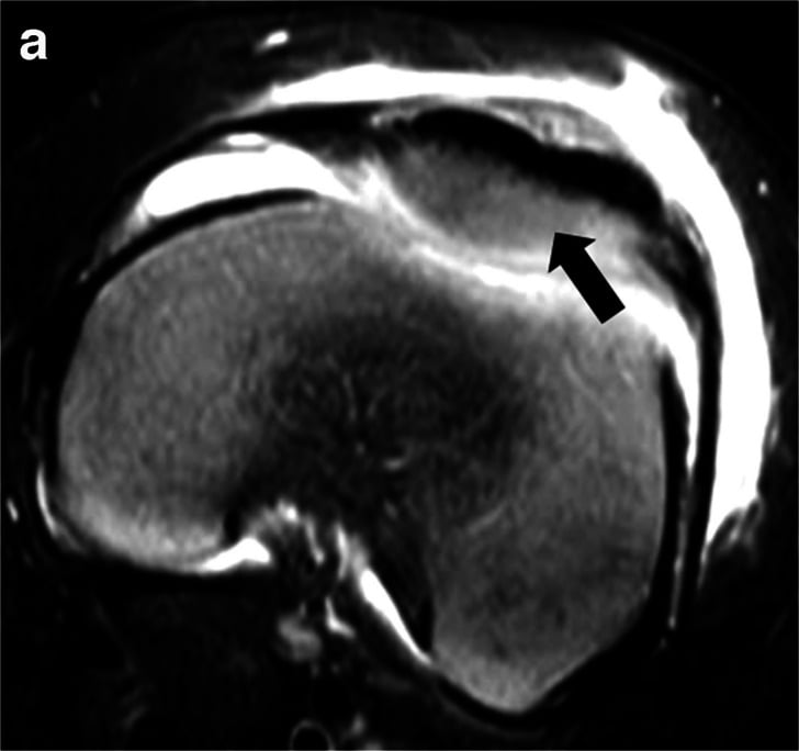
What drainage does. Washing out the joint removes the bacterial load and the proteolytic enzymes, reduces intra-articular pressure, prevents a biofilm establishing on the cartilage, and lets the surgeon see and debride the joint directly.
Why antibiotics alone fail. Antibiotics cannot penetrate an established biofilm, do not remove the enzymes already released, and cannot reduce the mechanical effect of raised pressure. The delay while they are given alone allows irreversible cartilage damage.
Microbiology
Staphylococcus aureus is the most common organism, causing 50-70% of septic arthritis overall and 60-70% of adult cases. Streptococcal species account for 15-20%. Gram-negative organisms (E. coli, Pseudomonas) are more common in the elderly and immunocompromised. Mycobacterial infection has an atypical presentation and a chronic course.
The organism follows the patient's age and risk factors.
- Most Common Organism
- Group B Streptococcus
- Second Most Common
- E. coli, S. aureus
- Key Clinical Feature
- Can spread from osteomyelitis via transphyseal vessels
- Most Common Organism
- Kingella kingae
- Second Most Common
- S. aureus
- Key Clinical Feature
- Difficult to culture - fastidious organism
- Most Common Organism
- Staphylococcus aureus
- Second Most Common
- Streptococcus pyogenes
- Key Clinical Feature
- 70% of paediatric septic arthritis
- Most Common Organism
- Neisseria gonorrhoeae
- Second Most Common
- S. aureus
- Key Clinical Feature
- Polyarticular, migratory arthritis, skin lesions
- Most Common Organism
- Staphylococcus aureus
- Second Most Common
- Streptococcus species
- Key Clinical Feature
- 60-70% of adult septic arthritis
- Most Common Organism
- S. aureus, Pseudomonas
- Second Most Common
- Candida species
- Key Clinical Feature
- Unusual joints: sternoclavicular, sacroiliac
- Most Common Organism
- Gram-negative rods
- Second Most Common
- Fungi, atypical mycobacteria
- Key Clinical Feature
- Indolent course, multiple joints
- Most Common Organism
- S. aureus
- Second Most Common
- Coagulase-negative Staph
- Key Clinical Feature
- Acute presentation - DAIR candidate
- Most Common Organism
- Coagulase-negative Staph
- Second Most Common
- Propionibacterium
- Key Clinical Feature
- Low virulence biofilm - two-stage revision
Kocher Criteria for Paediatric Septic Hip
Four predictors, and the probability of septic arthritis rises with each one present. The probabilities below, including 99.6% with all four, are from the original derivation cohort.
- A history of fever, conventionally over 38.5°C (101.3°F). A measured temperature did not survive Kocher's multivariate model. Fever may be absent in the immunocompromised, low-grade fever is less predictive, and documented fever is the most reliable.
- Non-weight bearing, refusal to bear weight on the affected limb. It is the most specific clinical sign but is also present in transient synovitis, and inability to bear weight is worse than a limp.
- ESR of at least 40 mm/hr; exactly 40 counts. ESR is less specific than CRP and slower to rise and normalise, and a CRP over 20 mg/L is an alternative cut-off.
- Serum WCC over 12,000 per microlitre. It may be normal in early infection and is less reliable than the synovial fluid WCC.
- Probability of Septic Arthritis
- Under 0.2% (none) to 3% (one)
- Recommendation
- Observe, serial examination
- Probability of Septic Arthritis
- 40%
- Recommendation
- Urgent aspiration and synovial fluid analysis
- Probability of Septic Arthritis
- 93%
- Recommendation
- Strong indication for surgical drainage
- Probability of Septic Arthritis
- 99.6%
- Recommendation
- Emergency surgical washout required
The criteria were derived for the paediatric hip to distinguish septic arthritis from transient synovitis. They should NOT be used to diagnose septic arthritis in adults or other joints, or to exclude infection in high-risk patients. A child with one criterion still has a 3% risk, so clinical judgement remains essential. A child with all four needs urgent aspiration under anaesthesia: do not delay for MRI.
Classification
Septic arthritis is described by its route (above), the joint, the host, the pathogen (see Microbiology) and, at arthroscopy, the Gächter stage.
Native or prosthetic joint. Native joints have higher cure rates with appropriate treatment. Prosthetic joint infection is more complex and often requires removal of the implant.
The host. Immunocompetent patients have a better prognosis and are treated to standard protocols. The immunocompromised get atypical organisms and need prolonged treatment, and intravenous drug users present in unusual joints (see below).
Child or adult. In children the hip is more often involved, progression is rapid and there is a risk of avascular necrosis. In adults the knee is most common and the cartilage tolerates infection better.
Gächter stage. The arthroscopic appearance guides treatment. Stages I-II respond well to arthroscopic lavage; stages III-IV often require open surgery and carry a worse prognosis because of the cartilage damage.
- Findings
- Turbid fluid, hyperaemic synovium
- Treatment Implications
- Single arthroscopic lavage often sufficient
- Findings
- Purulent fluid, fibrin deposits
- Treatment Implications
- Arthroscopic lavage, may need repeat
- Findings
- Thick fibrin, compartmentalisation
- Treatment Implications
- May require open synovectomy
- Findings
- Cartilage destruction, osteolysis
- Treatment Implications
- Open debridement, poor prognosis
Diagnosis and Investigations
The aspirate is the most important test. A synovial WCC over 50,000/μL is highly suggestive, and one over 25,000/μL is already suspicious. The Gram stain is immediate, but culture may take 48-72 hours.
- Normal
- Clear, straw-coloured
- Crystal Arthritis
- Turbid, yellow
- Septic Arthritis
- Turbid, purulent
- Clinical Note
- -
- Normal
- Under 200
- Crystal Arthritis
- 2,000-75,000
- Septic Arthritis
- Usually over 50,000
- Clinical Note
- Overlap exists - over 90% PMN more specific
- Normal
- Under 25%
- Crystal Arthritis
- 50-75% or higher
- Septic Arthritis
- Over 90%
- Clinical Note
- Over 90% polymorphs highly specific for sepsis
- Normal
- Negative
- Crystal Arthritis
- Negative
- Septic Arthritis
- Positive in 50%
- Clinical Note
- Immediate result - guides empiric therapy
- Normal
- Sterile
- Crystal Arthritis
- Sterile
- Septic Arthritis
- Positive in 70-80%
- Clinical Note
- Gold standard - obtain before antibiotics
- Normal
- Similar to blood
- Crystal Arthritis
- Normal
- Septic Arthritis
- Less than half blood glucose
- Clinical Note
- Bacteria consume glucose
- Normal
- None
- Crystal Arthritis
- MSU or CPPD positive
- Septic Arthritis
- None (may coexist)
- Clinical Note
- Always check - gout can be septic too
Gout and pseudogout can precipitate septic arthritis or coexist with it, especially in the elderly and immunosuppressed. If the synovial WCC is over 50,000, the patient is systemically unwell or clinical suspicion is high, send culture, start antibiotics and treat as septic arthritis whatever the crystals show.
Blood tests. The serum WCC is often raised but may be normal in the immunocompromised. CRP is raised and is useful for monitoring response; ESR is raised too but slower to change. Blood cultures are positive in 30-50% and are always taken if the patient is febrile.
Special investigations include:
- PCR for fastidious organisms (Kingella, mycobacteria)
- Procalcitonin, with higher sensitivity and specificity than CRP
- Lactate, raised in septic compared with inflammatory arthritis
Plain radiographs are the first-line image. Acutely they show joint space widening from the effusion, soft tissue swelling and loss of the fat planes, and they exclude a fracture or foreign body; chronic infection brings joint space narrowing and erosions. A radiograph cannot rule out sepsis.
Ultrasound detects an effusion, including the hip effusion in a child, guides aspiration and differentiates effusion from synovitis. It is operator-dependent and cannot distinguish septic from aseptic fluid.
Differential Diagnosis of the Acute Monoarticular Hot Joint
- Typical Features
- Acutely hot, exquisitely painful joint; often febrile; refusal to move/bear weight
- Synovial Fluid
- Turbid/purulent; WCC usually over 50,000 with over 90% PMN; Gram stain positive in ~50%
- Key Discriminator
- Positive Gram stain or culture; very high WCC with PMN predominance - treat empirically if in doubt
- Typical Features
- Recurrent, often first MTP joint; rapid onset; tophi; hyperuricaemia
- Synovial Fluid
- WCC 2,000-75,000; negatively birefringent needle-shaped MSU crystals
- Key Discriminator
- Crystals on polarised microscopy - but crystals do NOT exclude coexisting sepsis
- Typical Features
- Older patient; knee/wrist; chondrocalcinosis on XR
- Synovial Fluid
- WCC 2,000-50,000; positively birefringent rhomboid CPPD crystals
- Key Discriminator
- Rhomboid crystals plus chondrocalcinosis
- Typical Features
- Post-enteric/urogenital infection; oligoarthritis; enthesitis; may have conjunctivitis/urethritis
- Synovial Fluid
- Inflammatory, sterile (WCC often 2,000-50,000)
- Key Discriminator
- Sterile fluid with antecedent GI/GU infection; HLA-B27 association
- Typical Features
- Trauma, anticoagulation, or haemophilia; rapid swelling
- Synovial Fluid
- Frankly bloody aspirate; may show fat (lipohaemarthrosis) if intra-articular fracture
- Key Discriminator
- Blood (and fat globules) on aspiration; coagulation/trauma history
- Typical Features
- Well child, low-grade or no fever; able to bear some weight; recent viral illness
- Synovial Fluid
- Sterile inflammatory effusion; lower synovial WCC
- Key Discriminator
- Few/no Kocher predictors; settles with rest and NSAIDs - but aspirate if uncertain
- Typical Features
- Known RA/psoriatic/JIA; polyarticular history
- Synovial Fluid
- Inflammatory; sterile (RA itself raises septic risk - have a low threshold to aspirate)
- Key Discriminator
- Pattern and prior diagnosis - but immunosuppressed RA patients are HIGH-risk for superimposed sepsis
Polyarticular Septic Arthritis: The Dangerous Exception
Septic arthritis is usually monoarticular. Roughly 10-20% is polyarticular, involving two or more joints, and that is the presentation most likely to kill the patient.
Why it kills. Polyarticular sepsis reflects overwhelming bacteraemia, so mortality is much higher: quoted around 30-50% in some series, against roughly 10% for monoarticular disease.
The host and the organism. It is almost always haematogenous and usually S. aureus, sometimes streptococci, pneumococcus or gonococcus. It is strongly associated with pre-existing joint disease, rheumatoid arthritis being the classic host, and with immunosuppression (diabetes, steroids, malignancy).
Find the source. Two or more septic joints means blood-borne seeding, so hunt for the focus of the bacteraemia, and treat every affected joint as well as the systemic source.
- Infective endocarditis above all: echocardiography, repeated blood cultures, a new murmur, peripheral stigmata
- Skin
- Intravascular lines
- Urinary tract
- Spine
A polyarticular flare in a known rheumatoid patient is easily mislabelled an "RA flare", yet RA patients are the highest-risk group for true and for polyarticular sepsis. A flaring, systemically unwell rheumatoid patient must be aspirated, not simply given more steroid.
Septic Arthritis of the Unusual Joints: Sternoclavicular and Sacroiliac
Sepsis of the sternoclavicular, sacroiliac, symphysis pubis and spinal joints is characteristic of intravenous drug users and the immunosuppressed. The organism is still most often S. aureus, but with a notably higher rate of Pseudomonas and other Gram-negatives in drug users. These joints get missed because they are deep, hard to examine and aspirate, and present insidiously.
- Sternoclavicular joint
- Anterior chest or medial-clavicle pain, swelling and tenderness; insidious
- Sacroiliac joint (pyogenic sacroiliitis)
- Buttock, low-back or hip pain, often unilateral, with fever; mimics mechanical back pain
- Sternoclavicular joint
- Localised SC swelling and tenderness
- Sacroiliac joint (pyogenic sacroiliitis)
- FABER/Patrick test, direct SI tenderness, pelvic compression
- Sternoclavicular joint
- CT or MRI to define a retrosternal/mediastinal abscess and osteomyelitis
- Sacroiliac joint (pyogenic sacroiliitis)
- MRI - bone-marrow oedema, fluid, abscess
- Sternoclavicular joint
- Abscess can track into the mediastinum (mediastinitis)
- Sacroiliac joint (pyogenic sacroiliitis)
- Easily mistaken for mechanical pain or hip pathology
- Sternoclavicular joint
- Aspiration and culture; often en-bloc joint resection with soft-tissue coverage for abscess or osteomyelitis
- Sacroiliac joint (pyogenic sacroiliitis)
- CT-guided aspiration; antibiotics, drain an abscess; surgery less often needed
Keep a high index of suspicion in these patients and use cross-sectional imaging early. The sternoclavicular joint's capacity for retrosternal and mediastinal spread is what raises the threshold from a simple washout toward joint resection.
Management
Treatment Algorithm
Aspirate the joint urgently, before antibiotics. Send the fluid for WCC, Gram stain and culture (aerobic, anaerobic, fungal), check for crystals, and document the gross appearance (purulent, cloudy, bloody). Take blood cultures if febrile.
Start antibiotics once cultures are obtained. The Australian eTG regimen is flucloxacillin 2g IV 6-hourly plus ceftriaxone 2g IV daily, which covers S. aureus, Streptococcus and Gram-negatives; empiric cover must include S. aureus. Adjust when cultures are available.
Arthroscopic or open washout within 24 hours. Irrigate copiously, debride fibrin and synovial debris, and take multiple tissue samples. The technique by joint is set out under Surgical Technique.
Repeat the washout if the joint is not improving, on the indications listed under Surgical Technique. Up to 3 washouts may be needed; consider open surgery if arthroscopy is failing.
Complete a total course of 2-6 weeks. The IV-to-oral switch, duration by joint and CRP monitoring are set out under Postoperative Care.
- Major criteria (either one confirms PJI): a sinus tract communicating with the prosthesis/joint, OR two positive cultures of the same organism from the joint.
- Minor criteria (a threshold combination confirms PJI when no major criterion is met): raised serum CRP and/or ESR; raised synovial fluid white-cell count; raised synovial polymorphonuclear percentage; positive synovial alpha-defensin (or leucocyte esterase); positive periprosthetic tissue histology (neutrophils per high-power field); and a single positive culture.
- The EBJIS framework adds practical categories (infection "confirmed / likely / unlikely") and emphasises multiple periprosthetic tissue samples (commonly several, with a threshold number positive), prolonged culture incubation (to catch Cutibacterium/low-virulence organisms), and sonication of the explant to dislodge biofilm and raise culture yield.
Why it matters: PJI is a composite diagnosis. A single negative aspirate does not exclude it, and the thresholds differ from native-joint septic arthritis: synovial WCC cut-offs for PJI are much lower, in the low thousands, not over fifty thousand.
Surgical Technique
Arthroscopic lavage is preferred for most joints. Through the standard portals for the joint, inspect it and document the state of the cartilage, irrigate copiously with a minimum of 9-12 litres of saline, debride fibrinous debris and synovitis, and take multiple tissue samples for culture.
The knee. Use the standard anteromedial and anterolateral portals, lavage every compartment including the suprapatellar pouch, and debride loculations and fibrinous adhesions. A posterolateral portal may be needed for complete access. Consider a synovial drain postoperatively.
Open arthrotomy is required for the hip and for some arthroscopic failures: a complete capsulotomy for drainage, copious irrigation and debridement, and closure in layers. Leaving a drain is controversial; most surgeons avoid one because of the risk of retrograde infection.
The hip is too deep for effective arthroscopic drainage, and arthroscopy is not recommended. An anterior (Smith-Petersen) or anterolateral (Watson-Jones) approach allows complete release of the capsule, debridement of fibrinous material and copious irrigation (minimum 9 litres). At the hip, leave the capsule open or close it loosely.
Small joints. Serial daily aspirations may be adequate for fingers and toes, but they must show a falling WCC and sterile cultures. Convert to surgical drainage if the joint is not improving.
When to go back. Repeat surgery is indicated by:
- Persistent fever beyond 48-72 hours after drainage
- A rising or plateaued CRP
- Ongoing joint swelling, effusion or erythema
- New positive cultures
Complications
Cartilage destruction. Enzymatic degradation by bacterial toxins and host proteases destroys the cartilage; once established it is irreversible and leads to arthritis. Early diagnosis and aggressive treatment are the prevention.
Systemic. Sepsis and septic shock occur in 5-10%, with multi-organ dysfunction, and metastatic infection can reach other joints or cause endocarditis.
By joint. In the child's hip the risk is avascular necrosis of the femoral head; in the knee, stiffness and secondary osteoarthritis; in the shoulder, rotator cuff damage and stiffness.
- Incidence
- 40-50%
- Prevention/Treatment
- Early drainage, minimise cartilage damage
- Incidence
- 20-30%
- Prevention/Treatment
- Early ROM, physiotherapy
- Incidence
- 10-20%
- Prevention/Treatment
- Urgent drainage, protect blood supply
- Incidence
- Variable
- Prevention/Treatment
- Long-term monitoring, corrective surgery
Postoperative Care
Antibiotics. Continue IV antibiotics for a minimum of 2 weeks and switch to oral when the patient is clinically improving and the CRP is falling, with culture-directed therapy once sensitivities are available. The total course is 2-6 weeks, depending on the joint and the response:
- Small joints (fingers, toes): 2 weeks
- Large joints (knee, elbow): 4 weeks
- Hip and spine: 6 weeks minimum
- Prosthetic joints: 12 weeks (DAIR) or staged revision
Monitoring. Assess the patient daily (pain, swelling, range of motion) and chart the temperature. Check the CRP every 2-3 days at first, then weekly; it should fall by 50% at 2 weeks. A CRP that fails to fall suggests inadequate drainage, a resistant organism or the wrong diagnosis, so consider repeat imaging and aspiration.
Rehabilitation. Start gentle passive range of motion as soon as it is tolerated, protect weight-bearing until the infection is controlled, and progress to strengthening once it has resolved. Full weight-bearing follows when the joint is pain-free and the infection controlled, sport and heavy activity 3-6 months after resolution, with long-term follow-up for secondary arthritis.
The traditional teaching of "weeks of IV antibiotics" for bone and joint infection has been overturned by the OVIVA trial (Oral Versus Intravenous Antibiotics), which is examinable and practice-changing:
- Design: a large multicentre randomised non-inferiority trial in adults with bone and joint infection (including native septic arthritis, osteomyelitis and prosthetic-joint infection), randomising patients in the first six weeks of treatment to continued IV versus an early switch to oral antibiotics, after initial surgical management as indicated.
- Result: oral antibiotics were non-inferior to IV for the primary endpoint of treatment failure at one year. Oral was not worse, and was associated with shorter hospital stay and fewer line-related complications (catheter problems, line infections).
- Caveats: it relied on specialist infection input/oral agents with good bone bioavailability (e.g. fluoroquinolones, often with rifampicin for staphylococcal implant infection) and assured oral absorption/compliance. It does not mean "oral for everyone from day one": source control and appropriate agent selection still apply.
Why it matters: it justifies an early IV-to-oral switch once the patient is improving with a suitable, well-absorbed oral agent, reducing line complications and hospital stay, a direct change from the old reflex of prolonged IV therapy.
Outcomes
Native joints. With appropriate treatment the cure rate is 85-95%, and with early aggressive treatment 50-60% make a full recovery. Mortality in adults is 5-15%, higher in the elderly and those with comorbidities, and 10-20% need a joint replacement in the long term.
Prosthetic joints. Two-stage revision succeeds in 85-95%. Amputation is needed in 1-3%, after failed treatment or with comorbidities.
The paediatric hip. The risk of avascular necrosis is higher with delay. Coxa magna is a common sequela, and 30-40% develop arthritis in the long term.
What predicts the result. Delayed treatment beyond 24 hours significantly worsens outcomes, and each 24-hour delay increases the risk of cartilage destruction and a poor functional outcome.
- Better Outcome
- Under 24 hours
- Worse Outcome
- Over 72 hours
- Better Outcome
- Young, healthy
- Worse Outcome
- Elderly, comorbid
- Better Outcome
- Streptococcus
- Worse Outcome
- S. aureus, Gram-negative
- Better Outcome
- Small joint
- Worse Outcome
- Hip, shoulder
Guidelines, Registries & Global Practice
Global Epidemiology
Native-joint septic arthritis affects approximately 4-10 per 100,000 person-years in high-income populations, rising to 30-70 per 100,000 in those with rheumatoid arthritis or a prosthetic joint. Incidence is increasing worldwide, driven by an ageing population, more joint replacement surgery, immunosuppression, diabetes and injecting drug use. Staphylococcus aureus is the leading pathogen globally, but the proportion of MRSA varies markedly by region (low in much of Scandinavia and the Netherlands, substantially higher in parts of the USA and Asia), which directly affects empirical antibiotic choice. In sexually active young adults, Neisseria gonorrhoeae remains an important cause where prevalence is high. Reported mortality is 7-15% and long-term joint dysfunction occurs in roughly a third of survivors despite treatment.
Side-by-Side Guidance
- Scope
- Hot swollen joint - adults
- Core Recommendations
- Aspirate before antibiotics; urgent synovial Gram stain, culture, cell count; empirical anti-staphylococcal cover then organism-directed; joint drainage (aspiration, arthroscopic or open)
- Evidence Strength
- Consensus / low-moderate (largely observational base)
- Scope
- Prosthetic joint infection
- Core Recommendations
- Defines DAIR vs one-/two-stage exchange; rifampin combination for stable staphylococcal implant infection; structured antibiotic duration
- Evidence Strength
- Guideline - mostly low-quality evidence, strong consensus
- Scope
- Periprosthetic joint infection definition
- Core Recommendations
- Standardised diagnostic criteria (synovial WCC, alpha-defensin, culture, histology) for consistent diagnosis and research
- Evidence Strength
- International consensus definition
- Scope
- Periprosthetic hip/knee infection
- Core Recommendations
- Diagnostic work-up (ESR/CRP screen, then aspiration with WCC/differential and culture); supports staged management of chronic PJI
- Evidence Strength
- Appraised guideline - variable evidence levels
- Scope
- Empirical antimicrobials
- Core Recommendations
- Flucloxacillin (di/cloxacillin) plus a third-generation cephalosporin empirically; vancomycin where MRSA likely; ceftriaxone if gonococcal; de-escalate on culture
- Evidence Strength
- National antimicrobial guideline
Shared across guidelines:
- Aspirate and culture BEFORE antibiotics whenever feasible (do not delay treatment in the septic patient)
- Empirical cover always includes anti-staphylococcal activity; add MRSA cover (e.g. vancomycin) where local MRSA prevalence is high
- Ceftriaxone where gonococcal or Gram-negative infection is likely
- De-escalate to organism-directed therapy on sensitivities
- Typical duration 2-6 weeks (longer for prosthetic joints); increasingly oral switch is supported by the OVIVA trial in bone and joint infection
Registry and system evidence:
- Arthroplasty registries (AOANJRR, NJR England/Wales, Swedish & Australian registries) track PJI as a leading cause of revision and benchmark infection rates
- Practice varies in first-line drainage: arthroscopic lavage vs open arthrotomy vs serial aspiration, with no high-level RCT establishing superiority for native joints
- Australian eTG guidance is purely clinical and antimicrobial, not administrative or reimbursement-based
- Time-to-drainage and antibiotic stewardship are common quality metrics
MCQ Practice Points
Q: What synovial fluid WCC threshold suggests septic arthritis? A: Over 50,000 cells per microliter with over 90% polymorphs - this combination has 92% specificity. However, overlap exists with crystal arthropathy - gout can have WCC over 50,000.
Q: What is the most common organism causing septic arthritis in adults? A: Staphylococcus aureus - causes 60-70% of adult septic arthritis. Exception: sexually active young adults where Neisseria gonorrhoeae is most common.
Q: A child has all 4 Kocher criteria present. What is the probability of septic arthritis? A: 99.6% probability - this mandates emergency surgical washout. The 4 criteria are: a HISTORY of fever, non-weight bearing, ESR of at least 40, WCC over 12.
Q: What is the time window for considering DAIR in prosthetic joint infection? A: Under 3 months from surgery OR acute hematogenous infection with symptoms under 3 weeks - beyond this window, biofilm is established and two-stage revision is required.
Exam Viva Scenarios
Practise clinical reasoning and management decisions out loud
“A 3-year-old boy presents with 2 days of right hip pain and refusal to walk. Temperature 39.2°C. Held in flexion, abduction, external rotation. Blood tests: WCC 14,000, CRP 65, ESR 48. Ultrasound shows hip joint effusion. How would you assess and manage this child?”
“A 68-year-old woman presents 6 weeks after primary total knee replacement with 1 week of increasing pain, swelling, and wound drainage. Temperature 38.1°C. Knee warm, effusion present, small amount of serous drainage from wound. CRP 145. The implant appears well-fixed on plain radiographs. Is she a candidate for DAIR or does she need two-stage revision?”
Key Pathophysiology
- Cartilage damage begins 6-12 hours
- PMN enzymes (collagenase, elastase) destroy matrix
- Elevated intra-articular pressure compromises blood supply
- Pannus formation leads to fibrous ankylosis
Kocher Criteria (Pediatric Hip)
- Fever over 38.5C, Non-weight bearing, ESR at least 40, WCC over 12
- 4/4 criteria = 99.6% probability septic arthritis
- 3/4 criteria = 93% probability
- 2/4 criteria = 40% probability - urgent aspiration
Synovial Fluid Analysis
- WCC over 50,000 with over 90% PMN suggests sepsis
- Gram stain positive in 50% - immediate result
- Culture gold standard - obtain before antibiotics
- Crystals can coexist with sepsis - do not exclude
Microbiology
- Adult: S. aureus (60-70%), N. gonorrhoeae in sexually active
- Child: S. aureus, Kingella kingae (under 4 years)
- Neonate: Group B Strep, E. coli, S. aureus
- IV drug user: S. aureus, Pseudomonas, Candida
Treatment
- Native joint: washout within 24h + 2-6 weeks antibiotics
- Hip requires open arthrotomy (too deep for arthroscopy)
- PJI acute: DAIR if under 3 months, well-fixed, under 3 weeks symptoms
- PJI chronic: Two-stage revision (90% success)
- eTG empiric: Flucloxacillin + ceftriaxone
Evidence Base
Kocher Criteria: Original Derivation Study
- Retrospective review (1979-1996) of children with an acutely irritable hip at a tertiary paediatric centre
- Four independent multivariate predictors identified by logistic regression: history of fever, non-weight-bearing, ESR at least 40 mm/hr, serum WCC over 12,000/mm3
- Predicted probability of septic arthritis: 0 predictors under 0.2%, 1 = 3.0%, 2 = 40.0%, 3 = 93.1%, 4 = 99.6%
- Combining variables gave excellent discrimination where single variables overlapped
Diagnostic Accuracy of Synovial Fluid WCC (Rational Clinical Examination)
- Systematic review of 14 studies (6242 patients, 653 with confirmed septic arthritis) of the clinical evaluation for non-gonococcal bacterial arthritis
- Synovial WCC likelihood ratios rise with the count: over 50,000/uL LR 7.7 (95% CI 5.7-11.0); over 100,000/uL LR 28.0 (95% CI 12.0-66.0); under 25,000/uL LR 0.32
- Synovial PMN at least 90% gives LR 3.4 (95% CI 2.8-4.2); under 90% lowers likelihood (LR 0.34)
- Joint pain (sens 85%), history of swelling (78%) and fever (only 57%) are the most frequent findings; no clinical sign reliably excludes infection
Time-Course of Cartilage Matrix Destruction in Infective Arthritis
- Rabbit model of E. coli and S. aureus infective arthritis with biochemical quantification of matrix loss
- Glycosaminoglycan (proteoglycan) loss is the earliest event - by 48 hours GAG was reduced ~20% (E. coli) and ~42% (S. aureus tibial plateau)
- By the 3rd week GAG loss reached up to 73%; significant collagen loss (~42%) lagged behind, appearing only at 3 weeks
- Proteoglycan depletion precedes collagen degradation - the biochemical basis of rapid cartilage softening
Early Antibiotics Limit Cartilage Loss in Experimental Septic Arthritis
- Rabbit S. aureus knee arthritis treated with ceforanide started at varying times after inoculation
- Starting antibiotics at 4-12 hours limited GAG loss to ~18%, whereas treatment at 1, 2 or 7 days failed to prevent loss of over half the GAG by 3 weeks
- Treatment at 1 day reduced overall collagen loss by 37% and the area of articular erosion
- Antibiotic prophylaxis (before infection established) completely prevented cartilage degradation