Over 400 Genetic Bone Disorders | Pattern Recognition | Radiographic Diagnosis
- Achondroplasia - FGFR3 mutation, rhizomelic shortening, champagne-glass pelvis, spinal stenosis
- Osteogenesis imperfecta - collagen type I defect, blue sclerae, fractures, bisphosphonate treatment
- Pattern recognition - look at bone density (decreased/increased) and shape (epiphyseal/metaphyseal/diaphyseal)
- Spinal complications - cervical instability, kyphosis, stenosis are major surgical considerations
- New treatments - vosoritide for achondroplasia, bisphosphonates for OI, enzyme replacement for MPS
- “Achondroplasia: FGFR3 mutation, autosomal dominant, normal intelligence, spinal stenosis risk
- “OI Type I: blue sclerae, hearing loss - Type II is lethal
- “Mucopolysaccharidoses show dysostosis multiplex on X-ray
- “Osteopetrosis: dense but brittle bones - osteoclast dysfunction
Overview and Epidemiology
Skeletal dysplasias are a heterogeneous group of over 400 genetic disorders of bone and cartilage development. Each is rare, but together they are a significant burden of genetic skeletal disease. Population-based birth data put the overall prevalence at about 3.0 per 10,000 births (roughly 1 in 3,300), rising to 20 per 10,000 among stillbirths; the older textbook figure of 1 in 5,000 predates population registry ascertainment. [1,2]
Nosology. The Nosology and Classification of Genetic Skeletal Disorders, revised periodically by the International Skeletal Dysplasia Society, currently recognises 461 conditions in 42 groups, organised on clinical, radiographic and molecular criteria. Grouped by the structure affected, the major categories are:
- Disorders of cartilage growth (achondroplasia group)
- Disorders of collagen synthesis (osteogenesis imperfecta)
- Lysosomal storage disorders (mucopolysaccharidoses)
- Disorders of mineral homeostasis (rickets, hypophosphatasia)
- Disorders of bone resorption (osteopetrosis)
The common viable dysplasias. Approximate birth prevalences, conventional estimates unless a population measurement is named:
- Achondroplasia (1:25,000)
- Osteogenesis imperfecta (1:15,000)
- Spondyloepiphyseal dysplasia (~1:100,000, conventional estimate)
- Multiple epiphyseal dysplasia (~1:100,000, conventional estimate - likely under-ascertained, as mild MED presents late)
- Cleidocranial dysplasia (~1:80,000 measured in the Utah population data - Stevenson 2012, Evidence Base; the oft-quoted 1:1,000,000 is an older textbook convention)
Which is "most common". Achondroplasia is the dysplasia usually named as the most common viable one. On the figures above osteogenesis imperfecta is commoner at birth, so the precise statement is that achondroplasia is the commonest cause of disproportionate short stature in survivors.
Pathophysiology
Molecular basis. Skeletal dysplasias result from mutations in several different pathways.
- Dysplasia
- Achondroplasia, thanatophoric dysplasia
- Effect
- Gain-of-function inhibits chondrocyte proliferation
- Dysplasia
- Osteogenesis imperfecta
- Effect
- Abnormal type I collagen synthesis
- Dysplasia
- SED congenita, Stickler syndrome
- Effect
- Abnormal type II collagen in cartilage
- Dysplasia
- Mucopolysaccharidoses
- Effect
- GAG accumulation in tissues
- Dysplasia
- Osteopetrosis
- Effect
- Osteoclast dysfunction - no resorption
- Dysplasia
- Cleidocranial dysplasia
- Effect
- Defective intramembranous ossification
Which ossification fails. Most short-limbed dwarfism is a disorder of endochondral ossification: the achondroplasia group through growth plate dysfunction, the epiphyseal dysplasias through the cartilage model. Disorders of intramembranous ossification affect the flat bones, and cleidocranial dysplasia, with its clavicle and skull involvement, is the example.
Combined disorders. Osteogenesis imperfecta affects every tissue that contains type I collagen, and osteopetrosis affects bone remodelling throughout the skeleton.
Classification
Radiographic diagnosis relies primarily on pattern recognition, and four features organise it. Is bone density decreased (osteopenic) or increased (sclerosing)? Which part of the bone is affected: epiphysis, metaphysis or diaphysis? Which limb segment is short: rhizomelic (proximal), mesomelic (middle) or acromelic (distal)? And is the spine involved, with platyspondyly or vertebral beaking?
Density first. Assessing density on the radiograph is the first step with an unknown dysplasia, because it immediately narrows the differential diagnosis.
- Dysplasia
- Osteogenesis imperfecta
- Feature
- Fragile bones, multiple fractures
- Dysplasia
- Hypophosphatasia
- Feature
- Rickets-like changes, deficient alkaline phosphatase
- Dysplasia
- Idiopathic juvenile osteoporosis
- Feature
- Dysplasia
- Osteopetrosis
- Feature
- Diffuse sclerosis, Erlenmeyer flask
- Dysplasia
- Pyknodysostosis
- Feature
- Sclerosis plus acro-osteolysis
- Dysplasia
- Osteopoikilosis
- Feature
- Spotty sclerosis (benign)
- Dysplasia
- Melorheostosis
- Feature
- Flowing candle-wax appearance
Clinical Presentation
Short stature. Short stature may be proportionate or disproportionate, and most skeletal dysplasias are disproportionate: either the limbs or the trunk are short. Measure arm span, sitting height and the upper-to-lower segment ratio, then name the pattern of shortening (Classification).
- Dysplasia
- Osteogenesis imperfecta Type I
- Significance
- Thin sclerae showing choroid vessels
- Dysplasia
- Achondroplasia
- Significance
- Large head with prominent forehead
- Dysplasia
- Achondroplasia
- Significance
- Gap between 3rd and 4th fingers
- Dysplasia
- Cleidocranial dysplasia
- Significance
- Absent/hypoplastic clavicles
- Dysplasia
- MPS (Hurler, Hunter)
- Significance
- GAG accumulation in soft tissues
- Dysplasia
- MPS I (Hurler/Scheie)
- Significance
- NOT in Hunter syndrome
Family history. Establish the pattern of inheritance (autosomal dominant, autosomal recessive or X-linked) and the parents' ages: increased paternal age is associated with new dominant mutations.
Development. Motor milestones are often delayed in the severe forms. Cognitive development is normal in most, the exceptions being MPS types I-III. Record the growth velocity and pattern.
Associated symptoms. Ask specifically about:
- Respiratory problems (thoracic involvement, obstructive sleep apnoea)
- Hearing loss (OI, MPS)
- Visual problems (corneal clouding in MPS)
- Joint pain and stiffness
- Pattern / Body Segment
- Rhizomelic short-limb
- Discriminating Feature
- Frontal bossing, trident hand, champagne-glass pelvis
- Confirmatory Test
- FGFR3 G380R mutation
- Pattern / Body Segment
- Mild rhizomelic short-limb
- Discriminating Feature
- Milder than achondroplasia, often normal facies
- Confirmatory Test
- FGFR3 (non-G380R) mutation
- Pattern / Body Segment
- Severe rhizomelic, lethal
- Discriminating Feature
- Narrow thorax, telephone-receiver femora
- Confirmatory Test
- FGFR3 mutation (lethal alleles)
- Pattern / Body Segment
- Short-trunk
- Discriminating Feature
- Platyspondyly plus epiphyseal change, odontoid hypoplasia
- Confirmatory Test
- COL2A1 mutation (congenita); TRAPPC2 (tarda)
- Pattern / Body Segment
- Short-trunk
- Discriminating Feature
- Normal intelligence, dysostosis multiplex, corneal clouding
- Confirmatory Test
- Urine GAG, GALNS enzyme assay
- Pattern / Body Segment
- Short-limb with deformity
- Discriminating Feature
- Blue sclerae, fractures, Wormian bones
- Confirmatory Test
- COL1A1/COL1A2 mutation
- Pattern / Body Segment
- Rhizomelic short-limb
- Discriminating Feature
- Normal facies/skull, marked epiphyseal/metaphyseal change
- Confirmatory Test
- COMP mutation
- Pattern / Body Segment
- Disproportionate, acquired
- Discriminating Feature
- Metaphyseal fraying/cupping, bowing, low/normal Ca
- Confirmatory Test
- ALP, phosphate, vitamin D, PHEX
Investigations
The skeletal survey is the cornerstone of diagnosis:
- Skull (AP, lateral)
- Spine (AP, lateral)
- Chest (AP)
- Pelvis (AP)
- Long bones (humerus, radius/ulna, femur, tibia/fibula)
- Hand (AP)
Reading the survey. Work through the features in Classification in order: density, the part of the bone affected, limb proportions, the spine. Two of them need measurement. For proportion, measure the humerus and femur lengths and calculate the rhizomelic ratio; for the spine, assess vertebral height and shape, the interpedicular distance, and any platyspondyly, beaking or stenosis.
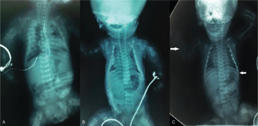
Genetic testing confirms the clinical and radiographic diagnosis, informs genetic counselling, allows prenatal diagnosis in subsequent pregnancies and gives access to specific treatments. The method depends on how clear the diagnosis is:
- Single-gene testing when the diagnosis is clear
- Skeletal dysplasia gene panels (50-500 genes)
- Exome or genome sequencing for atypical presentations
Laboratory tests are targeted to the suspected group:
- OI - bone markers, vitamin D and calcium, and COL1A1/COL1A2 genetic testing; collagen analysis on skin biopsy is historical
- MPS - urine GAG screening (dermatan sulfate, heparan sulfate), an enzyme assay in leucocytes for the specific enzyme of each MPS type, and genetic confirmation
- Metabolic bone disease - alkaline phosphatase (low in hypophosphatasia), calcium, phosphate, vitamin D and PTH
Further imaging. Each modality answers a specific question:
- MRI - foramen magnum stenosis in achondroplasia, spinal stenosis, cervical cord compression, and the brain if there are developmental concerns
- CT - cervical spine assessment, and 3D reconstruction for surgical planning
- Echocardiography - cardiac involvement in MPS; the aortic root in Marfan syndrome, a connective tissue disorder rather than a dysplasia
Antenatal Diagnosis and Prediction of Lethality
Many skeletal dysplasias are first detected on routine obstetric ultrasound, and the structured antenatal assessment, especially the prediction of lethality, is examinable.
The sonographic work-up.
- Measure all long bones: a femur length below roughly the 5th centile, or markedly short for gestation, flags a dysplasia
- Define the limb-segment pattern (rhizo-, meso- or acromelic)
- Assess bone mineralisation: a poorly ossified skull or spine suggests OI or hypophosphatasia
- Look for fractures, bowing and angulation
- Examine the hands and feet (polydactyly)
- Assess the thorax
Predicting lethality is the critical decision. Death is driven by pulmonary hypoplasia from a small chest, not by the limb shortening. Features predicting a lethal outcome:
- A small thoracic circumference
- A thoracic-to-abdominal circumference ratio below about 0.6
- A femur-length-to-abdominal-circumference ratio below about 0.16
- A narrow "bell-shaped" chest with short ribs
The lethal dysplasias to recognise.
- Thanatophoric dysplasia - the commonest lethal form; FGFR3, "telephone-receiver" femora and a narrow thorax
- Osteogenesis imperfecta type II - multiple intrauterine fractures, beaded ribs, a poorly mineralised calvarium
- Achondrogenesis - severe under-mineralisation
- Campomelic dysplasia - bowed long bones, with XY sex reversal
Confirmation and counselling. Targeted fetal molecular testing (amniocentesis or CVS with FGFR3/COL1 panels), 3D ultrasound or low-dose fetal CT of the skeleton, and multidisciplinary genetic counselling. The diagnosis guides delivery planning, and perinatal palliative care for the lethal forms. In a fetus with short limbs, measure the chest: thoracic size predicts survival.
Achondroplasia
Achondroplasia is caused by gain-of-function mutations in the FGFR3 gene. [3,4]
Genetics. The gene is FGFR3 (fibroblast growth factor receptor 3) on chromosome 4p16.3, and one mutation, G380R (glycine to arginine), accounts for 97% of cases. Inheritance is autosomal dominant, but 80% of cases are new mutations. Constitutive activation of FGFR3 inhibits chondrocyte proliferation in the growth plates.
FGFR3 mutations cause a spectrum of severity: Thanatophoric dysplasia (most severe, lethal) through SADDAN syndrome to Achondroplasia to Hypochondroplasia (mildest). All are gain-of-function mutations with varying degrees of receptor activation.
The head. Macrocephaly with frontal bossing, midface hypoplasia and a depressed nasal bridge. Intelligence is normal.
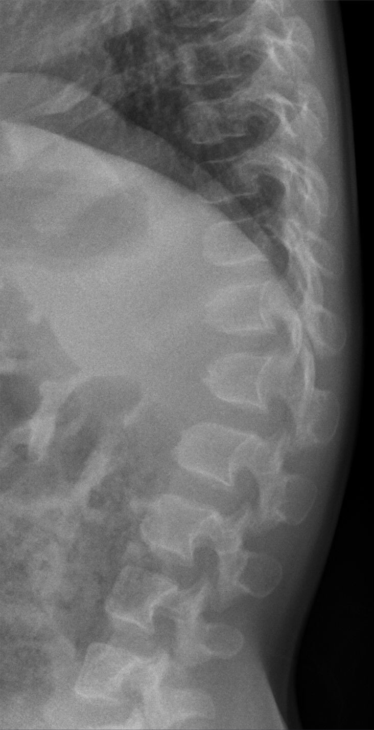
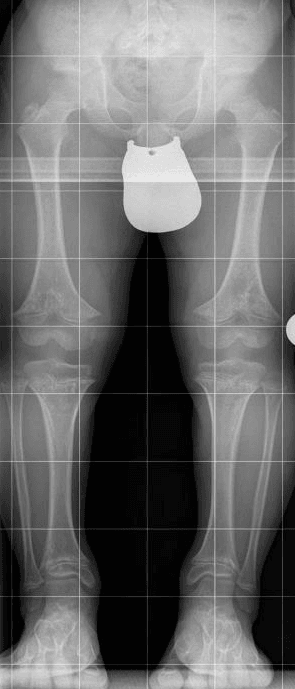
The skeleton. Rhizomelic shortening, the proximal limb segments most affected, with a trident hand and genu varum (tibial bowing). The spine has a thoracolumbar kyphosis in infancy; an exaggerated lumbar lordosis develops later.
- Finding
- Enlarged calvarium, small skull base, foramen magnum stenosis
- Clinical Significance
- Risk of cervicomedullary compression
- Finding
- Narrow interpedicular distance caudally, short pedicles, small canal
- Clinical Significance
- Spinal stenosis - major cause of morbidity
- Finding
- Champagne-glass shape, horizontal acetabular roofs, narrow sciatic notch
- Clinical Significance
- Characteristic diagnostic feature
- Finding
- Rhizomelic shortening, metaphyseal flaring, chevron deformity of femur
- Clinical Significance
- Affects endochondral ossification
- Finding
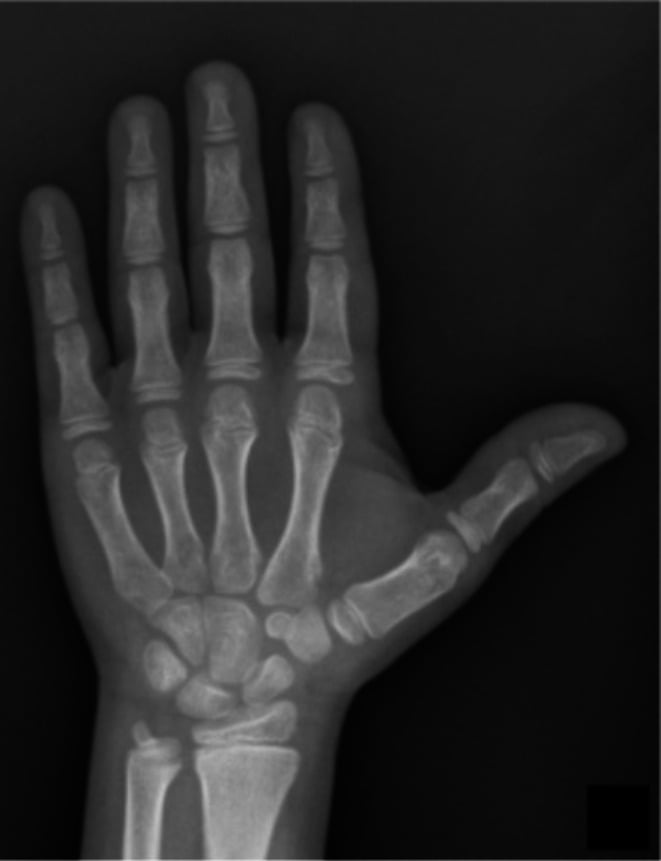
- Trident configuration, short tubular bones
- Clinical Significance
- Persistent gap between 3rd and 4th fingers
The spine. Foramen magnum stenosis is monitored in infancy and decompressed if symptomatic. Thoracolumbar kyphosis is braced, and fused if progressive. Spinal stenosis develops in adults and may need multilevel decompression.
The lower limb. Genu varum is corrected by guided growth or osteotomy, and avoiding obesity reduces mechanical stress. Limb lengthening is discussed under Management.
Vosoritide. A C-natriuretic peptide analogue that works by antagonising FGFR3 signalling, FDA approved in 2021. It has been shown to increase growth velocity in clinical trials (Evidence Base).
Osteogenesis Imperfecta
Osteogenesis imperfecta (OI) is a group of genetic disorders of type I collagen, producing bone fragility and other connective tissue manifestations. [5,6]
Genetics. The genes are primarily COL1A1 and COL1A2, which encode type I collagen, and Types I-IV are mostly autosomal dominant. Newer genes (CRTAP, LEPRE1, PPIB) cause recessive forms.
- Severity
- Mild
- Sclerae
- Blue
- Key Features
- Fractures after walking, normal stature, hearing loss common
- Severity
- Lethal
- Sclerae
- Dark blue
- Key Features
- Multiple intrauterine fractures, beaded ribs, perinatal death
- Severity
- Severe
- Sclerae
- Variable
- Key Features
- Progressive deformity, short stature
- Severity
- Moderate
- Sclerae
- White/blue
- Key Features
- Moderate fragility, normal sclerae in adults, dentinogenesis imperfecta
The classic triad.
- Blue sclerae - thin sclerae showing the choroidal vessels
- Bone fragility - multiple fractures, especially of the long bones
- Hearing loss - conductive, then sensorineural (otosclerosis)
Other features. Dentinogenesis imperfecta (opalescent teeth; dentin contains type I collagen), joint hypermobility, easy bruising, short stature in the severe forms and basilar invagination in Type III. Intelligence is normal.
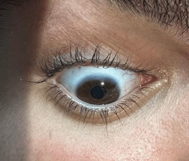
OI can be mistaken for non-accidental injury (NAI) due to multiple unexplained fractures. Key differentiators: blue sclerae, family history, Wormian bones on skull X-ray, osteopenia, and genetic testing. Always consider OI before diagnosing NAI in an infant with fractures.
Radiographic features.
- Generalised osteopenia
- Multiple fractures at various stages of healing
- Wormian bones (multiple small bones in the skull sutures)
- Codfish (biconcave) vertebrae
- Gracile long bones
- Popcorn calcification in the epiphyses (severe forms)
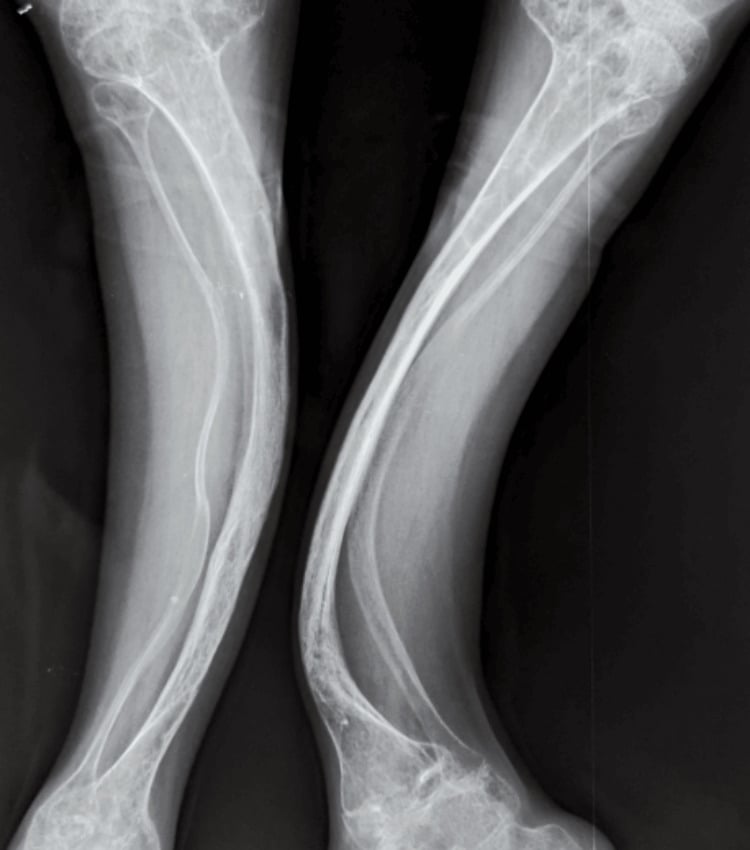
Bisphosphonates. Pamidronate and zoledronic acid reliably increase BMD, with consistent gains at the lumbar spine. Fracture reduction is reported by several trials but is not conclusively established on Cochrane review, which also found the evidence on pain, growth and functional mobility inconsistent. Counsel families accordingly, and do not quote a specific percentage for fracture reduction.
Supportive care. Calcium and vitamin D supplementation, and physiotherapy to strengthen muscles and prevent falls.
Surgery. Intramedullary rodding stabilises the long bones and prevents deformity, and telescoping rods (Bailey-Dubow, Fassier-Duval) grow with the child (Surgical Technique). Scoliosis is treated by spinal fusion, and basilar impression is decompressed if needed.
Mucopolysaccharidoses
The mucopolysaccharidoses (MPS) are lysosomal storage disorders caused by deficiency of the enzymes that degrade glycosaminoglycans (GAGs). The GAGs accumulate in tissues and cause progressive multisystem disease. [7]
- Name
- Hurler/Scheie
- Enzyme Deficiency
- Alpha-L-iduronidase
- Key Features
- Most severe, cognitive decline, corneal clouding
- Name
- Hunter
- Enzyme Deficiency
- Iduronate sulfatase
- Key Features
- X-linked, no corneal clouding, variable severity
- Name
- Sanfilippo
- Enzyme Deficiency
- Various heparan sulfate enzymes
- Key Features
- Behavioral issues, mild skeletal involvement
- Name
- Morquio
- Enzyme Deficiency
- N-acetylgalactosamine-6-sulfatase/GALNS (IVA) or beta-galactosidase/GLB1 (IVB)
- Key Features
- Normal intelligence, severe skeletal involvement, odontoid hypoplasia
- Name
- Maroteaux-Lamy
- Enzyme Deficiency
- Arylsulfatase B
- Key Features
- Normal intelligence, severe skeletal involvement
Dysostosis multiplex is the name given to the radiographic constellation of MPS:
- J-shaped sella turcica, from anterior extension of the sella
- Paddle-shaped (oar-shaped) ribs, widened
- Hook-shaped vertebrae with anteroinferior beaking
- Diaphyseal widening of the long bones, with expanded medullary canals
- Bullet-shaped metacarpals with proximal pointing
- A hypoplastic L1/L2 vertebra causing kyphosis
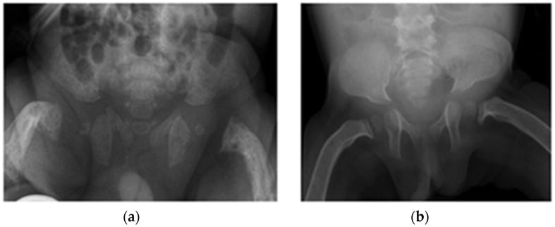
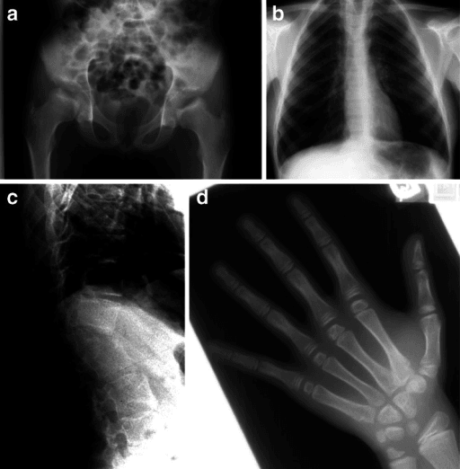
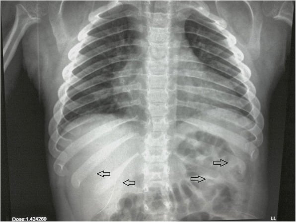
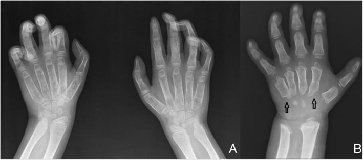
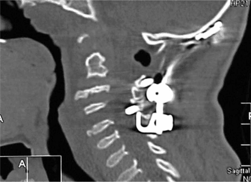
The cervical spine. Odontoid hypoplasia, especially in Morquio, brings atlantoaxial instability, and cervical stenosis also occurs. Occipitocervical fusion may be needed.
The thoracolumbar spine. A gibbus deformity at the thoracolumbar junction and progressive kyphosis may need spinal fusion.
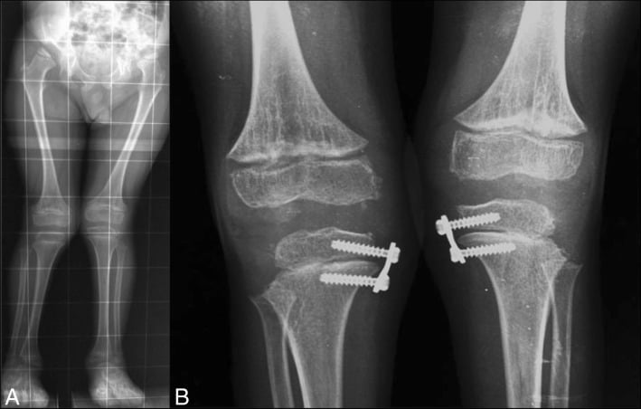
The lower limb. Genu valgum, especially in Morquio, and hip dysplasia, treated with guided growth or osteotomy.
Enzyme replacement. ERT is available for MPS I, II, IVA and VI: laronidase for MPS I, idursulfase for MPS II and elosulfase for MPS IVA. Earlier ERT produces better outcomes. It improves the soft tissue manifestations and can prevent progression if started early, but it does not reverse established skeletal changes.
Haematopoietic stem cell transplant is used in severe MPS I and is best performed early: Hurler syndrome patients transplanted before age 2 have significantly better cognitive outcomes. Gene therapy is under investigation.
Surgery. Address spinal instability early, and perform joint procedures as needed; some types need cardiac valve surgery.
Morquio syndrome (MPS IVA) has unique features: normal intelligence, severe skeletal involvement, odontoid hypoplasia with atlantoaxial instability. These patients need cervical spine precautions for any anaesthesia. Always obtain flexion-extension cervical spine imaging before surgery.
Other Important Dysplasias
Sclerosing dysplasias paradoxically have fragile bones despite their increased radiographic density, because bone remodelling is abnormal.
Osteopetrosis ("marble bone disease"). Osteoclast dysfunction means bone is not resorbed, so the skeleton is diffusely sclerotic but brittle. Failure of metaphyseal remodelling produces the Erlenmeyer flask deformity, and the spine shows sandwich vertebrae and a bone-within-bone appearance. The severe form causes pancytopenia and cranial nerve compression, and the severe infantile form is treated with bone marrow transplant.
Pyknodysostosis. Cathepsin K deficiency gives dense bones with acro-osteolysis (terminal phalangeal resorption), open fontanelles and micrognathia. Fractures are common despite the dense appearance. Toulouse-Lautrec reportedly had the condition.
A classic pattern-recognition dysplasia, and examiners reward the pentad of signs. Diastrophic dysplasia is autosomal recessive, caused by mutations in SLC26A2 (DTDST), the sulfate transporter, producing undersulfated cartilage proteoglycan. Intelligence is normal and the dwarfism is short-limbed (rhizomelic). The pathognomonic signs:
- "Hitchhiker thumb" - a proximally-set, abducted, hypermobile thumb with a short first metacarpal
- "Cauliflower ear" - acute cystic swelling of the pinna in infancy that later calcifies or ossifies
- Rigid, severe equinovarus (clubfoot) that resists casting
- Cervical kyphosis - the key spinal danger, which can be progressive with cord-compression risk and may need early posterior fusion
- Multiple joint contractures and dislocations (symphalangism, "gull-wing" interphalangeal deformity), with progressive scoliosis, early osteoarthritis and a cleft palate in many
The orthopaedic priorities follow: monitor and treat the cervical kyphosis, manage the recurrent or resistant clubfoot and contractures, address progressive scoliosis, and anticipate early joint degeneration. Contrast pseudoachondroplasia: COMP mutation, normal face and skull, normal at birth then growth failure around age two, marked epiphyseal and metaphyseal change, ligamentous laxity and C1-C2 instability.
Management
Management of a skeletal dysplasia is multidisciplinary, addressing both the medical and the orthopaedic needs, and is driven by the specific diagnosis and genotype.
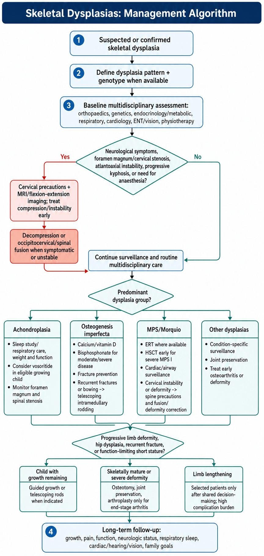
Medical care. The disease-specific drugs (vosoritide, bisphosphonates, enzyme replacement, HSCT) are covered under each condition. Growth hormone is not effective in most true skeletal dysplasias. Supportive care is vitamin D and calcium supplementation, CPAP for obstructive sleep apnoea and multimodal pain management, with the aim of optimising quality of life and addressing the systemic manifestations.
Monitoring. Regular growth measurements, sleep studies for at-risk patients, cardiac echo for MPS patients, and hearing and vision screening.
Spinal surgery. It needs careful preoperative assessment and often specialised techniques.
- Problem
- Foramen magnum stenosis
- Surgical Approach
- Posterior fossa decompression
- Problem
- Spinal stenosis
- Surgical Approach
- Multilevel laminectomy with/without fusion
- Problem
- Atlantoaxial instability
- Surgical Approach
- Occipitocervical fusion
- Problem
- Basilar invagination
- Surgical Approach
- Decompression and stabilization
- Problem
- Progressive kyphosis
- Surgical Approach
- Early posterior fusion
Before any anaesthesia, obtain flexion-extension cervical spine imaging. Morquio and SED are high-risk for atlantoaxial instability. Achondroplasia is high-risk too, but its craniocervical danger is foramen magnum stenosis rather than atlantoaxial instability.
Angular deformity. Guided growth (8-plates, staples) in the skeletally immature and corrective osteotomy in the skeletally mature, addressing both mechanical alignment and joint preservation.
The hip. Pelvic osteotomy for dysplasia, proximal femoral osteotomy for coxa vara or valga, and arthroplasty for end-stage arthritis, which is challenging in small stature.
Limb lengthening. Controversial, but it can improve function. It takes multiple procedures and has a high complication rate, so the patient's and family's goals need careful consideration.
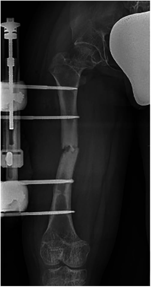
Fractures. In OI, intramedullary rodding with telescoping rods. In osteopetrosis the dense bone makes fixation difficult. Lower limb surgery must account for altered bone quality and anatomy.
Surgical Technique
Intramedullary Rodding in OI
Indications.
- Recurrent fractures (more than 2 per year in the same bone)
- Progressive bowing deformity
- Anticipated fracture through an osteopenic segment
The femur and tibia are the bones most commonly rodded, and in walking patients both femora and both tibiae are rodded.
Principles. Correct angular deformity with osteotomies if needed. The rod should span the entire bone length, stress risers at the rod ends are avoided, and the operation is combined with bisphosphonate therapy, which augments healing before and after surgery.
- Mechanism
- Fixed length
- Best For
- Non-ambulatory patients
- Consideration
- Needs replacement with growth
- Mechanism
- Telescopes at metaphysis
- Best For
- Growing children
- Consideration
- Complex, can malfunction
- Mechanism
- Telescopes at diaphysis
- Best For
- Growing children
- Consideration
- Newer design, better function
Rod choice. Fassier-Duval rods have become preferred for ambulatory OI patients. They provide stable fixation, accommodate growth, and have lower malfunction rates than earlier designs.
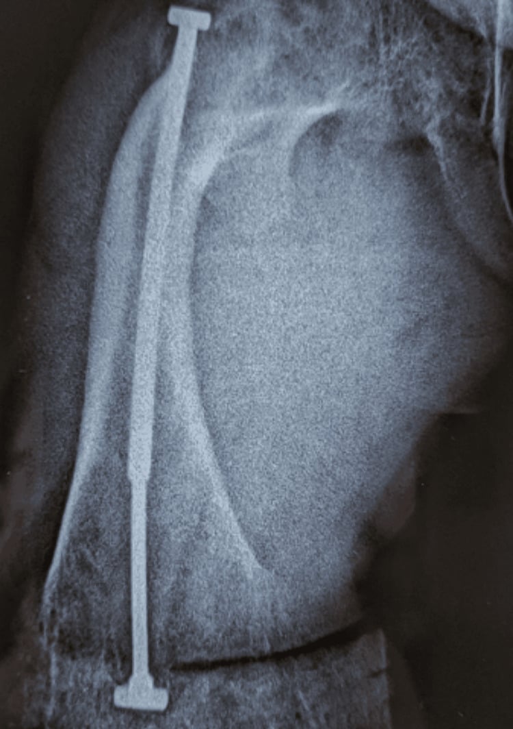
Deformity Correction Osteotomies
Multiple osteotomies are made through the apex of the deformity and the fragments threaded onto the rod, the "shish-kebab" technique. Periosteal stripping is kept to a minimum to preserve the blood supply.
Complications
Orthopaedic Complications
The spine. The spinal complications, and the dysplasias that carry them:
- Foramen magnum stenosis (achondroplasia) - brainstem compression
- Spinal stenosis - progressive neurological deficit
- Atlantoaxial instability (MPS, SED) - risk of cord injury
- Cervical kyphosis (diastrophic dysplasia) - may need early fusion
- Basilar invagination (OI) - cranial settling
- Progressive scoliosis - respiratory compromise
The limbs. Angular deformity (genu varum or valgum) progresses, and premature osteoarthritis follows in MED and SED. Pathological fractures occur in OI and, paradoxically, in osteopetrosis. Hip dysplasia and dislocation occur in MPS and the congenital dysplasias, and recurrent fractures can injure the growth plate.
The joints. Early-onset degenerative disease, joint contractures in MPS, and ligamentous laxity in OI, which overlaps with Ehlers-Danlos.
Medical Complications
- Complication
- Obstructive sleep apnoea, obesity
- Management
- Sleep study, CPAP, weight management
- Complication
- Hearing loss, respiratory failure (severe)
- Management
- Audiology, pulmonology input
- Complication
- Cardiac valve disease, cognitive decline
- Management
- Echo surveillance, HSCT if eligible
- Complication
- Pancytopenia, blindness, deafness
- Management
- BMT, supportive care
- Complication
- Respiratory failure (lethal)
- Management
- Perinatal palliative care
Surgical Complications
Fixation in abnormal bone. In OI, implants cut out and the bone refractures at the implant ends; in osteopetrosis, drilling is difficult and healing delayed. Telescoping rods in OI and staged treatment are the solution.
Spinal surgery. Dural ectasia in some syndromes, abnormal anatomy, difficult intubation (short neck, atlantoaxial instability) and an increased bleeding risk.
Postoperative Care
Immediately after surgery. Close neurological monitoring after spinal procedures, multimodal pain management, early mobilisation when safe, and DVT prophylaxis (mechanical, with chemical considered in adults).
After rodding or fracture fixation. Protected weight bearing initially, serial radiographs to confirm healing, and monitoring for implant complications.
- Mobilisation
- Day 1-2
- Weight Bearing
- PWB 6 weeks, WBAT 12 weeks
- Return to Activity
- 4-6 months
- Mobilisation
- Day 1-2
- Weight Bearing
- PWB 6 weeks, WBAT 12 weeks
- Return to Activity
- 4-6 months
- Mobilisation
- Collar 6-12 weeks
- Weight Bearing
- N/A
- Return to Activity
- 3-6 months
- Mobilisation
- Day 1
- Weight Bearing
- Full
- Return to Activity
- 6-12 weeks
Outcomes
Achondroplasia. Life expectancy is near normal with appropriate management. Foramen magnum decompression is highly effective when needed, spinal stenosis surgery improves quality of life, and vosoritide is showing promise for improved height outcomes.
Osteogenesis imperfecta. Rodding produces a marked reduction in fractures in the rodded bones; a ~90% reduction is the conventional figure from retrospective series, not a measured trial endpoint. Quality of life is significantly improved with modern management.
- Success Rate
- 85-95%
- Complications
- Rod migration, refracture
- Revision Rate
- 20-30% (growth)
- Success Rate
- 90%+
- Complications
- CSF leak, infection
- Revision Rate
- 5-10%
- Success Rate
- 80-90%
- Complications
- Nonunion, progression
- Revision Rate
- 10-15%
- Success Rate
- 70-80%
- Complications
- Instability, recurrence
- Revision Rate
- 15-20%
The percentages in this table are conventional estimates from retrospective series and clinical experience, not measurements from prospective cohorts - no large comparative outcome datasets exist for these procedures in skeletal dysplasia.
Guidelines, Registries & Global Practice
Global Epidemiology
Skeletal dysplasias are individually rare but collectively important. Population-based data from the Utah Birth Defect Network give a birth prevalence of about 3.0 per 10,000 births (rising to 20 per 10,000 stillbirths), with osteogenesis imperfecta (0.79/10,000), thanatophoric dysplasia (0.43/10,000) and achondroplasia (0.35/10,000) the commonest categories. See Stevenson DA et al. Am J Med Genet A 2012 (DOI). The International Skeletal Dysplasia Society 2019 Nosology recognises 461 disorders in 42 groups, with a causal gene now identified for 92% — Mortier GR et al. Am J Med Genet A 2019 (DOI). Achondroplasia (the commonest non-lethal dysplasia, roughly 1:25,000-1:30,000) and OI (roughly 1:15,000-1:20,000) account for the bulk of the orthopaedic caseload worldwide; incidence is broadly uniform across populations because most cases of achondroplasia arise from recurrent de novo FGFR3 G380R mutations linked to advanced paternal age.
Guidance Across Jurisdictions
- Scope
- Nosology & diagnosis
- Key Position
- 461-disorder nosology; radiographic group then targeted molecular testing
- Scope
- Achondroplasia lifetime care
- Key Position
- Structured foramen-magnum surveillance in infancy; multidisciplinary follow-up
- Scope
- Vosoritide & ERT commissioning
- Key Position
- Highly specialised commissioning with defined eligibility and stopping rules
- Scope
- Drug approval
- Key Position
- Vosoritide approved (FDA 2021, EMA 2021, TGA 2022); ERT approved for MPS I/II/IVA/VI
- Scope
- Orthopaedic surgery in MPS
- Key Position
- Early cervical assessment; flexion-extension imaging before any anaesthesia
Registry & Disease-Specific Networks
- CLARITY / international OI registries and the Brittle Bone Disease Consortium collect longitudinal fracture, bisphosphonate and rodding outcome data.
- Achondroplasia natural-history and post-marketing registries (e.g. the lifeline / industry-sponsored vosoritide cohorts) track growth, foramen magnum events and spinal stenosis.
- MPS patient registries (Hunter Outcome Survey, MPS I Registry) inform ERT eligibility, cardiac and skeletal surveillance, and long-term safety.
Practice Variation & Access
- Limb lengthening is performed selectively and is more common in parts of Europe and the Middle East than in the UK/Australasia, where it remains controversial.
- Vosoritide funding varies widely: reimbursed in several European systems and through specialised commissioning in the UK, but access elsewhere is often via managed-access or patient programmes.
- HSCT vs ERT sequencing for severe MPS I differs by centre and timing of diagnosis (newborn screening availability).
Across international fellowship examinations you must recognise common skeletal dysplasias radiographically, understand the orthopaedic complications of achondroplasia and OI, know the principles of telescoping-rod fixation in OI, and recognise cervical instability risks in MPS/SED. Classification by bone density and location is a common viva framework.
MCQ Practice Points
Q: What is the most common skeletal dysplasia and its genetic basis? A: Achondroplasia, caused by gain-of-function mutation in FGFR3 (fibroblast growth factor receptor 3). This mutation inhibits chondrocyte proliferation in the growth plate, causing rhizomelic (proximal) limb shortening with normal trunk length. Be precise about "most common": achondroplasia is the commonest non-lethal skeletal dysplasia and the commonest cause of disproportionate short stature in survivors. In population-based birth data that also capture stillbirths and terminations, osteogenesis imperfecta (0.79 per 10,000) and thanatophoric dysplasia (0.43 per 10,000) both outrank achondroplasia (0.35 per 10,000) - so the honest answer names the denominator.
Q: What is the life-threatening complication of achondroplasia in infancy? A: Foramen magnum stenosis causing cervicomedullary compression. The congenitally small foramen magnum compresses the brainstem and upper cervical cord, causing central apnoea, and is linked to the excess risk of sudden death in infancy. MRI screening is recommended in the first 2 years. Atlantoaxial instability is NOT the typical achondroplasia problem - it is the cervical danger in Morquio, SED, pseudoachondroplasia and Down syndrome; do not conflate the two.
Q: How do you distinguish spondyloepiphyseal dysplasia (SED) from multiple epiphyseal dysplasia (MED)? A: SED has short trunk with vertebral involvement (platyspondyly) plus epiphyseal abnormalities. MED has normal trunk height with only epiphyseal involvement. Both cause premature osteoarthritis, but SED patients are shorter and have spinal deformity.
Q: What orthopaedic complications are common in achondroplasia? A: Spinal stenosis (lumbar, may need multilevel decompression), foramen magnum stenosis (craniocervical), thoracolumbar kyphosis in infancy (often self-corrects), and genu varum (driven by relative fibular overgrowth). Hip and knee arthroplasty may be challenging due to anatomic variants. Atlantoaxial instability is not a typical achondroplasia feature - think Morquio, SED or pseudoachondroplasia for that.
Clinical Decision Scenarios
Practise clinical reasoning and management decisions out loud
“Classic presentation of achondroplasia or related FGFR3 disorder. The examiner wants systematic evaluation.”
“Important scenario - need to distinguish OI from NAI while ensuring child safety.”
“Tests knowledge of MPS complications and perioperative considerations.”
“Tests systematic approach to pattern recognition in skeletal dysplasias.”
“Tests knowledge of evidence-based management in skeletal dysplasia.”
CLASSIFICATION APPROACH
- Bone DENSITY first: osteopenic vs sclerosing
- LOCATION: epiphyseal, metaphyseal, or diaphyseal
- LIMB proportions: rhizomelic, mesomelic, acromelic
- SPINE: platyspondyly, beaking, canal size
ACHONDROPLASIA - STAMP
- S = Spinal stenosis (narrow canal, short pedicles)
- T = Trident hand (gap between 3rd and 4th fingers)
- A = Autosomal dominant, FGFR3 gain-of-function
- M = Macrocephaly, frontal bossing, midface hypoplasia
- P = Pelvis champagne-glass shaped
OSTEOGENESIS IMPERFECTA - BONED
- B = Blue sclerae (Type I)
- O = Osteopenia, multiple fractures
- N = Normal intelligence (distinguish from NAI)
- E = Ear - hearing loss (otosclerosis)
- D = Dentinogenesis imperfecta
OI TYPES
- Type I = Mild, blue sclerae, hearing loss - MOST COMMON
- Type II = Lethal, multiple intrauterine fractures
- Type III = Severe, progressive deformity, wheelchair
- Type IV = Moderate, white/normal sclerae in adults
DYSOSTOSIS MULTIPLEX (MPS)
- J-shaped sella turcica
- Paddle-shaped (oar) ribs
- Hook vertebrae with anteroinferior beaking
- Diaphyseal widening of long bones
- Bullet metacarpals with proximal pointing
MORQUIO SYNDROME (MPS IVA)
- NORMAL INTELLIGENCE - key differentiator
- Severe skeletal involvement
- Odontoid hypoplasia - cervical instability
- MUST get flex-ext C-spine before any anaesthesia
- ERT available (elosulfase alfa)
TREATMENT
- Achondroplasia: vosoritide (C-natriuretic peptide analogue) - FDA 2021
- OI: bisphosphonates (increase BMD), rodding procedures
- MPS: ERT for Types I, II, IVA, VI; BMT for Type I
- Spinal surgery for instability/stenosis
- Limb surgery for deformity correction
Evidence Base
Savarirayan R et al. Once-daily subcutaneous vosoritide in children with achondroplasia: phase 3 RCT. Lancet (2020)
- First disease-modifying therapy for achondroplasia (C-natriuretic peptide analogue)
- 1.57 cm/year increase in annualised growth velocity over placebo
- Final adult-height impact and long-term harms remain unknown
Dwan K et al. Bisphosphonate therapy for osteogenesis imperfecta. Cochrane Database Syst Rev (2016)
- Bisphosphonates increase BMD in children and adults with OI
- Fracture-reduction evidence is inconsistent and not conclusive
- Optimal agent, dose, duration and long-term safety remain undefined
Wraith JE et al. Enzyme replacement therapy for MPS I: randomised placebo-controlled trial of laronidase. J Pediatr (2004)
- ERT improves respiratory function and physical capacity in MPS I
- No reversal of established skeletal (dysostosis multiplex) changes
- Supports early treatment before irreversible bony deformity
Stevenson DA et al. Analysis of skeletal dysplasias in the Utah population. Am J Med Genet A (2012)
- Collective birth prevalence of skeletal dysplasias ~3 per 10,000
- OI and the FGFR3 chondrodysplasia group are the largest diagnostic categories
- Lethal forms cluster in stillbirths and terminations, biasing live-birth figures
Sillence DO, Senn A, Danks DM. Genetic heterogeneity in osteogenesis imperfecta. J Med Genet (1979)
- Original delineation of OI Types I-IV (Sillence classification)
- Type I: autosomal dominant, blue sclerae, presenile hearing loss
- Perinatal-lethal group (Type II) with crumpled femora and beaded ribs
Mortier GR et al. Nosology and classification of genetic skeletal disorders: 2019 revision. Am J Med Genet A (2019)
- 461 recognised skeletal dysplasias in 42 clinical/molecular groups
- A causal gene is now known for 92% of entities
- Reference framework for diagnosis and research in skeletal dysplasia
Suggested Reading
- Mortier GR, Cohn DH, Cormier-Daire V, et al. Nosology and classification of genetic skeletal disorders: 2019 revision. Am J Med Genet A. 2019;179(12):2393-2419. doi:10.1002/ajmg.a.61366
- Krakow D, Rimoin DL. The skeletal dysplasias. Genet Med. 2010;12(6):327-341. doi:10.1097/GIM.0b013e3181daae9b
- Horton WA, Hall JG, Hecht JT. Achondroplasia. Lancet. 2007;370(9582):162-172. doi:10.1016/S0140-6736(07)61090-3
- Savarirayan R, Tofts L, Irving M, et al. Once-daily, subcutaneous vosoritide therapy in children with achondroplasia: a randomised, double-blind, phase 3, placebo-controlled, multicentre trial. Lancet. 2020;396(10252):684-692. doi:10.1016/S0140-6736(20)31541-5
- Marini JC, Forlino A, Bachinger HP, et al. Osteogenesis imperfecta. Nat Rev Dis Primers. 2017;3:17052. doi:10.1038/nrdp.2017.52
- Dwan K, Phillipi CA, Steiner RD, Basel D, Cochrane Cystic Fibrosis and Genetic Disorders Group. Bisphosphonate therapy for osteogenesis imperfecta. Cochrane Database Syst Rev. 2016;10:CD005088. doi:10.1002/14651858.CD005088.pub4
- Muenzer J. Overview of the mucopolysaccharidoses. Rheumatology (Oxford). 2011;50 Suppl 5:v4-12. doi:10.1093/rheumatology/ker394
- Handa A, Nishimura G, Zhan MX, Bennett DL, El-Khoury GY. A primer on skeletal dysplasias. Jpn J Radiol. 2022;40(3):245-261. doi:10.1007/s11604-021-01206-5
- Cheung MS, Glorieux FH. Osteogenesis imperfecta: update on presentation and management. Rev Endocr Metab Disord. 2008;9(2):153-160. doi:10.1007/s11154-008-9074-4
- White KK, Sousa T. Mucopolysaccharide disorders in orthopaedic surgery. J Am Acad Orthop Surg. 2013;21(1):12-22. doi:10.5435/JAAOS-21-01-12
- Ireland PJ, Pacey V, Zankl A, et al. Optimal management of complications associated with achondroplasia. Appl Clin Genet. 2014;7:117-125. doi:10.2147/TACG.S51485
- Sillence DO, Senn A, Danks DM. Genetic heterogeneity in osteogenesis imperfecta. J Med Genet. 1979;16(2):101-116. doi:10.1136/jmg.16.2.101
- Wraith JE, Clarke LA, Beck M, et al. Enzyme replacement therapy for mucopolysaccharidosis I: a randomized, double-blinded, placebo-controlled, multinational study of recombinant human alpha-L-iduronidase (laronidase). J Pediatr. 2004;144(5):581-588. doi:10.1016/j.jpeds.2004.01.046
- Panda A, Gamanagatti S, Jana M, Gupta AK. Skeletal dysplasias: A radiographic approach and review of common non-lethal skeletal dysplasias. World J Radiol. 2014;6(10):808-825. doi:10.4329/wjr.v6.i10.808
- Shapiro JR, Germain-Lee EL. Osteogenesis imperfecta: effecting the transition from adolescent to adult medical care. J Musculoskelet Neuronal Interact. 2012;12(1):24-27.
Key Guidelines
- International Skeletal Dysplasia Society Nosology 2019
- International consensus statements on achondroplasia diagnosis and lifetime management; international consensus on thoracolumbar kyphosis in MPS I (Kuiper et al., Orphanet J Rare Dis 2019;14:17)
Additional Reading
- Spranger JW, Brill PW, Superti-Furga A, et al. Bone Dysplasias: An Atlas of Genetic Disorders of Skeletal Development. 4th ed. Oxford University Press; 2018.
- Bonafe L, Cormier-Daire V, Hall C, et al. Nosology and classification of genetic skeletal disorders: 2015 revision. Am J Med Genet A. 2015;167A(12):2869-2892.