One Plasma-Cell Lesion - Until Proven Otherwise
- Solitary plasmacytoma is a RARE localized proliferation of clonal (monoclonal) plasma cells with NO evidence of systemic disease; in bone it is a SOLITARY BONE PLASMACYTOMA (SBP), versus a SOLITARY EXTRAMEDULLARY PLASMACYTOMA (SEP) in soft tissue.
- Diagnosis requires a SINGLE lesion with biopsy-proven clonal plasma cells AND the EXCLUSION of multiple myeloma - normal marrow (no/limited clonal plasma cells), no other lesions on whole-body imaging, and no myeloma-related end-organ damage (no CRAB: hyperCalcaemia, Renal impairment, Anaemia, Bone lesions elsewhere).
- Workup to exclude occult MM: serum/urine protein electrophoresis and free light chains, FBC, calcium and renal function, BONE MARROW biopsy (with flow cytometry), and whole-body imaging - MRI and/or 18F-FDG PET (more sensitive than skeletal survey).
- RADIOTHERAPY is the treatment of CHOICE - it gives durable local control in SBP and is potentially curative in SEP; SURGERY is reserved for structural problems (impending/actual pathological fracture, spinal instability, neurological compromise), and the role of chemotherapy is debated.
- SBP has a HIGH rate of progression to multiple myeloma (substantially higher than SEP), so it needs LONG-TERM haematological surveillance; persistence of the paraprotein after treatment predicts progression.
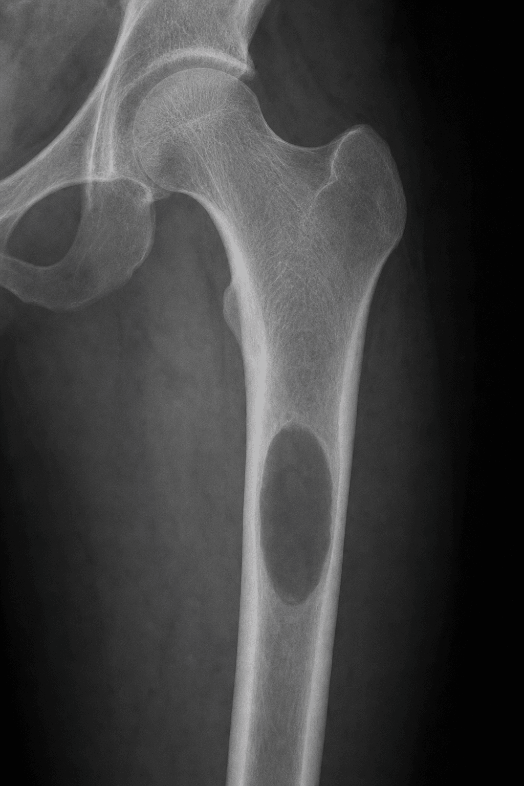
- Orthopaedically it presents as a painful lytic bone lesion (commonly axial - vertebrae - or proximal long bones) and may cause pathological fracture or spinal cord compression - so the orthopaedic surgeon often makes the diagnosis (biopsy) and manages the structural complications.
- “The exam trap: do NOT treat an apparent solitary plasmacytoma until you have EXCLUDED multiple myeloma (marrow biopsy + whole-body MRI/PET + paraprotein screen).
- “Treatment of choice is RADIOTHERAPY, not surgery - reserve surgery for fracture/instability/cord compression.
- “SBP progresses to MM far more often than SEP - it mandates long-term surveillance; a persistent paraprotein after RT is a bad sign.
A single biopsy-proven plasma-cell lesion, normal bone marrow, no other lesions on whole-body MRI/PET, and no CRAB end-organ damage. Treat with radiotherapy and survey long-term.
Clonal marrow plasma cells, additional lesions, or CRAB features mean the disease is systemic - it is multiple myeloma, needing systemic therapy, not localized RT alone. Mislabelling MM as solitary plasmacytoma undertreats the patient.
Definition & Spectrum
Solitary plasmacytoma is a rare neoplasm defined by a single, localized mass of clonal (monoclonal) plasma cells with no evidence of systemic disease. It is part of the plasma-cell neoplasm spectrum - the great majority (~95%) of which present as multiple myeloma. When the lesion is in bone it is a solitary bone plasmacytoma (SBP); when in soft tissue/extraosseous sites (commonly the head and neck) it is a solitary extramedullary plasmacytoma (SEP). SBP is more common than SEP and has a poorer prognosis because of its higher rate of progression to multiple myeloma, even though both respond well to treatment.
Presentation
- Bone pain at the lesion (often the spine, also pelvis/proximal long bones)
- Pathological fracture or vertebral collapse
- Spinal cord / nerve-root compression with a vertebral lesion
- Usually a slightly younger age than typical multiple myeloma; more common in men
- A solitary lytic ("punched-out") bone lesion, often well-defined
- MRI to define soft-tissue extent/cord involvement and screen the marrow
- Whole-body MRI or 18F-FDG PET-CT to confirm it is truly solitary (more sensitive than plain skeletal survey)
Diagnosis & Workup
Diagnosis of a solitary bone plasmacytoma requires all of:
- A single lesion of biopsy-proven clonal plasma cells.
- A normal bone marrow (no, or only minimal/limited, clonal plasma cells - more than a defined low threshold suggests myeloma).
- No other lesions on whole-body imaging (MRI/PET).
- No myeloma-related end-organ damage - i.e. no CRAB (hyperCalcaemia, Renal impairment, Anaemia, other Bone lesions). A small clonal marrow population with an otherwise solitary lesion is sometimes termed plasmacytoma with minimal marrow involvement and behaves more aggressively.
| 0 | 1 |
|---|---|
| Biopsy of the lesion | Confirm clonal plasma cells (the diagnosis) |
| Serum/urine protein electrophoresis + immunofixation | Detect and quantify a paraprotein (M-protein) |
| Serum free light chains | Sensitive clonality marker; prognostic if persistent |
| Bone marrow biopsy + flow cytometry | Exclude systemic marrow clonal plasma cells |
| FBC, calcium, renal function | Screen for CRAB (anaemia, hypercalcaemia, renal impairment) |
| Whole-body MRI and/or 18F-FDG PET-CT | Confirm the lesion is truly solitary (more sensitive than skeletal survey) |
Management
Definitive radiotherapy to the lesion is the standard treatment for solitary plasmacytoma - it provides durable long-term local control in solitary bone plasmacytoma and is potentially curative in the extramedullary form (which has a lower tendency to disseminate). Treatment volumes and dose follow specialist (e.g. ILROG) guidance. Surgery is not the primary treatment of the tumour itself; it is reserved for structural indications - impending or actual pathological fracture, spinal instability, or neurological compromise requiring decompression/stabilisation - usually combined with RT. The role of systemic chemotherapy is debated and not routine for true solitary disease.
Mainstay; durable local control (SBP), potentially curative (SEP). Plan with whole-body imaging to be sure the disease is truly solitary first.
For mechanical/neurological problems - fixation of (impending) pathological fracture, spinal stabilisation, cord decompression - then radiotherapy. Not a substitute for RT of the tumour.
Most patients with a solitary bone plasmacytoma will eventually progress to multiple myeloma (substantially more often than the extramedullary form), sometimes years later. Persistence of the paraprotein after radiotherapy predicts progression. Patients therefore need long-term haematological follow-up (serial paraprotein/free light chains, blood counts, calcium/renal function, and imaging as indicated) - reassurance that they are "cured" after RT is unsafe.
Evidence & Key Studies
Solitary plasmacytoma
- Solitary plasmacytoma is a localized clonal plasma-cell proliferation without systemic disease; solitary bone plasmacytoma is more common than the extramedullary form.
- Solitary bone plasmacytoma has poorer overall and progression-free survival than extramedullary disease because of its higher rate of evolution to multiple myeloma.
- Flow cytometry and MRI/18F-FDG PET refine diagnosis and exclude occult myeloma; radiotherapy is the treatment of choice, with the roles of surgery and chemotherapy still debated.
Radiation therapy for solitary plasmacytoma and multiple myeloma: guidelines from the International Lymphoma Radiation Oncology Group
- Definitive radiotherapy is the standard treatment for localized plasmacytoma; it provides long-term local control in solitary bone plasmacytomas and is potentially curative in extramedullary cases.
- Provides standardized work-up, target-volume and dose/fractionation recommendations for solitary plasmacytoma.
- Outlines the supportive (palliative) role of radiotherapy in multiple myeloma in the era of novel systemic agents.
According to PubMed, the SBP-versus-SEP distinction, progression risk and diagnostic refinements come from the cited Grammatico review, and the role of radiotherapy from the cited ILROG guideline. The diagnostic criteria (single lesion, normal marrow, no other lesions, no CRAB) follow standard plasma-cell-neoplasm definitions. (See also our Multiple Myeloma and Enneking Staging topics.)
Clinical Decision Scenarios
Practise clinical reasoning and management decisions out loud
“A 55-year-old man has a painful solitary lytic lesion in a vertebra; biopsy shows clonal plasma cells. How do you confirm the diagnosis of solitary plasmacytoma, and why does it matter?”
“How would you treat a confirmed solitary bone plasmacytoma, when is surgery indicated, and what do you tell the patient about prognosis?”
Mnemonics & Memory Aids
SOLITARY
Hook:SOLITARY: prove it is single, exclude myeloma, irradiate, and watch it long-term.
CRAB
Hook:No CRAB = compatible with solitary; any CRAB feature points to multiple myeloma.
Definition
- Single localized clonal plasma-cell tumour, no systemic disease
- Solitary bone (SBP) vs solitary extramedullary (SEP); SBP commoner, worse prognosis
- Part of the plasma-cell neoplasm spectrum (~95% present as MM)
Diagnosis
- Single biopsy-proven clonal lesion + normal marrow + no other lesions + no CRAB
- Workup: SPEP/UPEP + free light chains, FBC/Ca/renal, marrow biopsy, whole-body MRI/PET
- Whole-body MRI/PET more sensitive than skeletal survey
Management
- Radiotherapy = treatment of choice (durable local control; SEP potentially curative)
- Surgery only for fracture/instability/cord compression (+ RT)
- Chemotherapy role debated; not routine for true solitary disease
Prognosis
- High progression to multiple myeloma (SBP > SEP)
- Persistent paraprotein after RT predicts progression
- Needs long-term haematological surveillance