Intraoperative Neuromonitoring (IONM)
- Intraoperative neuromonitoring (IONM) detects IMPENDING neurological injury in REAL TIME during spinal surgery, allowing the team to intervene (release a correction, raise the blood pressure, remove implants) BEFORE the injury becomes permanent.
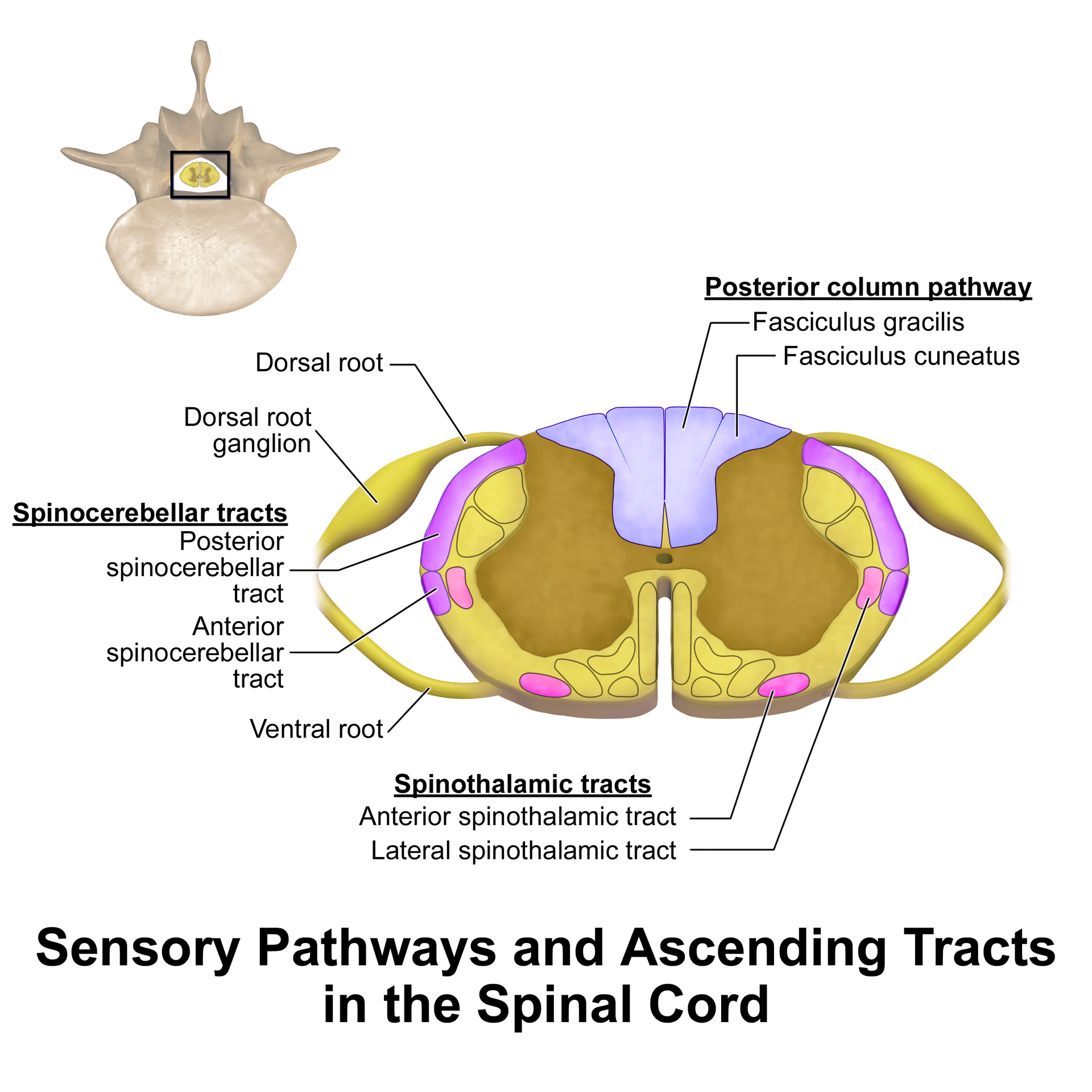
- SSEP (somatosensory evoked potentials) monitor the POSTERIOR (dorsal) COLUMNS - the SENSORY pathway; standard alarm criteria are roughly a 50% drop in AMPLITUDE or a 10% increase in LATENCY. Its key limitation is that it can MISS an isolated ANTERIOR-CORD / MOTOR injury.
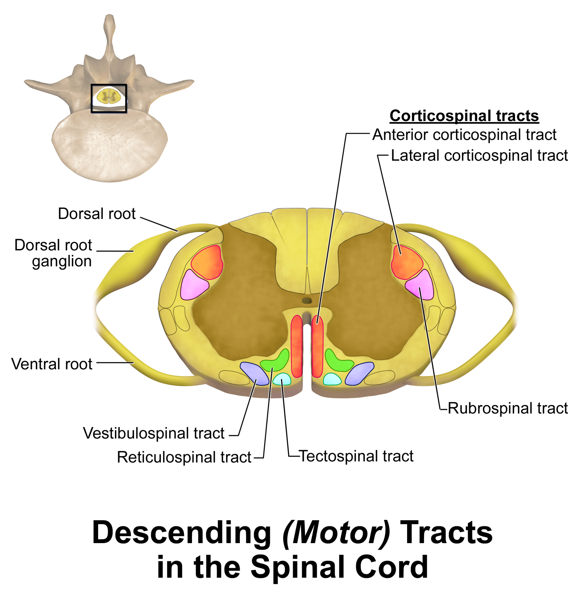
- MEP (transcranial motor evoked potentials) monitor the CORTICOSPINAL (MOTOR) tracts / anterior cord - more relevant to the motor deficits that matter most, and more immediate; they are VERY sensitive to inhalational anaesthetics and to NEUROMUSCULAR BLOCKADE.
- EMG has two forms: FREE-RUN EMG detects nerve-ROOT irritation continuously, and TRIGGERED (stimulated) EMG tests PEDICLE SCREWS - a LOW stimulation threshold to evoke a muscle response suggests a MEDIAL cortical breach with the screw close to a nerve root.
- MULTIMODAL monitoring (SSEP + MEP +/- EMG) is now standard because the modalities are complementary (SSEP misses motor, MEP misses sensory) - and ANAESTHESIA must be compatible: TOTAL INTRAVENOUS ANAESTHESIA (TIVA), AVOIDANCE/limitation of volatile agents, and NO long-acting muscle relaxants once MEP/EMG are needed.
- A SIGNAL CHANGE triggers a STRUCTURED RESPONSE - check anaesthetic/physiological factors (raise MAP/blood pressure, correct hypotension/anaemia/hypothermia, confirm not paralysed), then SURGICAL factors (release the correction/distraction, remove recently placed instrumentation, re-examine), and consider the STAGNARA WAKE-UP TEST to confirm gross motor function.
- “SSEP = sensory/posterior columns (alarm: 50% amplitude drop or 10% latency rise); MEP = motor/corticospinal (anterior cord) - use BOTH because each misses what the other monitors.
- “MEP and EMG demand anaesthesia compatibility: TIVA and NO muscle relaxants; volatile agents suppress MEP.
- “Triggered EMG with a LOW threshold = possible medial pedicle-screw breach; a signal change = run the anaesthetic-then-surgical checklist (raise BP, release correction) +/- wake-up test.
Monitors the dorsal (posterior) columns (sensory). Robust and continuous, but can MISS an isolated anterior-cord/motor injury (the cord's blood supply and the corticospinal tracts are anterior) - historically the cause of false-negative 'normal SSEP but woke up paraplegic'.
Monitors the corticospinal (motor) tracts / anterior cord - the function that matters most and the territory at risk in anterior spinal artery injury. More immediate, but sensitive to volatile anaesthetics and paralysis. Combine with SSEP for complete coverage.
Why Monitor & the Tracts at Stake
The purpose of IONM is to give the surgical team a real-time warning of an evolving neurological injury - from distraction/correction, direct trauma, implant malposition or ischaemia - so it can be reversed before it becomes permanent. Different modalities watch different tracts, and understanding the spinal-cord cross-section is the key: the posterior (dorsal) columns carry sensation (monitored by SSEP), while the corticospinal tracts carry motor output (monitored by MEP). Because these lie in different parts of the cord with different blood supplies, no single modality is sufficient.
The Modalities
Somatosensory evoked potentials stimulate a peripheral nerve (e.g. posterior tibial, ulnar) and record the response at the cortex/scalp (and sometimes spinal/peripheral). They monitor the dorsal-column-medial-lemniscus (sensory) pathway. Standard alarm criteria are about a 50% decrease in amplitude or a 10% increase in latency. SSEP is robust and relatively anaesthesia- tolerant, but signals are averaged (slightly delayed) and it does not monitor the motor tracts, so it can miss an anterior-cord injury.
| 0 | 1 | 2 | 3 |
|---|---|---|---|
| SSEP | Posterior columns (sensory) | ~50% amplitude drop or ~10% latency increase | Misses isolated motor/anterior-cord injury; averaged (delayed) |
| MEP | Corticospinal (motor) / anterior cord | Significant amplitude loss/threshold rise | Very sensitive to volatile agents & paralysis (need TIVA, no relaxant) |
| Free-run EMG | Nerve-root irritation | Bursts/neurotonic discharges | Non-specific; needs no muscle relaxant |
| Triggered EMG | Pedicle screw position | LOW stimulation threshold = medial breach | Threshold cut-offs vary; needs no relaxant |
| Wake-up test | Gross voluntary motor function | Patient cannot move feet on command | Single time point; interrupts surgery; risks (recall, extubation) |
Anaesthesia & Response to a Signal Change
For MEP (and EMG) to work, the anaesthetic must preserve neuromuscular transmission and cortical excitability: use TOTAL INTRAVENOUS ANAESTHESIA (TIVA), avoid or minimise volatile (inhalational) agents (which suppress MEP), and avoid long-acting muscle relaxants once motor monitoring is running. Maintain stable physiology - adequate mean arterial pressure, normothermia and haemoglobin - because hypotension, hypothermia and anaemia themselves reduce signals and threaten cord perfusion.
A genuine, significant signal change is an emergency. Work through a checklist:
- Confirm it is real - check for technical/equipment causes and verify with the neurophysiologist.
- Anaesthetic/physiological - raise the mean arterial pressure (improve cord perfusion), correct hypotension, anaemia, hypothermia, hypocapnia, and confirm the patient is not paralysed or over-anaesthetised.
- Surgical - release/reduce the correction or distraction, remove recently placed instrumentation or grafts, decompress/inspect the cord, and reassess.
- Confirm - consider a Stagnara wake-up test; if a true deficit persists, treat (e.g. maintain MAP, consider steroids per local protocol) and plan further imaging/management.
Evidence & Practice
The use of IONM in scoliosis/deformity surgery has risen steadily, and MULTIMODAL monitoring (e.g. combined SSEP + EMG, increasingly with MEP) is used more often than single-modality monitoring, because the modalities are complementary. Risk factors for intraoperative signal changes include a pre-operative neurological deficit, severe kyphosis, a large curve magnitude, and significant cord shortening (e.g. during vertebral column resection). While definitive randomised proof that monitoring reduces deficits is hard to obtain (the highest-risk cases are the ones monitored), multimodal IONM allows potential injuries to be localised and managed in real time and is considered a valuable safety tool in deformity surgery.
Evidence & Key Studies
Neuromonitoring in spinal deformity surgery: a multimodality approach
- Multimodal intraoperative monitoring (SSEP, MEP, EMG) localises and manages potential neurologic injuries in real time and is a valuable tool for the safety of spinal deformity surgery.
- Risk factors for intraoperative signal changes include preoperative neurologic deficit, severe kyphosis, increased curve magnitude and significant cord shortening (e.g. vertebral column resection).
- Although no evidence-based algorithm exists for signal changes, structured strategies help prevent and address neurologic alarms.
Demographic trends in the use of intraoperative neuromonitoring for scoliosis surgery in the United States
- Use of intraoperative neuromonitoring for scoliosis surgery rose from 27% (2005) to 46.9% (2011); multimodal monitoring was used more commonly than unimodal (64.6% vs 35.4%).
- The most commonly used combination was SSEP + EMG, and the least used was MEP alone.
- Observed neurological-injury rates were similar with and without monitoring (1.8% vs 2.0%), but the authors caution this likely reflects that higher-risk surgeries preferentially use monitoring.
According to PubMed, the multimodal approach, the risk factors for signal changes and the real-time- management rationale come from the cited Laratta review, and the trends/modality-usage data from the cited Ajiboye study. The specific SSEP alarm criteria (50% amplitude / 10% latency), the SSEP-vs-MEP tract distinction, triggered-EMG pedicle-screw testing, the TIVA/no-relaxant requirement and the Stagnara wake-up test are standard, well-established neuromonitoring teaching. (See also our scoliosis and pedicle-screw fixation material.)
Clinical Decision Scenarios
Practise clinical reasoning and management decisions out loud
“During scoliosis correction, what neuromonitoring modalities would you use, what does each monitor, and why is multimodal monitoring preferred?”
“Midway through the correction the MEPs are lost. What anaesthetic conditions are required for monitoring, and how do you respond to this signal change?”
Mnemonics & Memory Aids
SAME
Hook:SAME: SSEP sensory, Anterior cord = MEP, Multimodal, EMG for roots/screws.
ALARM
Hook:On an ALARM: confirm + anaesthesia, lift the BP, fix anaemia/temp, release the correction, then wake-up test.
Modalities & what they monitor
- SSEP = posterior columns (sensory); alarm ~50% amplitude drop or ~10% latency rise
- MEP = corticospinal (motor)/anterior cord; immediate; anaesthesia-sensitive
- EMG: free-run (root irritation) + triggered (low threshold = medial pedicle-screw breach)
Why multimodal
- SSEP misses motor/anterior-cord injury; MEP misses sensory
- Multimodal (SSEP + MEP +/- EMG) is now standard
- Risk factors for signal change: preop deficit, severe kyphosis, large curve, cord shortening (VCR)
Anaesthesia
- TIVA; avoid/minimise volatile agents (suppress MEP)
- No long-acting muscle relaxant once MEP/EMG running
- Maintain MAP, normothermia, Hb (perfusion)
Response to a signal change
- Confirm real (technical/neurophysiologist); check anaesthesia/paralysis
- Raise MAP; correct hypotension/anaemia/hypothermia
- Release correction/distraction, remove recent instrumentation; Stagnara wake-up test; treat if true deficit