Principles of Transfer | Radial: Wrist/Finger/Thumb Extension | Median: Opposition/LOAF | Ulnar: Claw/Intrinsic/Grip
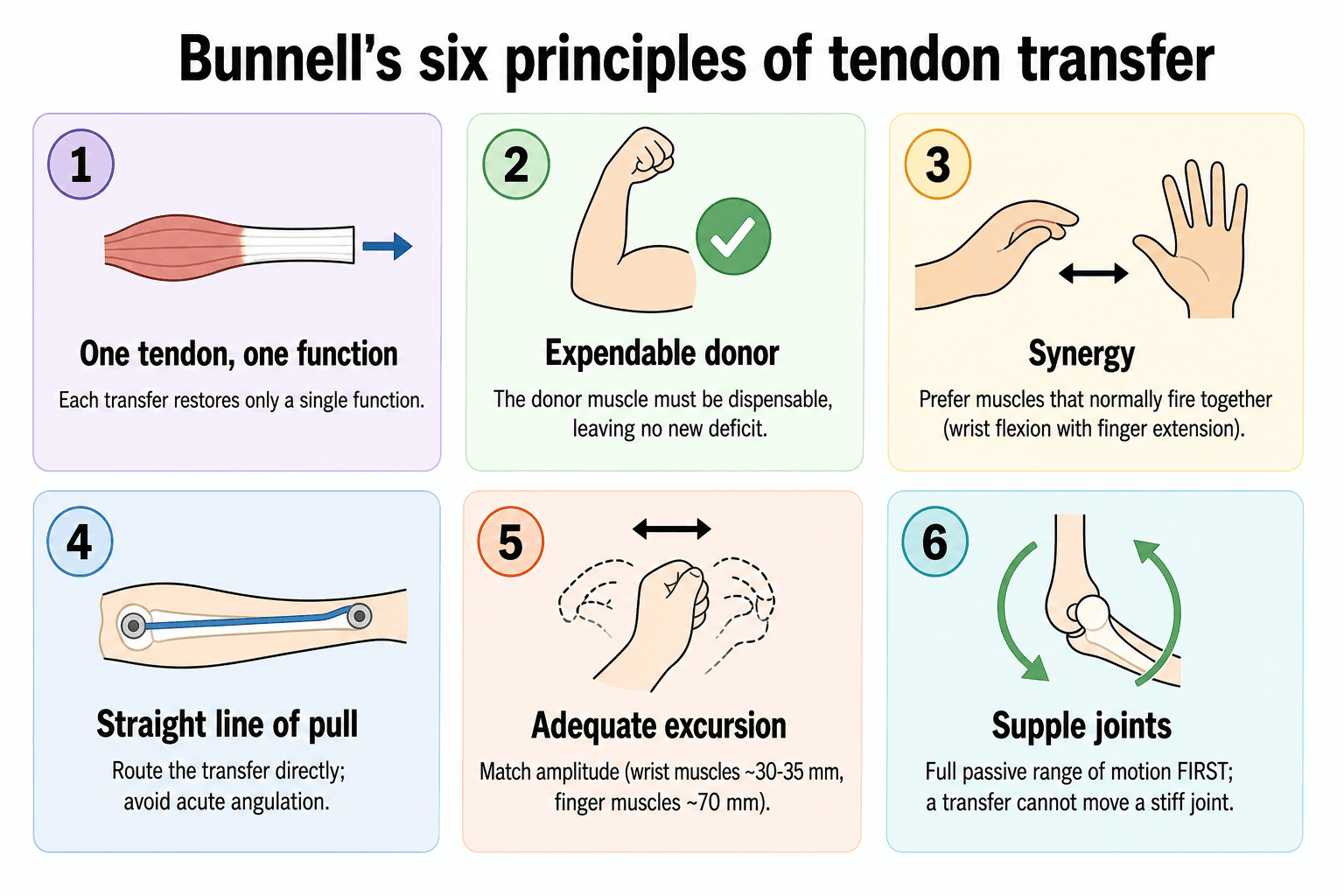
- The six principles of tendon transfer must be recited in any viva answer: one tendon one function, expendable donor, synergy, straight line of pull, adequate excursion, and supple joints before transfer - examiners test whether candidates understand these fundamentals before naming specific transfers
- Pronator teres to ECRB is the universal first transfer for radial nerve palsy - it restores balanced wrist extension and must be done before finger and thumb extension transfers
- For low median nerve palsy (opponensplasty), the FDS of the ring finger is the most commonly used donor - routed around the palmar fascia or through a pulley to recreate the opponents pull in a pronation-abduction direction
- Claw correction in ulnar palsy aims to restore the intrinsic plus position (MCP flexion with IP extension) - without this the patient has a paradoxical inability to extend the IP joints despite intact extensor tendons
- Tendon transfers fail in stiff joints - passive full range of motion must be achieved before any transfer is performed; Bunnell's dictum: 'the tendon transfer is only as good as the joint it moves'
- “Start any viva by reciting the six principles of tendon transfer - examiners want to hear them before you discuss specific procedures
- “For radial nerve palsy, always present transfers in order: (1) wrist extension, (2) thumb extension, (3) finger extension - this is the logical sequence of functional priority
- “For ulnar nerve claw correction, distinguish static procedures (tenodesis, capsulodesis) from dynamic transfers (FDS lasso, Brand) - dynamic transfers are preferred for active power grip
- “The Camitz opponensplasty uses the palmaris longus with a distal fascial extension and is reserved for severe thenar wasting where grip strength is less critical
Tendon Transfers for Radial, Median and Ulnar Nerve Palsy
Tendon transfers are never performed until passive full range of motion is achieved in all joints the transfer will cross. A transferred tendon cannot overcome a fixed contracture. If a patient has stiff PIP joints in extension from a chronic claw hand, therapy and serial splinting (or even joint release) must precede any intrinsic replacement transfer. Examiners will fail an answer that omits this prerequisite.
- Key functional deficits
- No wrist extension, no finger extension, no thumb extension, no supination from supinator
- Standard transfer set
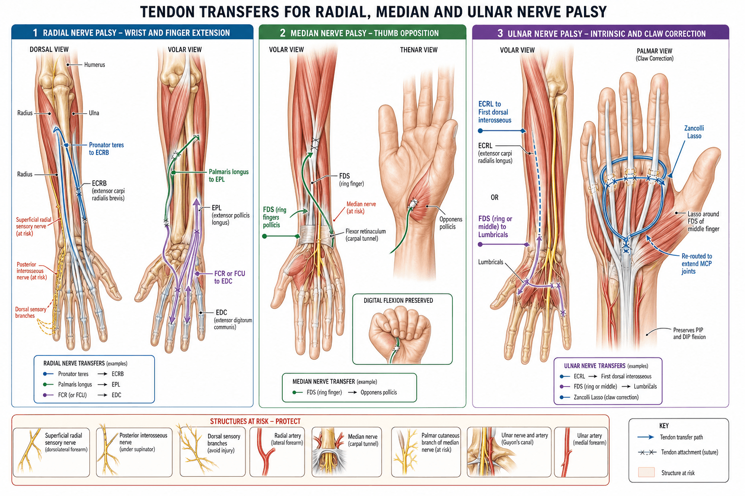
- PT to ECRB (wrist extension), FCR to EPL (thumb extension), PL to EDC (finger extension)
- Key functional deficits
- Wrist extension weak with radial deviation (ECRL intact), no finger or thumb extension
- Standard transfer set
- PT to ECRB for balanced wrist extension, FCR to EPL, PL to EDC
- Key functional deficits
- Loss of opposition (opponens pollicis, APB, superficial FPB), LOAF muscles, radial palm sensation
- Standard transfer set
- Opponensplasty: FDS ring finger (Royle-Thompson) or EIP (Burkhalter)
- Key functional deficits
- Claw hand, weak key pinch (adductor pollicis), loss of power grip, Wartenberg sign
- Standard transfer set
- Claw correction: Zancolli FDS lasso or Stiles-Bunnell; adductor pollicis: ECRB or BR with tendon graft
- Key functional deficits
- Total intrinsic loss: no opposition, claw all four fingers, no grip strength, flat thenar and hypothenar eminences
- Standard transfer set
- Opponensplasty plus four-finger intrinsic replacement (Brand extensor carpi split to lateral bands); staged if needed
Overview and Principles
Tendon transfer is the surgical rerouting of an expendable muscle-tendon unit from its normal insertion to a new insertion to restore a lost motor function following irreversible nerve injury. In the hand, it is the definitive treatment for radial, median and ulnar nerve palsies that have not recovered following nerve repair, grafting, or observed spontaneous regeneration.
The modern practice of tendon transfer in the upper limb was established by Sterling Bunnell, who codified the principles that remain the foundation of hand surgery. The goal is not to recreate normal anatomy but to restore functional grasp, release, pinch and opposition using remaining functional motors.
The six principles of tendon transfer (Bunnell):
- One tendon, one function: each transferred tendon restores only one function. A transfer split between two different functions produces weak and unpredictable movement in both.
- Expendable donor: the donor muscle must be genuinely dispensable. Removing it must not create a new functional deficit. For example, the FDS of the ring finger can be sacrificed because the FDP of the same finger provides independent flexion.
- Synergy: muscles that normally contract together should be preferentially used. A wrist flexor transferred to restore finger extension fires naturally during the grasp-release cycle because wrist flexion and finger extension are synergistic.
- Straight line of pull: the transfer must run as directly as possible from origin to new insertion. Acute angulations waste power and excursion.
- Adequate excursion: the donor tendon must have sufficient amplitude of glide to produce the required range of motion. Wrist-level muscles (FCR, ECRL, ECRB, PL, BR) have approximately 30-35 mm excursion; finger-level muscles (FDS, FDP) have approximately 70 mm.
- Supple joints: full passive range of motion must be present before transfer. A transferred tendon cannot mobilise a stiff joint - Bunnell's dictum.
Tendon transfers are typically performed 3 to 6 months after nerve injury if clinical and electrophysiological recovery is absent, or immediately in cases of irreparable nerve root avulsion (e.g., C5-C6 upper trunk brachial plexus injury). In isolated nerve palsies with a clear irreparable deficit (e.g., high radial nerve laceration with a long gap), early transfer may be considered at 3 months. Maintaining passive joint mobility with therapy and splinting during the waiting period is essential.
Relevant Anatomy and Biomechanical Principles
- Radial nerve (C5-C8, posterior cord): supplies all extensor compartments of the forearm. Above the supinator: ECRL, brachioradialis, supinator (via PIN after piercing supinator). The posterior interosseous nerve (PIN) branches at the supinator arcade of Frohse and supplies ECRB, ECU, EDC, EIP, EDQ, EPL, APL, EPB. The superficial radial nerve is purely sensory.
- Median nerve (C5-T1, lateral and medial cords): motor to PT, FCR, PL, FDS (all four), FDP to index and middle (via AIN: FPL, pronator quadratus, FDP index and middle). In the hand: LOAF (Lateral two lumbricals, Opponens pollicis, Abductor pollicis brevis, superficial head of Flexor pollicis brevis).
- Ulnar nerve (C8-T1, medial cord): motor to FCU, FDP to ring and little. In the hand: hypothenar (opponens digiti minimi, abductor digiti minimi, flexor digiti minimi), all palmar and dorsal interossei, medial two lumbricals, adductor pollicis, deep head of FPB.
- Tensioning: the transfer must be sutured at appropriate tension. Bunnell recommended suturing with the wrist and digit in the position the transfer would produce, then adjusting so the resting posture is correct. Overtension restricts motion; undertension produces a lag.
- Pulleys and routing: the natural flexor and extensor sheaths, retinacula and fascial slings act as pulleys. When routing a transfer, surgeons may create artificial pulleys (e.g., Guyon's canal for opponensplasty, or the distal edge of the dorsal retinaculum for EDC transfers) to maintain the line of pull.
- Excursion matching: the donor and recipient excursions should be matched as closely as possible. If the donor has less excursion than required, a tenodesis effect (wrist motion amplifying digit motion) can compensate - approximately 10 degrees of wrist flexion produces about 1-2 cm of apparent finger extension.
Patient Assessment and Pre-operative Planning
Assessment before tendon transfer:
- Document the precise nerve deficit using the Medical Research Council (MRC) grading for each muscle group. Distinguish high from low palsy for each nerve, as the transfer set differs.
- Assess joint mobility in every joint the transfer will cross. Use the Bunnell principle: if you cannot passively achieve full range, the transfer will fail. Institute intensive therapy, dynamic splinting, and serial casting before surgery.
- Evaluate donor muscles for strength (MRC grade 4 or better is required), excursion, and expendability. Test each proposed donor independently.
- Assess soft tissue status: scars, skin grafts, and contractures along the planned route may interfere with gliding. Staged soft tissue cover or scar release may be needed.
- Electrodiagnostic studies (EMG and nerve conduction): confirm the absence of reinnervation at the appropriate interval. Serial studies at 3-month intervals document recovery or its absence.
- Sensory assessment: a tendon transfer restores motor function only. Protective sensation (at minimum) in the target territory is needed for functional use of the reconstructed hand. If sensation is absent, consider sensory nerve reconstruction or educate the patient in protective strategies.
Even a perfectly executed tendon transfer will be functionally useless if the patient has no sensation in the reconstructed digits. Median nerve palsy with absent thumb sensibility produces a thumb that the patient does not use, regardless of how good the opposition transfer is. Always address sensory recovery as part of the reconstruction plan.
Surgical Technique
Standard transfer set (Jones / modified):
The three transfers are performed through longitudinal forearm incisions, routing tendons subcutaneously or through the interosseous membrane.
1. Pronator teres (PT) to ECRB - wrist extension The PT is harvested from its insertion on the radius with a strip of periosteum to lengthen it. It is rerouted and woven (Pulvertaft weave) into the ECRB tendon proximal to the dorsal retinaculum. The wrist should rest in approximately 20-30 degrees of extension when tensioned. This is the most important transfer as balanced wrist extension is prerequisite for effective finger and thumb extension via tenodesis.
2. Flexor carpi radialis (FCR) to EPL - thumb extension and retroposition The FCR is divided at its insertion on the second metacarpal base, withdrawn proximally, and routed around the radial border of the forearm subcutaneously to the EPL tendon proximal to Lister's tubercle. Alternatively, it may be passed through the interosseous membrane. Tension is set so the thumb rests in retroposition when the wrist is in neutral. This restores active thumb extension and the ability to release large objects.
3. Palmaris longus (PL) to EDC - finger extension The PL is harvested with a distal fascial extension (or a tendon graft if short), withdrawn proximally and routed subcutaneously to the EDC tendons over the dorsum of the hand. A single weave into all four EDC tendons restores MCP extension. If PL is absent (approximately 15% of the population), brachioradialis (BR) or FDS of the middle finger may be used.
Pearls: tension the PT-ECRB transfer first with the wrist in extension, then the FCR-EPL with the wrist in neutral, then the PL-EDC. Check that passive wrist flexion produces full digital extension (tenodesis effect) and that the patient can actively extend the wrist and digits simultaneously.
An overtensioned transfer produces a contracture; an undertensioned transfer produces a lag. The general rule is to tension the transfer with the part in the position the transfer is meant to produce (e.g., PT-ECRB tensioned with the wrist in 20-30 degrees of extension). Always check the tenodesis effect after tensioning: passive wrist flexion should produce full digital extension, and passive wrist extension should allow full digital flexion. Adjust tension before closing.
Structures at Risk and Complications
- Adhesions and restricted glide: the most common complication. Transferred tendons passing through scarred tissue beds or subcutaneous tunnels may develop adhesions that restrict excursion. Meticulous tissue handling, early protected mobilisation, and avoiding surgery through heavily scarred zones reduce this risk.
- Rupture of the transfer: occurs when the Pulvertaft weave is insufficient (fewer than three passes), when the tendon is of poor quality, or when early active motion is started too aggressively. Re-exploration and re-weaving is required.
- Overtension or undertension: overtension creates a fixed contracture (e.g., a wrist held in excessive extension); undertension produces a persistent lag. Both are avoided by careful intra-operative tensioning and checking the tenodesis effect.
- Loss of donor function: if a non-expendable donor is used, the patient trades one deficit for another. For example, harvesting FCR for EPL transfer in a patient who also needs FCR for wrist flexion will weaken wrist flexion. Planning transfers that use truly expendable donors avoids this.
- Nerve injury during routing: the superficial radial nerve, lateral antebrachial cutaneous nerve, and dorsal sensory branch of the ulnar nerve are all at risk during tendon harvest and routing. Identification and protection prevents neuroma formation.
- Infection: as with any surgical procedure, superficial or deep infection can jeopardise the transfer. Prophylactic antibiotics and meticulous wound management are standard.
- Persistent claw or inadequate correction: in ulnar palsy, inadequate correction occurs when the joint was stiff before surgery, when the transfer was undertensioned, or when the FDS lasso slips. Revision with an alternative transfer may be needed.
- Synergy mismatch: a transfer that violates the synergy principle (e.g., using an extensor to replace a flexor function without considering the relearning required) produces a movement pattern that is difficult to rehabilitate and may never become automatic.
Post-operative adhesions limiting tendon glide account for the majority of poor outcomes after tendon transfer. Tenolysis may be required at 3-6 months if active glide is significantly less than passive glide. The key preventive strategy is early protected mobilisation in a supervised hand therapy programme.
Splinting and Orthoses While Awaiting Recovery
While a nerve is recovering (or when transfer is declined or contraindicated), a palsy-specific orthosis does two jobs that the viva expects you to state: it prevents the fixed deformity that would doom a future transfer (Bunnell's supple-joint prerequisite) and it substitutes the lost function in the interim.
- Orthosis
- Dynamic wrist- and MCP-extension outrigger, or a static volar cock-up (wrist-extension) splint
- Dual purpose
- Holds the wrist extended to restore grip through tenodesis and prevents a wrist/finger flexion contracture
- Orthosis
- Anti-claw orthosis (lumbrical bar / knuckleduster / figure-of-eight) blocking MCP hyperextension
- Dual purpose
- Lets the long extensors extend the IP joints (restores the intrinsic-plus cascade) and prevents PIP flexion contracture
- Orthosis
- Opposition (C-bar / thumb web-space) splint holding the thumb in palmar abduction
- Dual purpose
- Positions the thumb for pinch and prevents a first web-space adduction contracture
Even if you are certain a patient will need a transfer, the interim orthosis is not optional: a clawed hand left unsplinted develops fixed PIP flexion contractures, and a radial palsy left in flexion develops a wrist and first-web contracture - either of which violates the supple-joint principle and makes the eventual transfer fail. The splint buys function now and protects the operation later.
Post-operative Rehabilitation and Motor Re-education
The rehabilitation is as important as the operation - a technically perfect transfer fails without structured therapy and motor relearning. It runs in three phases, and the synergy principle chosen at surgery determines how easily the patient relearns the movement.
- Timing
- About 3 to 4 weeks (up to 4 to 6)
- Goal
- Splint in the position the transfer produces (wrist extended for the radial set, intrinsic-plus for claw correction) so the weave heals without tension or rupture
- Timing
- From about week 3 to 4
- Goal
- Protected active motion; teach the patient to fire the donor through its original action (for example, 'pronate to extend the wrist' after PT-to-ECRB), aided by biofeedback, electrical stimulation and mirror therapy
- Timing
- From about week 6 to 12 onward
- Goal
- Progressive resistance and task-specific practice; return to work and sport typically by 3 to 6 months
A synergistic transfer (a wrist flexor used to restore finger extension, which naturally co-contract during the grasp-release cycle) is relearned quickly and becomes automatic; a non-synergistic transfer needs deliberate cortical retraining and may never be fully effortless. This is why synergy is chosen at surgery and reinforced in therapy. Throughout, early protected glide is the key defence against adhesions - the commonest cause of a disappointing result.
Guidelines, Registries and Global Practice
- ASSH (American Society for Surgery of the Hand) and BSSH (British Society for Surgery of the Hand): no single formal guideline exists for tendon transfers; practice follows the principles established by Bunnell, Brand, Riordan and Zancolli as described in standard hand surgery texts. The ASSH manual and BSSH education curricula teach the six principles as the non-negotiable framework.
- FESSH (Federation of European Societies for Surgery of the Hand): European hand surgery training mandates competency in tendon transfers for all three nerve palsies. The FESSH examination typically includes a viva station on tendon transfer principles and specific transfer selection.
- Nerve transfer vs tendon transfer debate: in recent years, nerve transfers (e.g., median-to-radial nerve transfer for radial palsy, anterior interosseous nerve-to-ulnar motor nerve transfer) have emerged as alternatives or adjuncts to traditional tendon transfers. Nerve transfers restore original muscle function but must be performed within 6-12 months of injury for best results. Current practice at major centres often combines nerve transfers (for reinnervation of critical muscles) with tendon transfers (for functions that cannot be restored by nerve transfer).
- Leprosy-endemic regions (India, Brazil, parts of Africa and Southeast Asia): tendon transfers for combined nerve palsy are performed in enormous numbers by specialised centres (e.g., the Leprosy Mission hospitals, LKH Karigiri). The Brand transfers were developed in this setting and remain the standard of care. Surgeons in these regions have some of the world's highest volumes and best-reported outcomes for intrinsic replacement.
- High-income countries: the aetiologic spectrum shifts to traumatic nerve injuries (lacerations, fractures, gunshot wounds) and compressive neuropathies. Nerve transfers are increasingly performed alongside or instead of tendon transfers, particularly in brachial plexus reconstruction.
- Paediatric practice: in obstetric brachial plexus injury and congenital conditions, tendon transfers are adapted for growth (e.g., using the Steindler flexorplasty for elbow flexion, or modified opponentsplasties in the growing child). Timing is critical to prevent fixed deformities.
- Post-operative rehabilitation is as important as the surgery itself. A structured hand therapy programme typically involves 4-6 weeks of protected immobilisation followed by progressive active and then resisted exercises. Biofeedback and electrical stimulation of the transferred muscle may aid motor relearning. Total rehabilitation takes 3-6 months.
Memory Aids
DONSYSPrinciples of Tendon Transfer - DONSYS
Hook:DONSYS: Donor expendable, One function, Not across stiff joints, Synergy, Yield/excursion, Straight pull - the six non-negotiable principles
STWRadial Nerve Palsy Transfers - STW (Superhero Three Ws)
Hook:PT-ECRB, FCR-EPL, PL-EDC: the Superhero Three that rescues the wrist, thumb and fingers in radial palsy
CLAWSUlnar Nerve Claw Correction - CLAWS
Hook:CLAWS: Capsulodesis, Lasso, Anterior (Brand), Wartenberg correction, Stiles-Bunnell - the five claw correction options from static to dynamic
Clinical Decision Scenarios
Practise clinical reasoning and management decisions out loud
“A 35-year-old carpenter sustained a closed mid-shaft humeral fracture 8 months ago, which was treated conservatively in a brace. The fracture has healed, but he has a persistent radial nerve palsy with no clinical or electrophysiological evidence of recovery. He has a wrist drop with no active finger or thumb extension. Sensation is intact over the dorsoradial hand. How would you manage this?”
“A 42-year-old musician presents with a claw deformity of the ring and little fingers 10 months after a laceration at the wrist that severed the ulnar nerve. The nerve was repaired primarily. EMG shows no reinnervation of intrinsic muscles. She has hyperextension at the ring and little MCP joints with PIP and DIP flexion, weak key pinch, and loss of power grip. How would you reconstruct her hand?”
The six principles (DONSYS)
- Donor expendable - must not create a new deficit
- One tendon, one function - never split between two functions
- Not across a stiff joint - Bunnell's dictum: achieve full passive ROM first
- Synergy - prefer muscles that contract together with the lost function
- Yield (adequate excursion) - match donor excursion to recipient requirement
- Straight line of pull - avoid acute angulation; use pulleys if needed
Radial nerve palsy transfers
- PT to ECRB: restores balanced wrist extension (most critical transfer)
- FCR to EPL: restores thumb extension and retroposition (routed around radial border)
- PL to EDC: restores finger MCP extension (if PL absent, use BR or FDS middle)
- Tension in order: wrist extension first, then thumb, then fingers
- Check tenodesis effect: passive wrist flexion produces digital extension
Median nerve palsy transfers
- Low palsy: opponentsplasty only (FDS ring = Royle-Thompson, EIP = Burkhalter, PL = Camitz)
- FDS ring: routed around FCU pulley at pisiform to APB insertion
- EIP: harvested dorsally, routed around ulnar border to thumb
- Camitz: PL with palmar fascia, for severe thenar wasting in low-demand patients
- High palsy: add FPL restoration (BR or FDS to FPL) and FDP side-to-side tenodesis
Ulnar nerve palsy transfers
- Claw correction (dynamic): Zancolli FDS lasso (workhorse), Stiles-Bunnell, Brand ECRB split
- Zancolli lasso: FDS divided at PIP, looped around A1 pulley, restores MCP flexion
- Brand: ECRB prolonged with four-tailed graft through carpal tunnel to lateral bands
- Static: volar plate capsulodesis, tenodesis (for stiff joints or poor donors)
- Wartenberg sign: suture ulnar slip of EDQ to EDC of ring finger
Timing and prerequisites
- Wait 3-6 months for nerve recovery unless irreparable injury confirmed
- Passive full ROM mandatory before any transfer (therapy, splinting, serial casting)
- Donor muscles must be MRC grade 4 or better
- Protective sensation in target territory is required for functional use
- Electrodiagnostic confirmation of no reinnervation before proceeding
Complications
- Adhesions: commonest cause of poor outcome; may require tenolysis at 3-6 months
- Rupture: insufficient weave, poor tendon quality, early aggressive mobilisation
- Overtension: fixed contracture; undertension: persistent lag
- Loss of donor function: using a non-expendable donor trades one deficit for another
- Nerve injury during routing: protect superficial radial, LABC, dorsal ulnar sensory nerves
Evidence Base
- Riordan codified the standard tendon transfer sets for each nerve palsy, describing the PT-to-ECRB, FCR-to-EPL and PL-to-EDC combination for radial palsy that remains the benchmark. His principles of synergistic transfer selection and straight-line routing are still taught in every hand surgery training programme worldwide.
- Brand pioneered the use of ECRB prolonged with a four-tailed tendon graft routed through the carpal tunnel to the lateral bands of each finger for intrinsic replacement in combined ulnar and median nerve palsy. In hundreds of leprosy patients, this transfer reliably corrected the claw deformity and restored functional grasp. Brand also emphasised the importance of excursion matching and the use of the tenodesis effect.
- Modification of the classic Brand transfer using ECRB to lateral bands provides reliable claw correction with good functional outcomes in ulnar nerve palsy.
- Riley, Mann and Burkhalter described the extensor pollicis longus (EPL) opponensplasty - rerouting EPL around the ulnar border of the wrist to restore thumb opposition in median nerve palsy - as a reliable alternative to FDS opponensplasty. Using an expendable extensor as the donor preserves the flexor cascade and avoids the PIP extension lag that can follow FDS harvest.