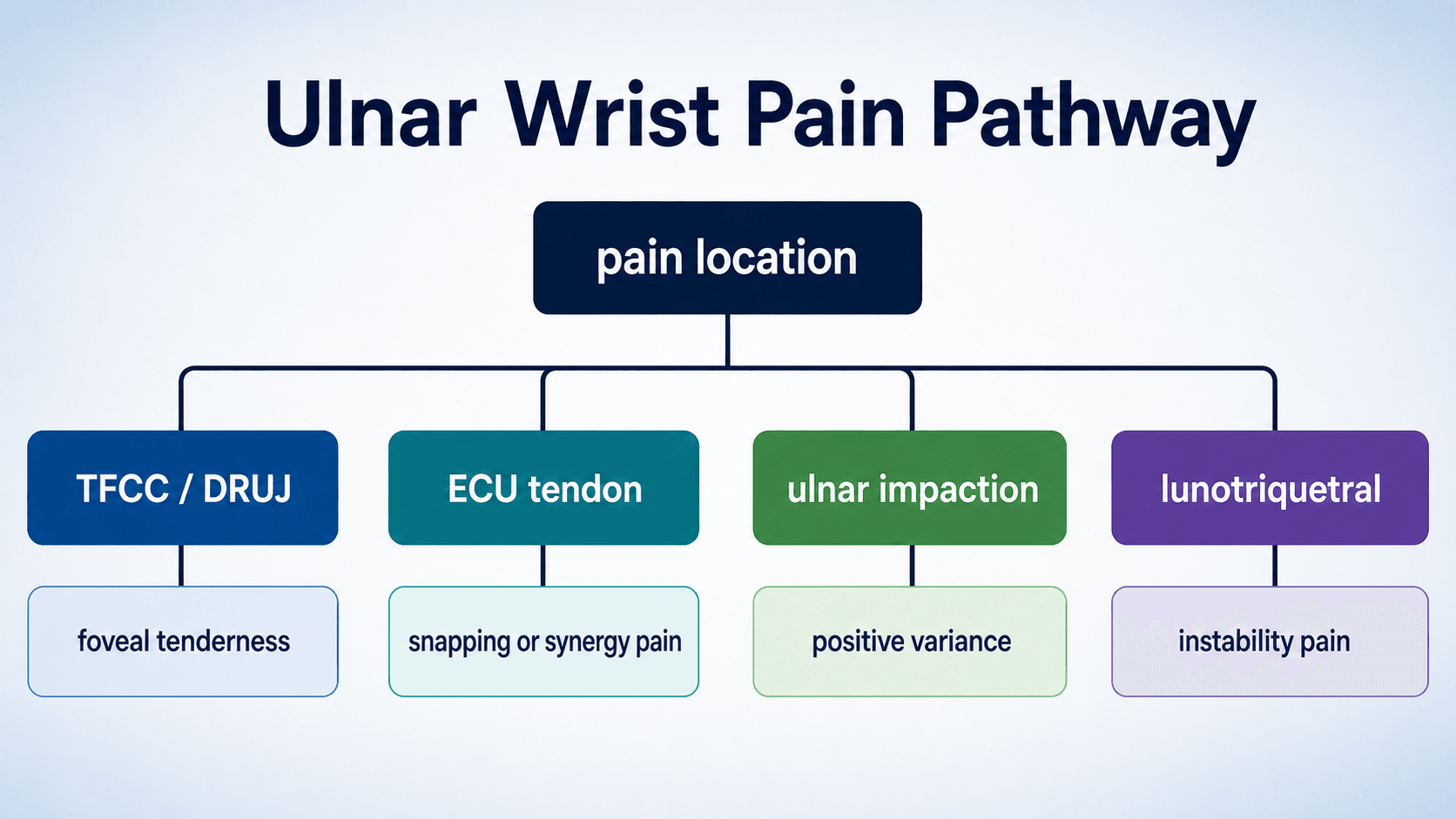
TFCC and Ulnar-Sided Wrist Pain
Localise the pain, test the DRUJ, then match treatment to tear biology and ulnar load
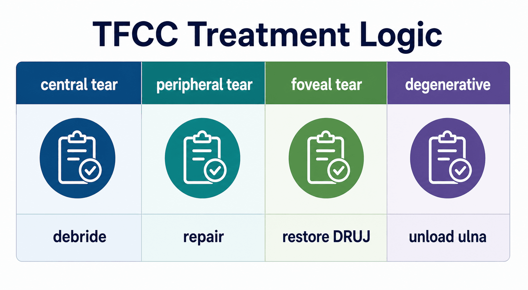
Treatment Logic
Critical Must-Knows
- Ulnar-sided wrist pain is a localisation problem; not every painful ulnar wrist is a TFCC tear.
- The TFCC includes the articular disc, dorsal and palmar radioulnar ligaments, ulnocarpal ligaments, meniscus homologue and ECU subsheath.
- DRUJ stability depends mainly on the deep foveal fibres of the radioulnar ligaments.
- Palmer classification describes the lesion; treatment also depends on DRUJ stability, reparability, cartilage status and ulnar variance.
- A central stable tear, a repairable peripheral tear and an unstable foveal tear are different clinical problems.
Clinical Pearls
- "Pain with a positive foveal sign suggests TFCC or ulnotriquetral pathology, but it is not a standalone diagnosis.
- "Test DRUJ translation in neutral, pronation and supination, and always compare the opposite wrist.
- "A positive MRI without matching clinical symptoms should not automatically lead to surgery.
- "Debridement alone is a poor answer for a symptomatic foveal tear with DRUJ instability.
- "If ulnar impaction is the driver, the treatment must address ulnar overload, not just the torn disc.
The key mistake is treating the MRI instead of the patient
TFCC signal change is common, especially with age and ulnar loading. The decision to operate should come from symptoms, examination, DRUJ stability, imaging concordance, cartilage status and patient goals.
At a Glance Table
Fast Clinical Sorting
| Pattern | Likely source | Confirm with | Management implication |
|---|---|---|---|
| Deep foveal tenderness with pain on rotation | TFCC peripheral or foveal tear | Foveal sign, DRUJ ballottement, MRI or arthroscopy | Assess stability before deciding debridement versus repair |
| Prominent ulnar head, clunk or painful translation | DRUJ instability | Ballottement in neutral, pronation and supination; CT if incongruent | Foveal repair, reconstruction or salvage depends on cartilage and chronicity |
| Snapping tendon over ulnar head | ECU subluxation or tendinopathy | ECU synergy test, resisted extension-ulnar deviation, dynamic ultrasound | Treat ECU pathology, not TFCC alone |
| Ulnocarpal load pain with positive ulnar variance | Ulnar impaction | PA neutral wrist radiograph, grip view, MRI marrow signal | Consider ulnar unloading when non-operative care fails |
| Pain over lunotriquetral interval | Lunotriquetral ligament injury | LT shear or ballottement test, MRI arthrogram or arthroscopy | Different reconstruction pathway from TFCC |
| Pisiform tenderness and pain with resisted FCU | Pisotriquetral or FCU pathology | Pisotriquetral grind, carpal tunnel view, local injection | Avoid mislabelling as TFCC |
TIDEUlnar Wrist Differential
| T | TFCC and DRUJ Foveal pain, rotation pain and instability. |
| I | Impaction Ulnocarpal overload, positive ulnar variance and lunate or triquetral changes. |
| D | Dynamic tendons ECU subluxation, ECU tendinopathy and FCU or pisotriquetral pain. |
| E | Extra causes Fracture, inflammatory disease, ganglion, nerve pain and referred pain. |
| T | TFCC and DRUJ Foveal pain, rotation pain and instability. | D | Dynamic tendons ECU subluxation, ECU tendinopathy and FCU or pisotriquetral pain. |
| I | Impaction Ulnocarpal overload, positive ulnar variance and lunate or triquetral changes. | E | Extra causes Fracture, inflammatory disease, ganglion, nerve pain and referred pain. |
Hook:TIDE keeps the ulnar wrist differential broad.
CPFUTFCC Treatment Biology
| C | Central Stable central tears are usually debrided if symptomatic. |
| P | Peripheral Repairable vascular peripheral tears may heal after repair. |
| F | Foveal Foveal detachment must restore DRUJ stability. |
| U | Ulnar overload Degenerative impaction needs unloading when load remains the driver. |
| C | Central Stable central tears are usually debrided if symptomatic. | F | Foveal Foveal detachment must restore DRUJ stability. |
| P | Peripheral Repairable vascular peripheral tears may heal after repair. | U | Ulnar overload Degenerative impaction needs unloading when load remains the driver. |
Hook:Central, peripheral, foveal and ulnar overload are four different treatment problems.
NPSDRUJ Instability Check
| N | Neutral Start with the wrist neutral and compare the other side. |
| P | Pronation Some instability is position-dependent and only appears in pronation. |
| S | Supination Repeat in supination and document pain, translation and endpoint. |
| N | Neutral Start with the wrist neutral and compare the other side. |
| P | Pronation Some instability is position-dependent and only appears in pronation. |
| S | Supination Repeat in supination and document pain, translation and endpoint. |
Hook:Test the DRUJ in Neutral, Pronation and Supination.
Overview and Epidemiology
The triangular fibrocartilage complex is the main soft-tissue stabiliser and load-bearing structure on the ulnar side of the wrist. It sits between the distal ulna, sigmoid notch, proximal carpus and ECU subsheath. A painful TFCC lesion may be traumatic, degenerative or part of a wider ulnar-sided wrist problem.
Patients usually present with ulnar wrist pain, pain with gripping or forearm rotation, clicking, weakness, or a feeling that the distal radioulnar joint is unstable. The history must separate a single traumatic event from chronic overload. A fall on an extended wrist with forearm rotation suggests traumatic TFCC injury. Repetitive axial loading, racquet sport, gymnastics, manual work, positive ulnar variance or distal radius malunion suggest ulnocarpal overload.
TFCC abnormalities may also be incidental. Degenerative fraying increases with age and load. The clinical question is therefore not "is there a TFCC signal change?", but "is this lesion responsible for this patient's pain or instability, and what mechanical problem needs to be treated?"
Anatomy and Biomechanics
The TFCC is not one structure. It is a composite stabilising system. The articular disc carries load between the carpus and ulna. The dorsal and palmar radioulnar ligaments are the key stabilisers of the DRUJ. Their deep fibres insert into the ulnar fovea and are critical for stability. The ulnocarpal ligaments connect the ulna and TFCC region to the lunate and triquetrum. The meniscus homologue and ECU subsheath complete the ulnar soft-tissue complex.
The central disc is relatively avascular and has limited healing potential. The peripheral ulnar side is more vascular and is more suitable for repair. This is the biological reason central stable tears are treated differently from peripheral and foveal tears.
The TFCC also behaves as a load regulator. Positive ulnar variance increases ulnocarpal load and can produce degenerative disc wear, lunate or triquetral chondromalacia and lunotriquetral ligament involvement. Negative or neutral variance does not exclude TFCC injury, but it changes the likelihood that ulnar overload is the main driver.

TFCC Structures and Clinical Meaning
| Structure | Role | Clinical implication |
|---|---|---|
| Central articular disc | Ulnocarpal load-bearing surface | Poor healing potential; symptomatic stable central tears are usually debrided rather than repaired |
| Deep foveal radioulnar fibres | Primary DRUJ stabiliser | Foveal detachment produces instability and usually needs restoration if symptomatic |
| Peripheral capsule and superficial fibres | Peripheral support and soft-tissue continuity | Repairable if tissue quality is good and cartilage is preserved |
| Ulnocarpal ligaments | Support lunate and triquetrum | Distal avulsions may mimic isolated TFCC pain but behave as carpal stabiliser injuries |
| ECU subsheath | Stabilises ECU tendon at the ulnar wrist | Snapping ECU can coexist with TFCC symptoms and needs specific treatment |
Pathophysiology
TFCC pathology can be grouped into four practical mechanisms.
Acute traumatic tear
Usually follows axial load, rotation or traction. The important question is whether the tear is central and stable, peripheral and repairable, or foveal with DRUJ instability.
Degenerative ulnar overload
Ulnar impaction produces progressive wear of the TFCC, chondral change in the lunate or triquetrum and sometimes lunotriquetral ligament involvement.
Post-fracture or malunion pattern
Distal radius malunion, radial shortening, sigmoid notch incongruity or ulnar styloid base nonunion can create ulnar-sided pain and DRUJ dysfunction.
Associated soft-tissue pathology
ECU instability, FCU or pisotriquetral disease, LT injury, inflammatory synovitis and ganglion can coexist with or mimic TFCC symptoms.
A stable tear is not the same problem as an unstable tear
A patient with a stable central perforation and a patient with a foveal detachment may both report ulnar wrist pain. The first may need debridement after failed non-operative care. The second needs restoration of the DRUJ stabiliser if instability is symptomatic and cartilage is salvageable.
Classification Systems
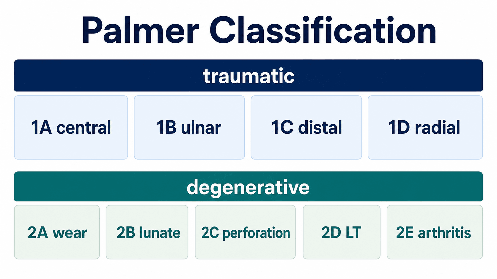
Palmer Classification
| Type | Lesion | Clinical meaning | Typical treatment direction |
|---|---|---|---|
| 1A | Central traumatic perforation | Stable disc tear with limited healing potential | Debridement if symptomatic after non-operative care |
| 1B | Ulnar-sided avulsion with or without styloid fracture | May involve peripheral or foveal attachment; assess DRUJ stability | Repair or foveal refixation when symptomatic and repairable |
| 1C | Distal ulnocarpal ligament avulsion | Carpal stabiliser injury rather than simple disc tear | Repair or reconstruction when unstable or persistently symptomatic |
| 1D | Radial-sided avulsion | Radial attachment injury; may be technically difficult | Repair selected symptomatic unstable tears |
| 2A to 2E | Degenerative spectrum | Progressive wear, chondromalacia, perforation, LT involvement and arthritis | Debridement, wafer, ulnar shortening or salvage depending stage and load |
Clinical Assessment
History should define the mechanism, chronicity and load demand. Ask about fall, twisting injury, racquet or bat sport, gymnastics, manual work, distal radius fracture, previous surgery, inflammatory arthritis, clicking, snapping, instability, weakness, pain with push-up from a chair, and symptoms during pronation or supination. Record hand dominance, work demands, sport and what the patient needs the wrist to do.
Examination starts with comparison. Look for swelling, ulnar head prominence, scars, distal radius malunion, ECU snapping, hypothenar wasting and range restriction. Palpate in a structured way: fovea, ECU groove, FCU and pisiform, lunotriquetral interval, ulnocarpal joint, hook of hamate and DRUJ. Then test motion, grip, DRUJ stability and tendon-specific provocative manoeuvres.
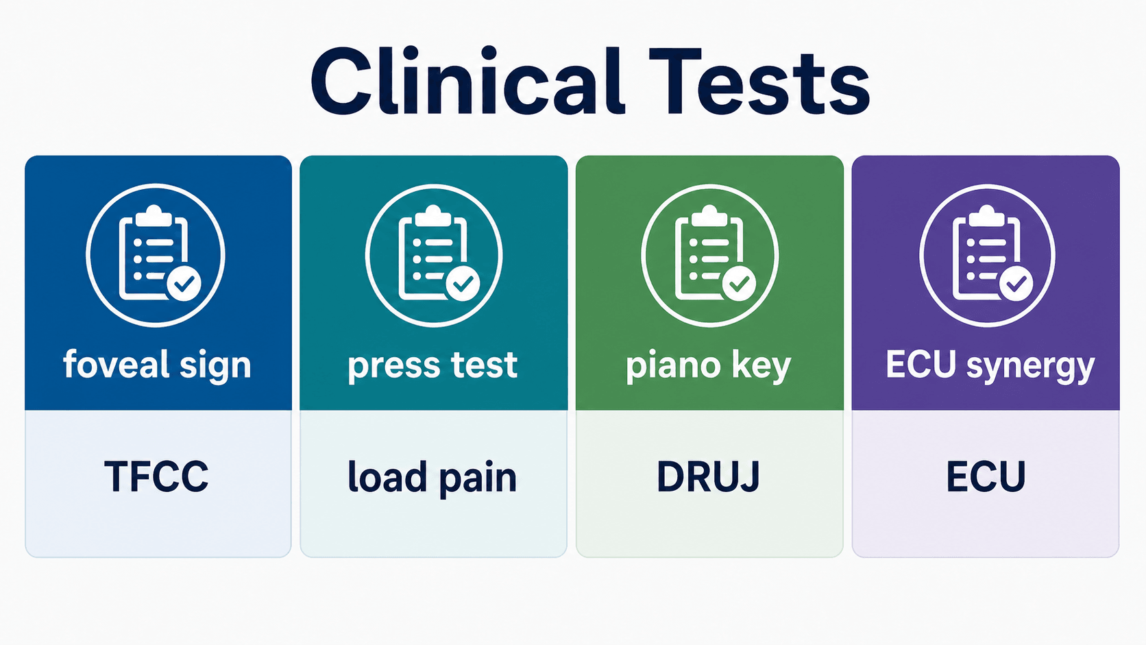
Clinical Tests
| Test | How to do it | Positive finding | What it suggests |
|---|---|---|---|
| Ulnar fovea sign | Press in the soft spot between ulnar styloid, FCU, volar ulnar head and pisiform | Localised deep tenderness | TFCC peripheral/foveal or ulnotriquetral pathology |
| DRUJ ballottement | Translate distal ulna dorsally and volarly relative to radius in neutral, pronation and supination | Painful excess translation or poor endpoint | DRUJ instability or foveal detachment |
| Piano key sign | Press prominent distal ulna volarly and observe rebound | Excess mobility or pain | DRUJ instability, but compare with the other side |
| Ulnocarpal stress test | Axial load with ulnar deviation and forearm rotation | Ulnar-sided load pain | TFCC tear or ulnar impaction; not specific alone |
| Press test | Patient pushes up from chair using the hand | Reproduction of ulnar wrist pain | Load-sensitive TFCC or ulnocarpal pathology |
| ECU synergy test | Resisted thumb radial abduction or resisted wrist extension-ulnar deviation | ECU groove pain or snapping | ECU tendinopathy or subsheath pathology |
| LT shear or ballottement | Stress lunate and triquetrum against each other | Painful clunk or local LT pain | Lunotriquetral ligament injury |
Red flags and alternative diagnoses
Do not miss infection, inflammatory arthritis, occult fracture, malignancy, acute DRUJ dislocation, acute Essex-Lopresti pattern, vascular insufficiency or ulnar nerve symptoms. Numbness, coldness, progressive swelling, fever, severe night pain or acute deformity should change the pathway.
Investigations
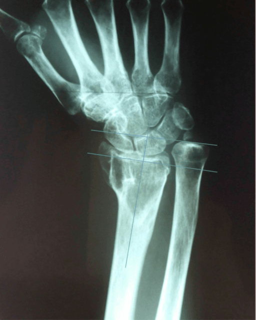
Start with good radiographs. Standard PA and lateral wrist films should be obtained with reproducible positioning. Look for distal radius malunion, ulnar styloid base nonunion, DRUJ arthritis, ulnar variance, carpal alignment, lunate or triquetral cystic change and occult fracture signs. A clenched-fist or pronated grip view can accentuate dynamic ulnar variance in selected cases.
MRI is useful when the clinical picture matches TFCC or ulnar-sided soft-tissue pathology. MR arthrography may improve detection of full-thickness communication, but imaging must be interpreted with symptoms and examination. CT is valuable for DRUJ congruity, sigmoid notch deformity, malunion, ulnar styloid base nonunion and occult fracture. Dynamic ultrasound is useful for ECU subluxation.
Arthroscopy remains the most direct method of assessing tear location, cartilage, trampoline test, hook test, peripheral reparability and combined intra-articular pathology. It is both diagnostic and therapeutic, but should not be used as a substitute for a thoughtful preoperative diagnosis.
Investigation Choices
| Investigation | Best for | Limitations |
|---|---|---|
| Plain radiographs | Variance, malunion, styloid nonunion, arthritis, carpal alignment | Can miss soft-tissue tears and occult fractures |
| MRI | TFCC signal, marrow oedema, ECU, LT ligament, occult pathology | False positives occur; resolution and reader expertise matter |
| MR arthrogram | Full-thickness communication and intra-articular tear assessment | Invasive; still must be clinically correlated |
| CT | DRUJ congruity, sigmoid notch, malunion and fracture | Limited soft-tissue information |
| Dynamic ultrasound | ECU subluxation and tendon pathology | Operator dependent; not a full TFCC assessment |
| Wrist arthroscopy | Direct tear assessment, cartilage and treatment | Invasive; should answer a defined clinical question |
Management Decisions
Management is based on symptoms, chronicity, DRUJ stability, tear location, tissue quality, cartilage status, ulnar variance and patient demand. Most stable, non-acute presentations deserve a period of non-operative care unless there is clear mechanical instability or a time-sensitive associated injury.
Non-operative care is appropriate for many stable TFCC and ulnar-sided wrist pain presentations. The usual elements are activity modification, splinting or cast immobilisation, anti-inflammatory measures when appropriate, hand therapy, proprioceptive retraining, ECU control when relevant, and graded return to load.
Non-Operative Plan
| Element | Practical detail | Reason |
|---|---|---|
| Immobilisation | Wrist or Munster-style immobilisation depending on rotation pain and DRUJ symptoms | Reduces painful forearm rotation and ulnocarpal load |
| Therapy | Range, proprioception, grip progression, ECU control and gradual loading | Restores function without provoking recurrent synovitis |
| Injection | Consider diagnostic or therapeutic injection when diagnosis remains uncertain | Can localise pain generator and reduce inflammation |
| Review point | Escalate if persistent mechanical symptoms, instability or load-limiting pain remains | Avoids prolonged ineffective treatment for repairable instability |
Surgical Technique
The operative plan should be written before the patient enters theatre: diagnostic arthroscopy only, debridement, peripheral repair, foveal repair, ulnar unloading, correction of malunion, ECU stabilisation or a combined procedure. Position the patient supine with the arm on a hand table. Use traction for wrist arthroscopy, protect superficial sensory nerves around portals, and confirm portals with surface anatomy before incision.
Arthroscopy Setup and Diagnostic Sequence
| Step | Purpose | Pitfall |
|---|---|---|
| Position and traction | Provide stable access to radiocarpal and midcarpal compartments | Excess traction can distort assessment and injure soft tissues |
| Portal planning | Common radiocarpal portals include 3-4, 4-5 and 6R or 6U depending target | Protect dorsal sensory branches and ECU region |
| Systematic inspection | Assess cartilage, SL, LT, TFCC disc, peripheral rim and synovitis | Do not focus only on the MRI-reported area |
| Probe tests | Trampoline assesses central tension; hook test assesses foveal attachment | A stable central tear and foveal detachment need different operations |
| Treat the lesion | Debride, repair, refix or unload according to the mechanical diagnosis | Debridement that destabilises the rim can worsen symptoms |
Procedure Principles
| Procedure | Core steps | Technical priorities |
|---|---|---|
| Central debridement | Diagnostic arthroscopy, define stable rim, shave unstable central flap, preserve peripheral attachments | Remove only unstable tissue; avoid enlarging into the vascular/stabilising rim |
| Peripheral repair | Freshen tear edge if needed, pass sutures, capture capsule/TFCC rim, secure outside capsule or with all-inside device | Protect dorsal sensory nerve branches; avoid overtightening |
| Foveal repair | Confirm instability, prepare foveal footprint, pass sutures through deep fibres, fix to fovea with transosseous tunnels or anchor | Reduction of the deep stabiliser matters more than cosmetic disc appearance |
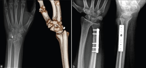
| Ulnar shortening osteotomy | Preoperative variance plan, diaphyseal osteotomy, compression plating, confirm length and rotation | Avoid nonunion, hardware irritation and over-shortening |
| Wafer procedure | Arthroscopic or open distal ulnar dome resection in selected overload cases | Preserve DRUJ cartilage and avoid excessive resection |
Describe the operation as a sequence of decisions
For TFCC surgery, a strong answer is not just a list of portals. State the diagnosis, why the lesion is symptomatic, whether the DRUJ is stable, what you will inspect arthroscopically, what finding will change treatment, and how you will protect the stabilising rim.
Postoperative Care
Postoperative care depends on what was done. Debridement is usually followed by early motion once wounds settle. Peripheral repair or foveal repair needs protection from rotation and load while the repair heals. Ulnar shortening osteotomy needs bone-healing precautions and hardware surveillance.
Rehabilitation Principles
| Procedure | Protection | Progression |
|---|---|---|
| Arthroscopic debridement | Short period of dressing or splint comfort | Early wrist and forearm motion, then graded strengthening |
| Peripheral repair | Immobilise wrist and limit forearm rotation initially | Protected motion, then progressive strengthening and load |
| Foveal repair | Stronger rotation protection, often above-elbow or Munster-style immobilisation early | Delay forceful pronation-supination until healing is secure |
| Ulnar shortening osteotomy | Protect weight bearing until osteotomy healing is evident | Monitor union and plate irritation; return to heavy load only after union |
Patients should be warned that grip strength, rotation comfort and load tolerance recover gradually. Persistent ulnar wrist pain after surgery may reflect wrong diagnosis, untreated ulnar impaction, DRUJ arthritis, ECU pathology, neuroma, stiffness, nonunion after osteotomy or complex regional pain.
Complications
Diagnostic failure
The commonest failure pattern is treating a visible TFCC tear while missing the real pain generator: ulnar impaction, ECU instability, LT injury, distal radius malunion, DRUJ arthritis or inflammatory disease.
Instability after inadequate treatment
Debridement without recognising foveal detachment can leave ongoing DRUJ instability.
Surgical morbidity
Portal nerve irritation, stiffness, infection, tendon irritation, persistent synovitis and complex regional pain can occur after wrist arthroscopy.
Osteotomy problems
Ulnar shortening can cause delayed union, nonunion, plate irritation, refracture after plate removal, DRUJ symptoms or overcorrection.
Failure Analysis
| Persistent symptom | Consider | Next step |
|---|---|---|
| Persistent rotation pain | DRUJ instability, incongruity or arthritis | Repeat focused examination, CT and review arthroscopy findings |
| Persistent load pain | Ulnar impaction or uncorrected positive variance | Reassess radiographs and MRI for overload pattern |
| Snapping after TFCC treatment | ECU subsheath instability | Dynamic ultrasound and ECU-specific examination |
| Diffuse pain and stiffness | Complex regional pain, over-immobilisation or inflammatory pathology | Early recognition, therapy and pain strategy |
Outcomes and Prognosis
Outcomes are best when diagnosis, tear biology and mechanics match the operation. Stable central tears often improve after debridement when the symptoms are truly from the tear and there is no untreated overload. Peripheral and foveal repairs do best when tissue is repairable, cartilage is preserved, instability is corrected and rehabilitation is followed. Degenerative overload outcomes depend on correcting the load problem while avoiding overtreatment in low-demand or minimally symptomatic patients.
Poorer prognostic factors include chronic instability, DRUJ arthritis, marked cartilage loss, distal radius malunion that is not corrected, workers compensation or high-load occupational demands, smoking when osteotomy is performed, and prolonged pain sensitisation.
Controversies and Areas of Uncertainty
Much of TFCC practice rests on case series and expert classification rather than randomised data, so several decisions remain genuinely contested.
Wafer versus ulnar shortening osteotomy
For mild positive variance with a central degenerative perforation, the arthroscopic wafer avoids hardware and nonunion but gives limited correction; ulnar shortening corrects variance more reliably but carries roughly a 4 percent nonunion risk and frequent hardware irritation. There is no high-level trial that settles which is superior, and selection is variance-, cartilage- and surgeon-driven.
Open versus arthroscopic repair
Systematic reviews report broadly equivalent outcomes, with scant evidence to mandate either approach. The real determinant is whether the lesion is a peripheral wall tear or a deep foveal detachment, not the access route.
Ulnar styloid fracture fixation
A styloid tip fracture rarely needs fixation. The debate is the styloid base fracture with DRUJ instability: some restore stability by fixing the base, others by repairing the foveal TFCC directly. The unifying principle is to treat the stabiliser, not the radiograph.
When MRI signal is incidental
Degenerative TFCC change and even perforation are common with age and ulnar loading and are frequently asymptomatic. There is no agreed threshold at which an imaging finding alone justifies surgery; concordance with symptoms, examination and stability remains the arbiter.
Reconstruction versus arthroplasty for the chronically unstable, arthritic DRUJ
For the irreparable, arthritic DRUJ, the balance between soft-tissue reconstruction, traditional salvage (Darrach, Sauve-Kapandji, hemiresection) and constrained DRUJ arthroplasty is shifting toward implant arthroplasty in suitable patients, but durability data are still maturing. Match the procedure to age, demand, bone stock and soft-tissue quality rather than defaulting to a single salvage.
Evidence Base
Palmer classification: the original descriptive framework
- Built from anatomic, biomechanical and 10 years of clinical experience using examination, radiographs, arthrography, arthroscopy and arthrotomy.
- Separates traumatic lesions (Class 1, classified by location: 1A central, 1B ulnar, 1C distal, 1D radial) from degenerative lesions (Class 2, graded 2A to 2E).
- Degenerative spectrum links TFCC wear with ulnar head, ulnocarpal and lunotriquetral disease.
Foundational anatomy: the TFCC as a composite load-bearer and DRUJ stabiliser
- Across 61 specimens the TFCC was a homogeneous complex of disc, dorsal and volar radioulnar ligaments, meniscus homologue, ulnar collateral ligament and ECU sheath.
- Perforations were present in 53 percent of specimens, and every perforated wrist showed lunate or distal ulna cartilage erosion.
- The TFCC both cushions the ulnar carpus and is a major DRUJ stabiliser; excision risks ulnolunate abutment and instability.
Ulnar fovea sign: a validated bedside localiser
- In 272 consecutive arthroscopy patients the fovea sign detected foveal disruption or ulnotriquetral ligament injury with 95.2 percent sensitivity and 86.5 percent specificity.
- Ulnotriquetral tears typically had a stable DRUJ; foveal disruptions typically had an unstable DRUJ.
- A simple, reproducible test that points to two distinct ulnar-sided pain sources.
Systematic approach to ulnar-sided wrist pain
- Ulnar-sided wrist pain has a broad anatomical differential with small structures and subtle imaging findings.
- TFCC, DRUJ, ECU, FCU, lunotriquetral ligament and ulnocarpal overload must be separated clinically.
- A structured physical examination should localise the structure before imaging is interpreted.
Guidelines, Registries & Global Practice
TFCC injury is one of the commonest causes of ulnar-sided wrist pain worldwide and is frequently associated with distal radius fractures: an ulnar styloid or peripheral TFCC injury accompanies a large proportion of displaced distal radius fractures, although most do not produce symptomatic DRUJ instability and do not require fixation. Because the TFCC is not an implant, there is no dedicated arthroplasty-style registry; the strongest pooled data come from systematic reviews rather than national joint registries. The principles below hold across FRCS (Tr and Orth), FRACS, EBOT/FEBOT, ABOS, DNB/MS and SICOT practice.
There is no single international society guideline that mandates one TFCC pathway. The points of genuine consensus and divergence between major bodies are summarised below.
Society and Group Positions on TFCC and Ulnar Wrist Pain
| Body | Emphasis | Practical recommendation |
|---|---|---|
| IFSSH / EWAS (international hand-surgery and wrist-arthroscopy groups) | Arthroscopy-based, treatment-oriented classification (Atzei classes 1 to 5; Herzberg disc-reins-wall scheme) | Define the unstable structure arthroscopically, then debride, repair the wall, reinsert the fovea or reconstruct accordingly |
| BOA / BSSH (UK) | Stability-led decision making and non-operative first for stable tears | Confirm DRUJ stability and symptom concordance before surgery; hand-therapy-led rehabilitation |
| AAOS / ASSH (US) | Distal radius fracture guidance notes routine ulnar styloid fixation is not required | Fix the styloid or repair the foveal TFCC only when the DRUJ is unstable after radius fixation |
| AO Foundation | Restore radial length and alignment to protect the DRUJ | Correct distal radius malunion or sigmoid notch incongruity as a driver before shortening a normal ulna |
Practice varies by resources rather than by principle. In high-resource settings, MRI, MR arthrography and diagnostic wrist arthroscopy are widely available, and foveal repair, ulnar shortening osteotomy and constrained DRUJ arthroplasty are routine. In limited-resource settings, diagnosis leans more heavily on a structured examination, plain radiographs for ulnar variance and malunion, and a diagnostic or therapeutic ulnocarpal injection, with arthroscopy reserved or replaced by open techniques. Across all settings, the documentation that justifies surgery is the same: the functional limitation, failed non-operative measures, imaging concordance with the symptomatic side, and objective DRUJ instability.
Clinical Scenarios
Use these scenarios to practise clinical reasoning and management decisions
"A 24-year-old tennis player has six months of ulnar wrist pain after a twisting injury. MRI reports a TFCC tear. How do you assess and manage this?"
"A 55-year-old manual worker has chronic ulnar-sided wrist pain, positive ulnar variance and MRI marrow signal in the lunate with TFCC degeneration."
"A 38-year-old has a clunking, painful DRUJ four months after a plated distal radius fracture. Radial length and alignment are restored on radiographs, but the ulnar head translates and the fovea sign is positive. How do you proceed?"
TFCC and Ulnar Wrist Pain Clinical Summary
Clinical summary
Assessment
- •Localise the pain: fovea, ECU, ulnocarpal, lunotriquetral, pisotriquetral or DRUJ.
- •Compare DRUJ translation with the other wrist in neutral, pronation and supination.
- •Record load pain, clicking, snapping, instability and functional demand.
- •Do not rely on MRI without concordant symptoms and examination.
Imaging
- •PA and lateral radiographs first.
- •Assess ulnar variance, malunion, styloid base, DRUJ arthritis and carpal alignment.
- •MRI or MR arthrogram helps when the clinical question is soft-tissue pathology.
- •CT is useful for DRUJ congruity, malunion and occult fracture.
Treatment
- •Stable central tear: non-operative care, then debridement if persistent.
- •Repairable peripheral tear: repair when symptoms persist.
- •Foveal tear with instability: restore foveal attachment.
- •Degenerative ulnar impaction: unload the ulna or correct the malunion.
Do Not Miss
- •ECU instability.
- •Lunotriquetral ligament injury.
- •Distal radius malunion.
- •DRUJ arthritis.
- •Inflammatory, infectious, vascular or nerve pathology.
"The safest approach is to localise the ulnar wrist pain, define DRUJ stability, identify ulnar overload, then choose treatment according to tear biology and mechanics."
References
- 1.
- 2.
- 3.
- 4.
- 5.
- 6.
- 7.
- 8.
- 9.
- 10.