1-5% primary THA, posterior most common - Lewinnek safe zones, larger heads, dual mobility
- Lewinnek safe zones: inclination 40±10°, anteversion 15±10°, combined 25-45°
- Posterior dislocation most common (75-80%) - hip flexed, adducted, internally rotated
- First dislocation: closed reduction + hip brace, identify cause
- Recurrent: revision surgery - liner exchange, larger head, dual mobility, component revision
- Prevention: optimal positioning, head 36mm or larger, dual mobility in high risk
- “50% of dislocations occur within 3 months - early risk period
- “Posterior approach has 2-3x higher dislocation rate than anterior/anterolateral
- “CT scan best for assessing component version and impingement
- “Dual mobility cups indicated for revision, cognitive impairment, and neuromuscular disease
Total Hip Arthroplasty: Dislocation
Overview
THA dislocation is complete loss of contact between the femoral head and the acetabular component. It is a devastating complication for the patient's function and satisfaction and for healthcare costs, and dislocation or instability is among the leading indications for early revision in joint replacement registries worldwide (AOANJRR, NJR, AJRR). Examiners expect a systematic assessment of component position and soft-tissue tension.
How often. Primary THA dislocates in 1-5% of cases, the rate varying with approach and surgeon experience and running higher in the hands of first-time surgeons. Revision THA dislocates in 10-25%, 25% or more where there was previous instability and higher still after multiple revisions, and dislocation is a major risk factor for further revision.
When. Half of all dislocations (50%) occur in the first 3 months, 30% between 3 and 12 months and 20% after the first year.
What it costs the patient. Severe pain and disability, an emergency presentation and a closed reduction under sedation, then a high risk of recurrence. Add psychological trauma and fear of movement, the potential for revision surgery and reduced satisfaction scores.
Risk Factors
The summary groups the risk factors as the 4 P's: patient, procedure, position and power.
Patient. Age over 70, female sex (lower muscle mass, anatomical factors), BMI at either extreme (under 20 or over 35), and frailty, a significant independent risk factor. The rough multipliers often attached to these factors - two to three times for age, three to four for cognitive impairment, one and a half to two for female sex - are conventional teaching estimates rather than figures derived from any study cited on this page. Quote them as direction of effect, and if an examiner presses for a number, say where it comes from or decline to give one.
Neuromuscular and cognitive disease - the highest risk of any group. Parkinson's disease, prior stroke with residual deficit, cerebral palsy and other neuromuscular disorders, cognitive impairment, dementia or delirium, and alcohol or substance abuse and psychiatric disorders through non-compliance. The evidential catch: the randomised trial that establishes the large-head benefit deliberately excluded patients with dementia and neuromuscular disease, so the strongest evidence for the commonest prophylactic manoeuvre does not cover the patients you most want it for.
Medical and anatomical. Previous ipsilateral hip surgery, developmental dysplasia (DDH), prior femoral fracture with altered anatomy, inflammatory arthropathy through soft-tissue quality, and abductor muscle damage or denervation, a critical loss of stability.
Procedure. The posterior approach carries 2-3x the dislocation rate of the anterior and anterolateral approaches. Its rate is higher without soft-tissue repair and falls to 1-2% with enhanced repair; revision carries 10-25% whatever the approach.
- Dislocation Rate
- 2-5% (historically higher)
- Peak Risk Period
- First 6 weeks
- Protective Strategies
- Posterior soft tissue repair, larger heads ≥36mm, combined anteversion optimization
- Dislocation Rate
- 0.5-2% (lowest rate)
- Peak Risk Period
- First 3 months
- Protective Strategies
- Preserve anterior capsule, avoid excessive ER, table positioning awareness
- Dislocation Rate
- 1-3% (intermediate)
- Peak Risk Period
- First 3 months
- Protective Strategies
- Repair gluteus medius, avoid abductor damage, optimize tension
- Dislocation Rate
- 10-25% (any approach)
- Peak Risk Period
- First 3 months
- Protective Strategies
- Consider dual mobility, constrained liners in high risk
Position. Cup malposition outside the Lewinnek zone is covered above. On the femoral side, excessive anteversion or retroversion, an undersized stem and varus positioning, which reduces offset and tension, all add risk.
The head. The randomised comparison on this page found 28mm heads dislocated at 5.4% against 1.3% for 36mm at one year, about a fourfold difference, and larger still among primaries alone (4.4% against 0.8%), so 28mm is largely abandoned. A 32mm head carries intermediate risk. A low head-neck ratio adds impingement, and modular necks risk malposition if misassembled.
Power. Inadequate soft-tissue repair, especially of the posterior capsule and external rotators, and loose soft tissues from prior surgery. Failure to restore offset, a leg-length discrepancy in either direction, and unrecognised intraoperative instability. Abductor damage from superior gluteal nerve injury or detachment, and pre-existing abductor weakness.
Pathophysiology
What Holds a Hip In
Bone. The native acetabulum covers 170-180° of the femoral head. A THA cup typically covers 150-170°, coverage traded for range of motion, and the impingement-free arc is set by component design and positioning.
Static soft tissues. The joint capsule is the primary restraint to dislocation, reinforced by its ligaments:
- Iliofemoral ligament (Y-ligament of Bigelow) - the strongest; prevents hyperextension
- Ischiofemoral ligament - prevents internal rotation in extension
- Pubofemoral ligament - prevents abduction and external rotation
- Acetabular labrum - deepens the socket, and is not present in THA
Dynamic soft tissues. The anterior fibres of gluteus medius and minimus prevent anterior dislocation, the short external rotators (piriformis, obturators, gemelli) reinforce the posterior capsule, and iliopsoas stabilises anteriorly. Each depends on adequate muscle tension and activation.
Component factors. Larger heads increase the jump distance and the impingement-free arc, and neck-cup impingement is the most common cause of mechanical dislocation. Both, with head-neck ratio and liner design, are taken apart under Mechanism by Direction.
Position and tension. Cup inclination governs superior and medial coverage, cup anteversion governs anterior and posterior coverage, and femoral anteversion works with cup version, so stability depends on the two combined. Leg length provides soft-tissue tension, and lateral offset restores the abductor moment arm and tensions the abductors.
Safe Zones and Spinopelvic Mobility
The Lewinnek zone. Cup inclination 40° ± 10° (30-50°) and anteversion 15° ± 10° (5-25°). Outside these ranges the risk of dislocation rises 2-4x: in Lewinnek's original series it was 6.1% against 1.5%, roughly fourfold, though on only 9 dislocations.
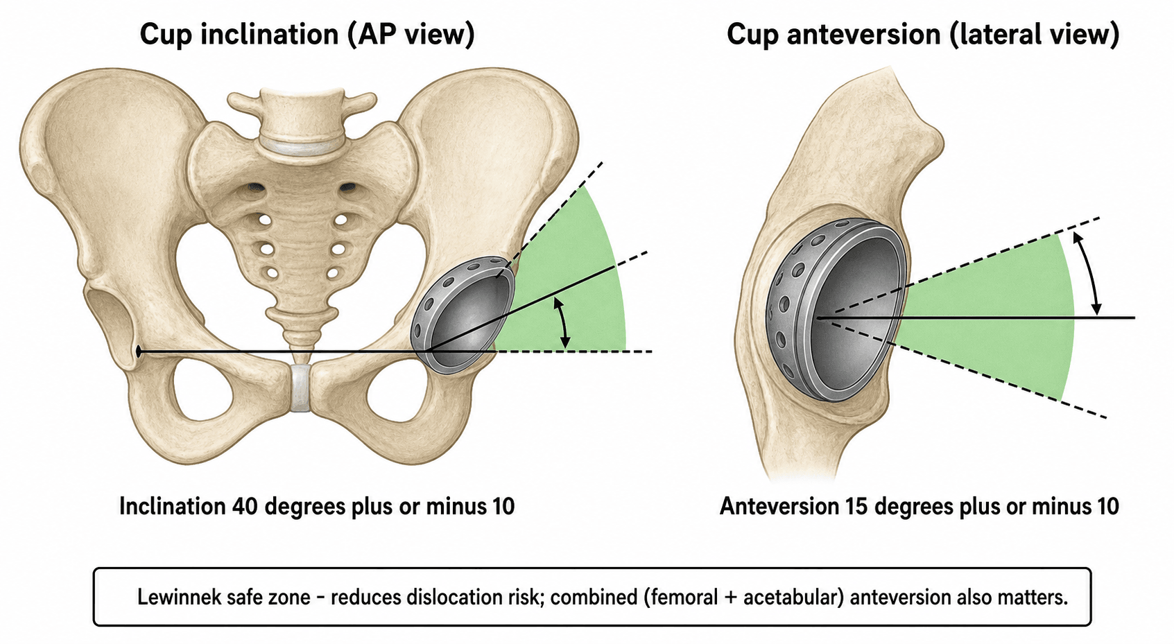
Beyond the zone. Inclination over 55° is high risk, inclination under 25° impinges, and a retroverted cup carries a very high posterior risk.
Combined anteversion. Cup anteversion plus femoral anteversion, widely regarded as the most critical parameter. The target is quoted as 25-45°, or more widely 25-50°. Below 25° the hip is at risk of posterior instability and above 45° of anterior instability, and below 20° or above 50° the risk is high. McKibbin's traditional combined index is acetabular plus femoral anteversion of 37° ± 10°.
EXAMINER FAVOURITE: "A patient dislocates despite cup position within Lewinnek safe zones - why?" Answer: safe zones are statistical concepts, not absolute. Other factors matter: combined anteversion, spinopelvic mobility / functional safe zone, impingement, soft tissue tension, patient factors (compliance, neuromuscular disease), and component design (head size, offset). A substantial proportion of dislocations occur in the "safe zone" - in the navigated series cited on this page, 14.2% of hips inside the Lewinnek zone lay outside the functional safe zone, and the best predictor of that was femoral mobility, not cup position.
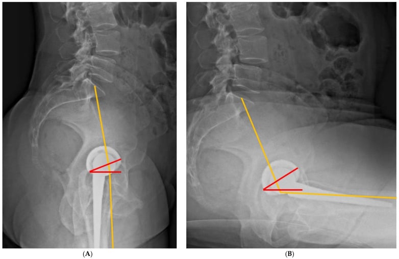
Measuring spinopelvic mobility. "Spinopelvic mobility" is a phrase until you attach a film to it. Get standing and seated lateral radiographs of the lumbar spine, pelvis and hip and read the sacral slope on each. Normally the pelvis rotates posteriorly on sitting and the sacral slope falls by roughly 20°, which opens the functional anteversion of the cup and lets the femur flex without impinging.
The stiff spine. A change of less than about 10° defines the stiff spine, and that is the dangerous pattern. Its commonest cause is previous lumbar fusion, with risk rising as more levels are fused and highest with fusion to the sacrum.
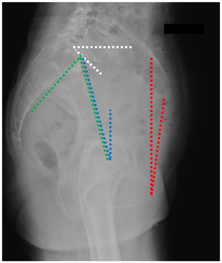
Which way the stiff spine dislocates. A stiff kyphotic spine leaves the pelvis retroverted on standing and unable to retrovert further on sitting, so the hip must supply the whole flexion arc and levers out posteriorly. A stiff spine fused in lordosis keeps the pelvis anteverted, which closes functional anteversion and predisposes to anterior instability in extension and external rotation. The hypermobile spine is the mirror problem and far less dangerous.
Why ask about the back in a hip clinic. A fused lumbar spine is one of the strongest identifiable risk factors for dislocation, and it is identifiable in clinic before the operation. That is the reason to plan a dual mobility bearing or a patient-specific cup target rather than a standard one.
Classification
By Direction and Timing
Direction is the first descriptor: posterior, anterior, or the rare superior or inferior dislocation, with the mechanisms described under Pathophysiology. Timing is the second, and it points toward the cause:
- Early (under 3 months) - usually component malposition, soft-tissue laxity, technique, compliance or incomplete healing. Usually a single episode, and often responds to conservative care
- Intermediate (3-12 months) - borderline component position, emerging abductor dysfunction, variable compliance
- Late (over 1 year) - consider polyethylene wear, component loosening or migration, soft-tissue attenuation and new neurological problems
- Recurrent (2 or more dislocations) - component malposition, wear or abductor dysfunction. Late and recurrent dislocations often require surgical revision
Clinical Presentation
History. Sudden severe hip pain after a precipitating movement or fall, a sensation that "something went out", and inability to move the leg or bear weight, with previous episodes if it is recurrent. The precipitating movement points to the direction: sitting to standing, a low chair, tying shoes or getting out of a car for posterior; stepping backward, arising from bed or external rotation for anterior; a fall or impact for traumatic.
Examination. The posture of the limb gives the direction away:
- Posterior (most common) - hip flexed, adducted and internally rotated; leg shortened 1-3cm; greater trochanter prominent; severe pain with movement; unable to straight-leg raise
- Anterior - hip extended, abducted and externally rotated; leg may appear lengthened or neutral; femoral head occasionally palpable in the groin
CRITICAL SAFETY: Always perform and document a complete neurovascular examination before attempted reduction. Sciatic nerve injury occurs in 10-20% of posterior THA dislocations. Document specific testing of ankle dorsiflexion (common peroneal) and plantarflexion (tibial), plus sensation in the dorsum and plantar foot. Assess the femoral nerve in anterior dislocations and document pulses. Repeat the examination after reduction.
Investigations
Plain Radiography
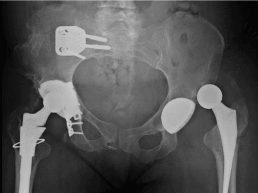
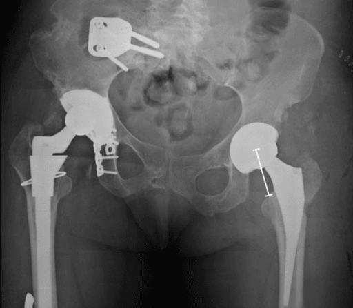
AP pelvis - essential. Both hips for comparison. Confirm the direction of dislocation and measure cup inclination, the angle between the plane of the cup opening and the inter-teardrop line. Compare with the immediate post-operative films, and look for migration, loosening, polyethylene wear, periprosthetic or acetabular wall fracture and heterotopic ossification.
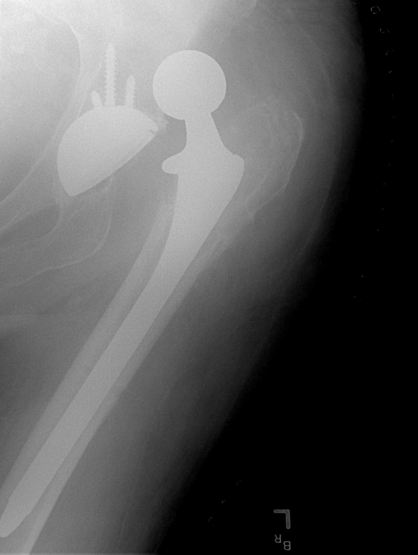
Lateral hip - cross-table or frog lateral. Estimates cup anteversion by the ellipse method, with significant error; CT is the gold standard and more accurate. The lateral also shows anterior and posterior wall integrity and the profile of the femoral component.
Post-reduction films - mandatory. Confirm a concentric reduction, with no subluxation and an intact Shenton's line, look for new iatrogenic fractures, and document the final component relationship as a baseline.
Computed Tomography
CT is often essential for recurrent dislocation or revision planning, and for recurrent instability it gives the precise 3D measurement of acetabular and femoral version needed to calculate combined anteversion. Protocol: thin (1-2mm) cuts of the pelvis and proximal femur to below the lesser trochanter, with 3D reconstruction.
- Acetabulum - inclination on coronal reconstructions; anteversion on axial cuts at the superior dome, the angle between the acetabular opening plane and the AP pelvic axis; bone stock
- Femur - neck or stem version, the angle between the neck axis and the posterior femoral condyles
- Combined anteversion - acetabular plus femoral anteversion, judged against the target above
- Impingement - anterior (psoas tendon, anterior rim) and posterior (ischium, posterior wall, component-on-component), with software-based detection where available
- 3D reconstruction - complex anatomy (DDH, revision), surgical planning, bone defects and consent
Other Tests
- Bloods and aspiration - not routine. If infection is suspected before revision, ESR, CRP and aspiration (cell count, culture, alpha-defensin)
- MARS MRI - rarely needed; assesses abductor integrity or soft-tissue pathology
- Neuromuscular - formal neurology consultation for a persistent sciatic deficit, and physiotherapy assessment of abductor function
Management
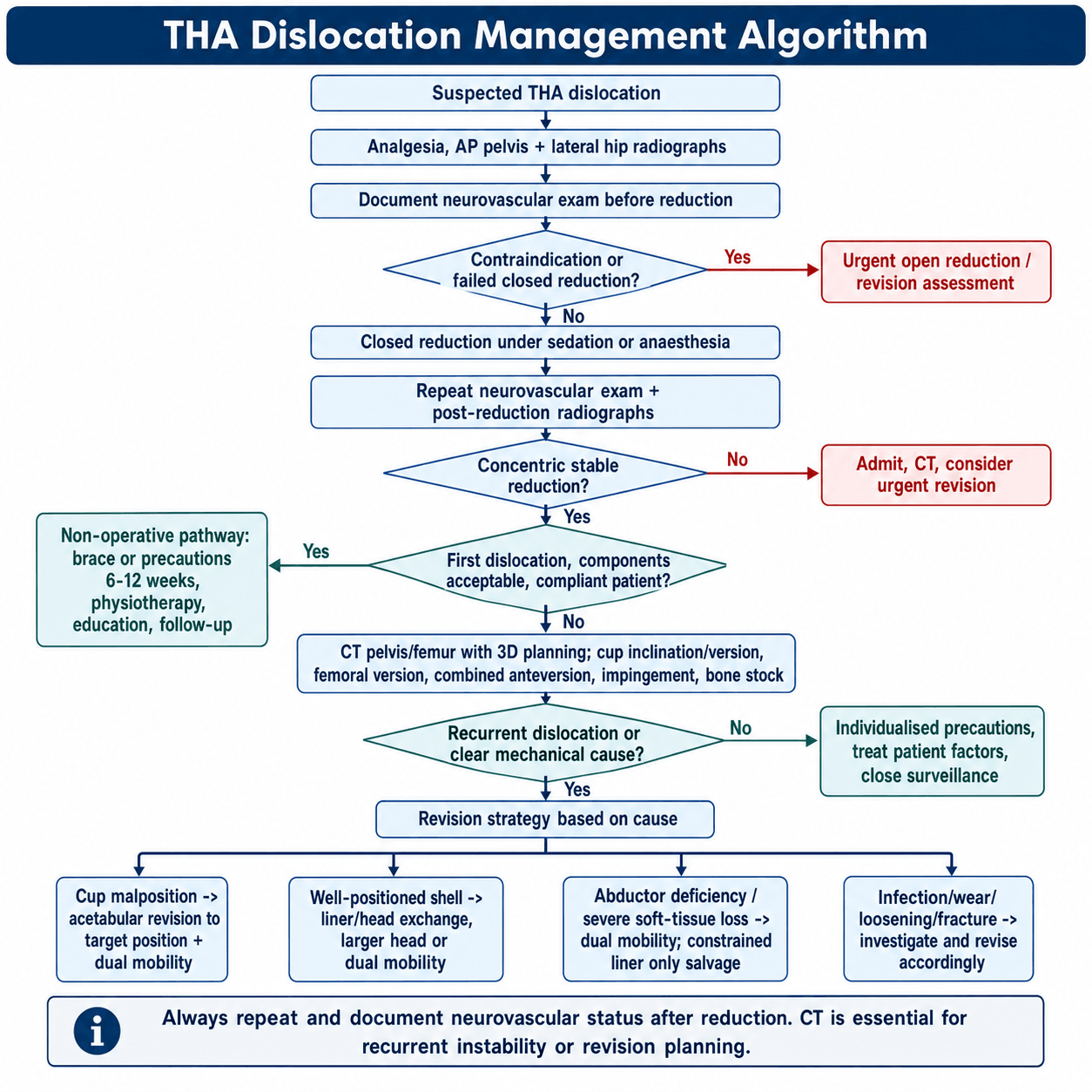
Immediate Management of Acute Dislocation
In the emergency department. Analgesia, with IV opioids and procedural sedation for the reduction. Neurovascular examination and documentation, AP pelvis and lateral hip radiographs, and nil by mouth in preparation for sedation. Contact the orthopaedic surgeon, and review the operative notes and post-operative films.
Closed reduction. Indicated for all acute THA dislocations unless contraindicated, and performed as soon as feasible, ideally within 6 hours. Contraindications are an associated acetabular or femoral fracture requiring fixation, gross component loosening and suspected infection.
- Allis method (posterior) - adequate sedation or GA, patient supine. An assistant stabilises the pelvis with downward pressure on the ASIS; the surgeon flexes hip and knee to 90° and applies inline traction with gentle internal rotation and rocking. A palpable clunk indicates reduction; test stability and take post-reduction films
- Stimson technique - patient prone, hip off the edge of the bed flexed to 90°, gravity-assisted with gentle downward pressure on the calf
- Anterior reduction - inline traction with the hip extended and gentle internal rotation; direct pressure on the head may assist. More difficult than posterior
After reduction. Repeat and document the neurovascular examination, take AP pelvis and lateral hip films to confirm a concentric reduction, test the safe arc of motion and document the direction of instability. Fit a hip abduction brace, teach strict precautions and review at 1-2 weeks. A recurrent dislocation needs CT to identify the cause, and revision is planned if malposition is found: the cause must be identified, not just the symptom treated.
POST-REDUCTION MONITORING: After closed reduction, the patient MUST have a repeat neurovascular exam documented. New or worsening sciatic nerve deficit may indicate nerve entrapment and requires urgent repeat imaging (MRI) and possible exploration or open reduction. Do not discharge until stability is confirmed and the patient can safely mobilise with precautions.
Admit or discharge. A first dislocation with a stable reduction and good support may be discharged. Admit for recurrent dislocation, persistent instability, a new neurovascular deficit or failed closed reduction.
Direction-specific precautions.
- Posterior - avoid flexion beyond 90°, adduction past the midline and internal rotation; no low chairs or couches; elevated toilet seat; sleep supine or on the contralateral side. Minimum 6 weeks, often 12
- Anterior - avoid extension beyond neutral and external rotation; no reaching backward while standing. 6-12 weeks
Surgical Technique
Principles of Revision for Instability
Planning. CT with 3D reconstruction gives precise cup inclination and version and femoral version, the combined anteversion, the malpositioned component or components and the bone stock, from which the target positions are planned. Have a full revision system available: dual mobility components, constrained liners as backup, larger heads, and augments for bone defects. The approach is usually that of the index operation, most commonly posterior, extended if needed.
Indications. Recurrent instability with a well-positioned, stable acetabular shell (inclination 30-50°, anteversion 5-25°), no wear or osteolysis, and a combined anteversion correctable by head size or a femoral change. Before operating, confirm shell stability and exact position on CT, verify the liner type and modularity, and document the shell manufacturer and size.
Technique. Use the previous approach, take down the capsular repair, expose and confirm shell stability, then remove the liner with dedicated tools, protecting the threads and taper, and inspect the shell. Select the largest head possible (36-40mm) and consider an elevated or lipped liner, oriented posteriorly for posterior instability, or a dual mobility liner if the shell is compatible and the patient is high-risk.
Seating and testing. Clean the taper and locking mechanism, ensure correct orientation, impact fully and verify the locked position. Assess the femoral component and taper and exchange the head for a larger size if appropriate. Perform a meticulous posterior soft-tissue repair, then test stability through the full range; if it is unstable, convert to dual mobility or a constrained liner.
Pitfalls. Intraoperative shell loosening, liner-shell incompatibility and an inadequate available head size.
Complications
Recurrent Dislocation
The numbers. Recurrence follows 25-50% of first dislocations, 60-75% after a second and over 80% after a third. Two or more episodes define recurrent dislocation.
Why hips keep dislocating. Non-compliance, neuromuscular or cognitive decline, unrecognised component malposition, failure to address the underlying cause, inadequate soft-tissue repair, an inappropriate head size and uncorrected impingement.
Management. Non-operative treatment rarely succeeds. Work up systematically with CT, and revise, typically after the second dislocation: two or more dislocations usually mandate revision. Correct the underlying cause and add an enhanced stability construct, dual mobility preferred.
Sciatic Nerve Injury
What it looks like. Usually a neuropraxia, with a permanent deficit in 1-2%. The common peroneal division is most affected: foot drop, numbness over the dorsal foot and first web space, and a high-steppage gait. The tibial division causes plantarflexion weakness and plantar numbness.
Management. Gentle reduction, an ankle-foot orthosis for foot drop, and physiotherapy, with EMG and nerve conduction studies at 3-4 weeks if the deficit persists. Neuropraxia recovers in 80-90% over 6-12 months; consider tendon transfer, posterior tibial for foot drop, if there is no recovery by 12-18 months.
Periprosthetic Fracture
Posterior wall. The most common fracture with posterior dislocation, and it may be occult on plain films, so it often needs CT. Small stable fragments are treated non-operatively; large fragments, over 25-30% of the wall, require ORIF and may need a larger head or dual mobility for stability.
Femur and iatrogenic fractures. Femoral periprosthetic fractures (Vancouver classification) are rare and may occur during reduction, so use a gentle technique. Iatrogenic fractures of the greater trochanter, acetabulum or femur during revision require intraoperative recognition and fixation.
Component Wear and Loosening
Recurrent dislocation causes repetitive trauma to the bearing, accelerating polyethylene wear and potential osteolysis, and constrained liners increase cup-bone interface forces and can accelerate loosening. Follow with serial radiographs for progressive radiolucent lines, migration, osteolysis or a change in position. Aseptic loosening requires revision, and malposition and wear are addressed at the same operation with larger heads, dual mobility or highly cross-linked polyethylene.
Intraprosthetic Dislocation in Dual-Mobility Constructs
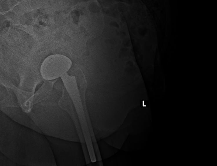
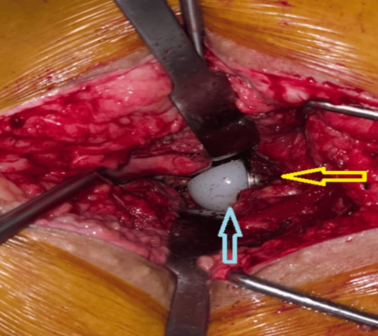
What it is. Dissociation of the small femoral head from the mobile polyethylene liner. It is rare, about 0.5-1%, but devastating, and it presents like a standard dislocation. Mechanically it is different: closed reduction can damage the liner or leave the dissociated components unreduced.
How to recognise and treat it. Suspect it when a dual-mobility hip has a new dislocation with an eccentric or abnormal head-liner relationship on AP and lateral films, and obtain CT when the relationship is unclear. Plan open reduction with liner and head exchange rather than repeated forceful closed reduction.
Psychological Impact
Fear of recurrence, anxiety about movement, depression, reduced quality of life and self-imposed restriction of activity are common. Manage with education, realistic expectations, psychological support where needed and a supervised, graded return to activity, with the emphasis on what the patient can do safely.
Postoperative Care
The first 6 weeks. Hip precautions individualised to risk: after primary posterior-approach THA, a systematic review of 7 studies and 6,900 patients found no difference in dislocation with or without routine precautions (2.2% against 2.0%; Crompton 2020, Acta Orthop), so formal restrictions, an abduction pillow or a brace are reserved for higher-risk patients. Neurovascular checks (especially after closed reduction), multimodal analgesia, VTE prophylaxis per protocol and early mobilisation with a walker. Discharge once mobilisation is safe, with assistive devices: elevated toilet seat, reacher, sock aid, long shoe horn.
Review. The first review at 1-2 weeks checks the wound and takes radiographs to exclude early dislocation; any prescribed precautions continue and the assistive device is weaned as strength improves. At week 6, clinical and radiographic review with a Trendelenburg assessment, then begin weaning the brace and gradually liberalise activity.
6-12 weeks and beyond. Progressive strengthening, low-impact activity (walking, swimming, cycling) and driving when safe. At week 12, a final short-term review: any formal precautions stop if the hip is stable, with lifelong awareness of extreme positions. At 3-6 months, unrestricted activity if stable, with maintenance abductor strengthening. From 1 year, annual clinical and radiographic surveillance for late dislocation, wear and loosening; lifelong follow-up is recommended for all THA.
After revision for instability. Revision patients have a higher recurrence risk and need enhanced follow-up:
- Hip abduction brace for 12 weeks, strictly
- Protected weight bearing for 6 weeks if there was bone work, for ingrowth; after a cementless cup revision, 6 weeks' protected weight bearing for ingrowth
- Hip precautions set individually by the cause of instability, the reconstruction and the patient's ability to follow them; the evidence against routine precautions (Crompton 2020) comes from primary posterior-approach THA, not revision for instability
- Serial radiographs at 6 weeks, 3 months, 6 months and 1 year, then annual follow-up
- Enhanced rehabilitation and close monitoring for recurrence and other complications
Red flags for urgent review.
- A sensation of instability or near-dislocation
- Acute dislocation
- New severe pain
- A change in leg length
- Inability to bear weight
- New neurological symptoms
Outcomes
Outcomes by Treatment Strategy
- Closed reduction and non-operative care (first dislocation) - 50-75% do not recur, and re-dislocation is quoted at 30-40%. Better with well-positioned components, a traumatic mechanism and good compliance; worse with malposition, an atraumatic mechanism, neuromuscular disease or cognitive impairment. Satisfaction varies and depends on recurrence
- Isolated liner exchange - with a well-positioned shell, success is quoted at both 60-80% and 70-85%, lower if the shell is borderline, with recurrent instability in 15-30%. Better with larger heads and dual mobility liners, which outperform standard liners; it fails if the underlying malposition is not addressed
- Acetabular revision - 80-90% success with dual mobility and 70-85% with a standard large head; re-dislocation 5-15% with dual mobility and 10-20% with standard bearings. Achieving the combined anteversion target is critical, navigation may improve positioning, and bone defects increase complexity and the risk of failure. Satisfaction is good if the cause is addressed
- Dual mobility - dislocation 0.5-2% in primary use and 2-5% in revision, markedly lower than standard bearings in high-risk groups; excellent stability, and the best outcomes for recurrent instability in current evidence
- Constrained liner - prevents dislocation mechanically in 80-90%, with re-dislocation quoted at 10-15% at 5 years, but reoperation for locking ring failure or loosening is quoted at 10-25% or 15-25%. Good stability with some functional limitation; less favourable than dual mobility in most series, so it remains a salvage option
Recurrent dislocations (3 or more), abductor deficiency, cognitive impairment, non-compliance with precautions, and neuromuscular disease all predict a poorer outcome and a higher chance of failure after intervention.
Prevention Strategies
Surgical Technique
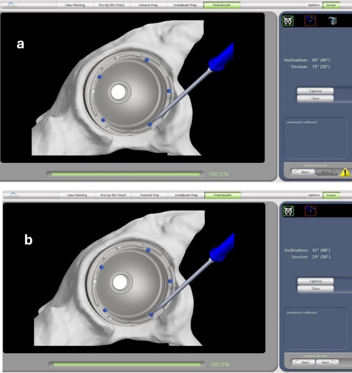
Component position - the most important. Target Lewinnek's 40° of inclination and 15° of anteversion, but prioritise combined anteversion, individualised to the femoral version. For the best stability, aim narrower than Lewinnek: inclination 35-45°, cup anteversion 15-20°, the middle of his range, and a combined anteversion of 35-40°, the middle of the target range, for a margin of safety. Use navigation where available. On the femoral side, restore native version, typically 10-15° of anteversion, combined with the cup for the total.
Head size. A minimum of 32mm in modern practice and never 28mm: 36mm or larger for most patients, and 36-40mm or dual mobility for the high-risk, 40mm if the acetabulum allows.
Soft tissues by approach.
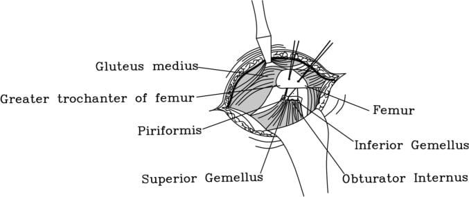
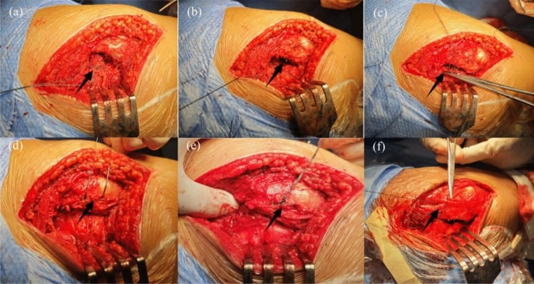
- Posterior - meticulous repair of the posterior capsule and short external rotators to the greater trochanter is mandatory and substantially reduces dislocation. Capsular repair with heavy suture (No. 2 or 5), the rotators to bone through transosseous tunnels or anchors; consider capsular plication if lax, and a trochanteric slide in complex revisions
- Anterolateral - anatomical gluteus medius repair, avoid superior gluteal nerve injury, restore abductor tension
- Direct anterior - preserve the anterior capsule, avoid excessive external rotation, avoid anterior notching
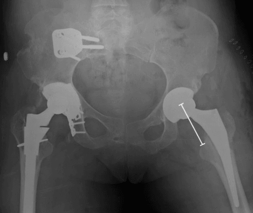
Offset and leg length. Restore native offset to within ±5mm; lateral offset is critical, tensioning the abductors for dynamic stability. Avoid over-lengthening, which brings dissatisfaction and nerve injury, and shortening, which brings instability.
Test before closing. Intraoperative stability testing is essential. With trial components, confirm stability in 90° of flexion with internal rotation and adduction (posterior) and in extension with external rotation (anterior), and assess the impingement-free arc. Never accept instability at closure: modify the position or increase the head size there and then.
Pre-Operative Risk Stratification
Very high risk - consider enhanced-stability implants. These patients may need dual mobility, or a constrained liner, prophylactically:
- Prior THA dislocation in the same hip
- Neuromuscular disease (Parkinson's, stroke)
- Severe cognitive impairment or dementia
- Abductor deficiency or damage
- Revision for instability
- Tumour resection with soft-tissue loss
- Multiple prior hip surgeries
- Age over 80 with frailty
The plan is dual mobility (primary or revision), consideration of the direct anterior approach if the surgeon is experienced, a larger head if a standard bearing is used, and extended rehabilitation.
Moderately high risk - modify the technique.
- Age 70-80 (over 75 in the relative high-risk group whose technique is modified)
- Revision THA not for instability
- Previous hip surgery (fracture, prior arthroplasty)
- DDH with anatomical abnormalities
- Inflammatory arthropathy
- Morbid obesity
- Milder neuromuscular disease
- Posterior approach in a high-risk patient
The plan is a head of 36mm or more, meticulous soft-tissue repair and component position optimisation, extended precautions (12 weeks), an abduction brace and enhanced education.
Patient Education
Teach the precautions before the operation, by video or demonstration and in writing, set realistic expectations and plan for hazards at home. After it, give direction-specific precautions, mobilise early with an assistive device, strengthen the abductors, which is critical for dynamic stability, and assess home safety. High-risk patients, including those with neuromuscular disease or cognitive impairment, wear a hip abduction brace for 6-12 weeks. Awareness of high-risk positions is lifelong, as is the need for urgent review if instability is suspected.
Guidelines, Registries & Global Practice
Global Epidemiology and Registry Evidence
Instability/dislocation is consistently among the leading early indications for revision THA across major national registries (AOANJRR, NJR for England/Wales, AJRR, Swedish/Nordic registries). Registry data converge on several practice-changing signals:
- Approach: posterior approach carries a higher early dislocation/revision rate than direct anterior in most registries; the gap narrows substantially with enhanced posterior soft tissue repair.
- Head size: heads ≥36mm have lower dislocation than 28-32mm; 28mm is now rarely used in modern primary THA.
- Dual mobility: lowers revision for dislocation specifically, but international registry meta-analysis (Farey 2022, including AOANJRR, NJR, and four others) found no reduction in all-cause revision for hip-fracture THA and a possible infection trade-off — supporting selective rather than universal use.
- Re-revision burden: revision for instability carries a high re-revision rate, underlining that prevention at the index operation is paramount.
Side-by-Side Society Guidance
- Emphasis
- Patient-specific risk stratification; component positioning; larger heads and dual mobility for high-risk patients
- Emphasis
- Surgeon experience and approach selection; meticulous technique; registry surveillance via NJR
- Emphasis
- Restoration of offset, leg length, and soft tissue tension; intraoperative stability testing
- Emphasis
- Spinopelvic assessment (functional safe zone) in stiff-spine patients; dual mobility in defined high-risk groups
High- vs Limited-Resource Variation
- High-resource settings: CT and navigation/robotics for version planning; routine availability of dual mobility, constrained liners, augments, and modular revision systems; spinopelvic imaging for at-risk patients.
- Limited-resource settings: reliance on plain radiographs and mechanical alignment guides; emphasis on fundamentals — optimal positioning, ≥36mm heads where available, meticulous posterior soft tissue repair, and robust patient education/precautions, which deliver most of the achievable risk reduction at low cost.
Peri-operative Pharmacology (Global Principles)
- VTE prophylaxis: LMWH or oral anticoagulant (e.g. rivaroxaban, apixaban) with mechanical prophylaxis; duration commonly 10-14 days, extended up to ~35 days in higher-risk patients (follow local/society guidance).
- Antibiotic prophylaxis: a first-generation cephalosporin (e.g. cefazolin, weight-adjusted) within 60 minutes of incision, re-dosed for long cases or major blood loss; vancomycin/teicoplanin if MRSA risk or beta-lactam allergy; no proven benefit beyond 24 hours.
MCQ Practice Points
Q: What is the most common direction of THA dislocation and why? A: Posterior dislocation (75-80% of cases). This is associated with the posterior approach, which disrupts the short external rotators and posterior capsule. Risk activities include flexion greater than 90°, adduction, and internal rotation.
Q: What are the Lewinnek "safe zones" for acetabular cup positioning? A: Inclination 40° (±10°) and anteversion 15° (±10°). Cups outside these ranges have significantly higher dislocation rates. Combined anteversion (cup + stem) of 25-50° is the modern target to account for stem version.
Q: What is the mechanism of dual mobility cups in reducing dislocation? A: Dual articulation provides a larger effective head size (increased jump distance). The small inner bearing articulates within a larger polyethylene liner, which itself articulates with the metal shell, providing greater range of motion before impingement.
Q: When is a constrained liner indicated in revision for instability? A: Recurrent dislocation with adequate component positioning and soft tissue deficiency (abductor insufficiency, neurological deficit), as a salvage option. Constrained liners mechanically lock the head into the socket but increase stress at the bone-implant interface, risking loosening.
Exam Day Cheat Sheet
Incidence, Direction and Risk Factors
- Primary 1-5 per cent, revision 10-25 per cent (conventional ranges); posterior 75-80 per cent, anterior 15-20; about half within 3 months
- Patient: older age, cognitive impairment, neuromuscular disease
- Procedure: posterior approach, revision surgery
- Position: component malposition, impingement
- Power: soft-tissue laxity, abductor dysfunction
Cup Position and Combined Anteversion
- Lewinnek: inclination 40 plus or minus 10 degrees, anteversion 15 plus or minus 10
- Lewinnek showed an association with ANTERIOR dislocation only, and none with POSTERIOR - which is why safe-zone hips still dislocate
- Combined anteversion (cup plus femoral) target 25-45 degrees, ideally 35-40; one component can compensate for the other (cup 25 + femoral 15 = 40)
- Functional safe zone is spinopelvic and femoral mobility, not cup angle alone (Tezuka)
Acute Management and the First Dislocation
- Neurovascular examination BEFORE and after reduction, with post-reduction radiographs
- Closed reduction (Allis: flexion to 90 degrees, traction and internal rotation)
- Well-positioned components: non-operative, brace and precautions
- Malpositioned components: CT, then early revision
- Recurrence after a first dislocation is conventionally quoted at 25-50 per cent
Recurrent Dislocation and Its Consequences
- Two or more events is a SURGICAL problem; non-operative management fails. CT is mandatory to measure combined anteversion
- Liner exchange plus larger head if the cup is well positioned; cup revision with dual mobility is the usual answer; constrained liner is salvage only
- A cup at 55 degrees inclination is the primary problem - revise the cup
- Repeated events cause accelerated wear and loosening; sciatic neuropraxia and posterior wall fracture also occur
Head Size and Dual Mobility
- 36 mm versus 28 mm: 1.3 versus 5.4 per cent dislocation at one year (Howie RCT); larger heads increase jump distance and impingement-free arc
- Dual mobility: 28-32 mm head within a mobile polyethylene bearing, effective diameter around 50 mm; intraprosthetic dislocation uncommon in modern designs
- Registry caution: lower revision for dislocation but NO all-cause revision benefit in fracture THA, with a possible infection trade-off (Farey 2022)
Prevention
- Soft-tissue repair: posterior capsule and short external rotators to the greater trochanter - mandatory posteriorly (Pellicci: 4 per cent to 0)
- Thirty-six mm head minimum
- Anteversion combined 25-45 degrees
- Brace for selected high-risk patients
- Leg length and offset restoration
- Education on precautions
- High-risk patients get dual mobility
- Inclination 35-45 degrees
- Patient selection and approach choice
Registry Data and Exam Traps
- Instability is a leading cause of early revision (AOANJRR, NJR, AJRR), and re-revision after revision for instability is high
- TRAP: safe zones are not absolute - Lewinnek never showed a posterior association, and the functional safe zone is spinopelvic
- TRAP: combined anteversion matters more than either component alone
- TRAP: dual mobility beats a constrained liner where both are options, and recurrent instability always needs CT
ULTIMATE EXAM PEARL: If an examiner shows recurrent dislocation, your immediate thought process should be: (1) "This is a SURGICAL problem - non-operative management has failed"; (2) "I need a CT scan to measure combined anteversion and identify the malpositioned component"; (3) "Treatment is revision surgery to correct malposition PLUS dual mobility for enhanced stability." Demonstrating this systematic approach shows senior-level decision making. Never say "try another closed reduction and longer bracing" for recurrent dislocation - this shows poor understanding and will fail the viva.
Clinical Decision Scenarios
Practise clinical reasoning and management decisions out loud
“”
“”
“”
Evidence Base
Lewinnek Safe Zone
- Series of 300 THAs; 9 (3%) dislocated
- Cup anteversion 15° ± 10° and lateral opening (inclination) 40° ± 10° defined the 'safe range'
- Dislocation rate 1.5% within the safe range vs 6.1% outside it
- Greatest risk in the first 30 days; higher risk in hips with prior surgery
- THE CAVEAT THAT IS ALMOST ALWAYS DROPPED, AND IT UNDERMINES HOW THE ZONE IS TAUGHT: ANTERIOR dislocations were associated with increased cup anteversion, but there was NO SIGNIFICANT CORRELATION between cup-orientation angle and POSTERIOR dislocation. The safe zone was derived from a dataset in which cup position did not predict the commoner direction of dislocation at all
Large Femoral Heads and Dislocation - RCT
- Multicentre RCT of 644 patients randomised to 36mm vs 28mm metal-on-highly-cross-linked-polyethylene
- Overall 1-year dislocation: 1.3% (36mm) vs 5.4% (28mm), p=0.012
- Primary THA: 0.8% (36mm) vs 4.4% (28mm), p=0.024
- Revision subgroup 4.9% (2 of 41) vs 12.2% (5 of 41), NOT significant: difference 7.3% with a confidence interval running from MINUS 5.9% to plus 21.1%, p=0.273 - the interval includes the possibility that the larger head is worse in revision
- The absolute event numbers are small throughout: 4 dislocations of 299 against 17 of 316 overall, and 2 of 258 against 12 of 275 in primaries
Enhanced Posterior Soft Tissue Repair
- Two surgeons adopted an identical enhanced posterior soft tissue repair via the posterior approach
- Surgeon 1: dislocation fell from 4% (395 hips) to 0% (395 hips) after enhanced closure
- Surgeon 2: dislocation fell from 6.2% to 0.8% after enhanced closure
- Differences highly statistically significant
Functional Safe Zone - Why Lewinnek Is Not Always Predictive
- 320 navigated primary THAs; 92.5% were within the Lewinnek safe zone
- Of hips within Lewinnek, 14.2% fell outside the sagittal functional safe zone (combined sagittal index)
- These patients were considered at risk for dislocation despite 'normal' cup angles
- Best predictor of falling outside the functional safe zone was femoral/spinopelvic mobility, not cup position
Dual Mobility in Revision THA - Meta-analysis
- Systematic review and meta-analysis of comparative studies in revision THA
- Dual mobility had a significantly lower dislocation rate than fixed-bearing cups
- Dual mobility also showed better overall implant survivorship and lower aseptic loosening risk
- No significant difference in infection risk between groups
Dual Mobility for Femoral Neck Fracture - Meta-analysis
- 23 studies, 7,189 patients (mean age 77.8 years) treated with dual mobility THA for femoral neck fracture
- Dislocation significantly lower vs conventional THA (OR 0.26) and hemiarthroplasty (OR 0.27)
- Intraprosthetic dislocation rate only 0.04% (3 of 7,189)
- No increase in other complications
International Registry Meta-analysis - Dual Mobility for Hip Fracture
- Six arthroplasty registries (Australia, Denmark, Sweden, Netherlands, UK, USA): 15,024 dual mobility vs 97,200 conventional THAs for hip fracture
- Cumulative percent revision at 5 years was 4.3% (95% CI 4.2-4.5) for conventional THA against 4.7% (4.3-5.3) for dual mobility - the dual mobility figure is numerically HIGHER, though the intervals overlap
- No reduction in all-cause revision for dual mobility once between-registry differences were adjusted for (HR 0.96, 95% CI 0.86-1.06) at 5 years
- Lower proportion revised for dislocation (0.9% vs 1.4%) but higher proportion revised for infection (1.2% vs 0.8%)
- The confounding is measured, not merely suspected: in most registries dual mobility patients were significantly OLDER, had MORE comorbidities and more often had a posterior approach (p less than 0.001) - all of which independently raise revision risk and bias against dual mobility
- Dual mobility use rose to 21% of fracture THAs by 2019 (2,438 of 11,874), with wide between-country variation, and the authors call for an RCT powered on dislocation and subsequent revision