Tile / Young-Burgess Classification of Pelvic Ring Injuries
Tile tells you whether the ring is mechanically stable and therefore what fixation you need. Young-Burgess tells you how the ring failed and therefore what else to expect (blood loss, associated injuries, posterior lesion). Examiners expect you to know both, use them in parallel, and to be able to translate one to the other: an APC III is a Tile C; an LC I is usually a Tile A or B2-equivalent stability.
The Young-Burgess Classification — Mechanism of Failure
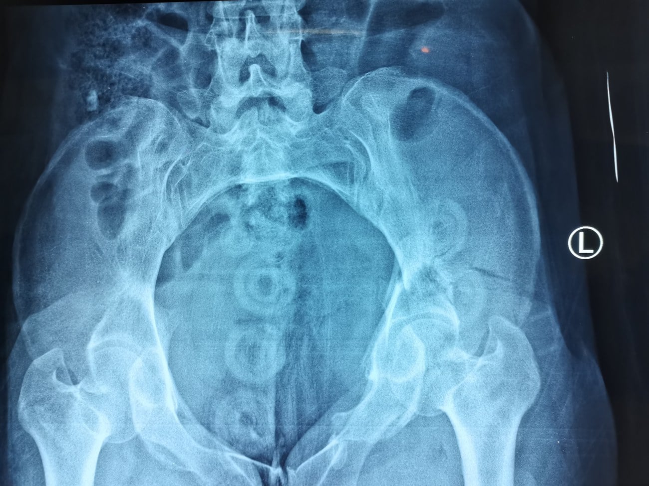
The Young-Burgess system, published in 1986 and refined in 1990, classifies pelvic ring injuries by the vector of the deforming force. It correlates closely with blood loss, associated injuries and mortality, and is the language of trauma resuscitation.
- Mechanism
- Anteroposterior compression, mild
- Radiographic Hallmark
- Symphysis diastasis less than 2.5 cm; intact SI
- Tile Equivalent
- Stable (Tile A equivalent)
- Resuscitation Profile
- Minimal blood loss; outpatient management possible
- Mechanism
- Anteroposterior compression, moderate
- Radiographic Hallmark
- Symphysis diastasis more than 2.5 cm; anterior SI diastasis, posterior SI intact
- Tile Equivalent
- Rotationally unstable (Tile B1)
- Resuscitation Profile
- Moderate bleeding; binder and anterior fixation
- Mechanism
- Anteroposterior compression, severe
- Radiographic Hallmark
- Symphysis diastasis more than 2.5 cm; complete SI disruption
- Tile Equivalent
- Rotationally and vertically unstable (Tile C)
- Resuscitation Profile
- High blood loss, often arterial; binder, ex-fix, angioembolisation
- Mechanism
- Lateral compression, mild
- Radiographic Hallmark
- Ipsilateral sacral compression fracture and horizontal pubic ramus fracture
- Tile Equivalent
- Stable (Tile A equivalent)
- Resuscitation Profile
- Lowest blood-loss profile of the four
- Mechanism
- Lateral compression, moderate
- Radiographic Hallmark
- Ipsilateral crescent (iliac) fracture with pubic ramus fracture
- Tile Equivalent
- Rotationally unstable (Tile B2)
- Resuscitation Profile
- Low to moderate blood loss
- Mechanism
- Lateral compression, severe
- Radiographic Hallmark
- LC-I or LC-II on one side with contralateral APC — the 'windswept' pelvis
- Tile Equivalent
- Rotationally unstable (Tile B3)
- Resuscitation Profile
- High rate of GU and neurovascular injury on the open-book side
- Mechanism
- Vertical shear (axial load, often fall from height)
- Radiographic Hallmark
- Vertical displacement of the hemipelvis through SI joint, sacrum or iliac fracture
- Tile Equivalent
- Rotationally and vertically unstable (Tile C)
- Resuscitation Profile
- Highest transfusion requirement; often arterial bleeding
- Mechanism
- Combined mechanism (often crush)
- Radiographic Hallmark
- Mixed pattern — APC and LC and / or VS components
- Tile Equivalent
- Variable; usually unstable
- Resuscitation Profile
- Variable, often high; treat by the worst component
APC · LC · VS · CMYoung-Burgess categories
Hook:APC and LC are graded I–III as severity rises; VS and CM are by definition unstable (Tile C-type).
The Tile Classification — Stability of the Posterior Ring
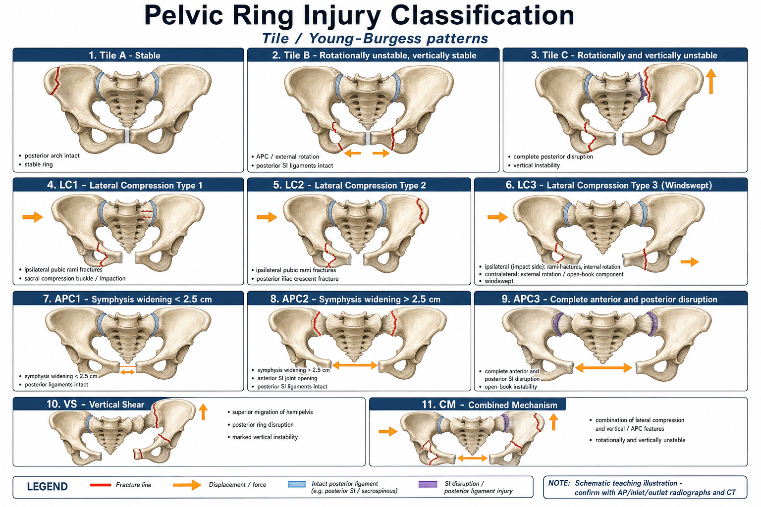
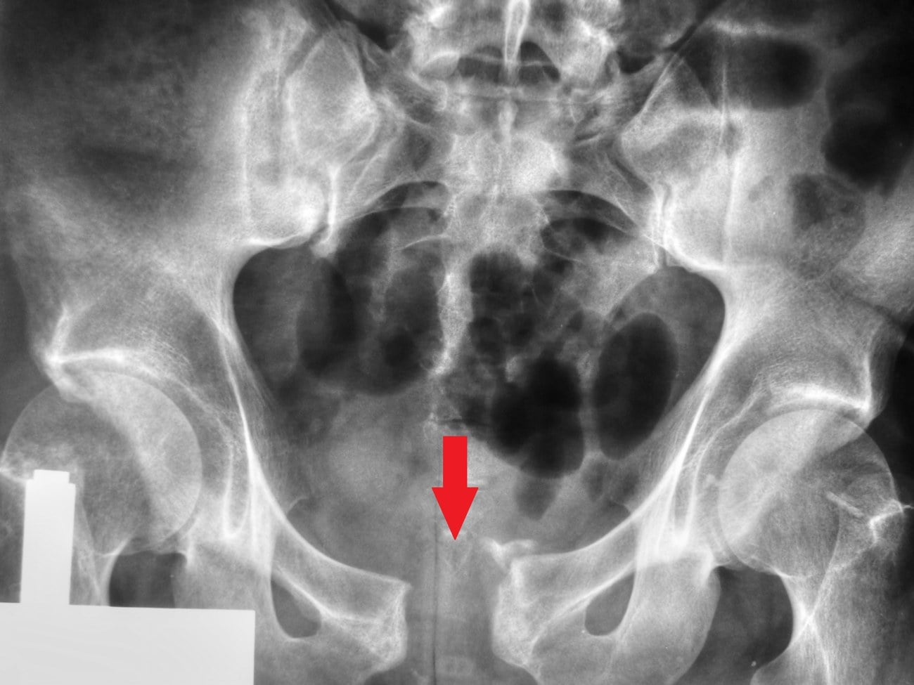
The Tile system, published in 1988, classifies pelvic ring injuries by the stability of the posterior osteoligamentous complex (the posterior arch, sacroiliac ligaments, sacrotuberous and sacrospinous ligaments). The three types predict the need for, and type of, definitive fixation.
- Stability
- Stable in all planes
- Posterior Lesion
- Intact posterior arch
- Anterior Lesion
- Avulsion (A1), iliac wing or anterior arch (A2), transverse sacrum or coccyx (A3)
- Typical Management
- Symptomatic — protected weight-bearing
- Stability
- Rotationally unstable, vertically stable
- Posterior Lesion
- Partial posterior SI disruption — anterior SI, sacrospinous and sacrotuberous ligaments torn; posterior SI intact
- Anterior Lesion
- Open book / external rotation (B1), LC ipsilateral (B2), LC contralateral / bucket-handle (B3)
- Typical Management
- Anterior fixation (external fixator or symphyseal plate) usually sufficient
- Stability
- Rotationally and vertically unstable
- Posterior Lesion
- Complete posterior SI disruption — all SI ligaments torn, often with vertical displacement through sacrum, SI joint or iliac wing
- Anterior Lesion
- Always significant — symphyseal diastasis or vertical ramus fracture (C1 unilateral, C2 bilateral, C3 with acetabular fracture)
- Typical Management
- Posterior fixation (iliosacral screws or trans-sacral plate), usually with anterior fixation as well
Alright · Bend · CollapseTile A / B / C
Hook:The dividing line is the posterior SI complex: intact (A), partially torn (B), completely torn (C).
The vertical stability of a Tile B injury is what makes it a B and not a C. A B2 lateral-compression pelvis will not displace proximally on a push-pull radiograph; a C will. If in doubt, a CT with inlet and outlet views (or examination under anaesthesia) separates B from C and dictates fixation.
Resuscitation and Initial Management
The Young-Burgess type drives the first 30 minutes of resuscitation more than the Tile type. A high-energy APC II, APC III, VS or CM is presumed to be bleeding from the presacral venous plexus, raw cancellous bone, or named arterial branches (superior gluteal, internal iliac, obturator).
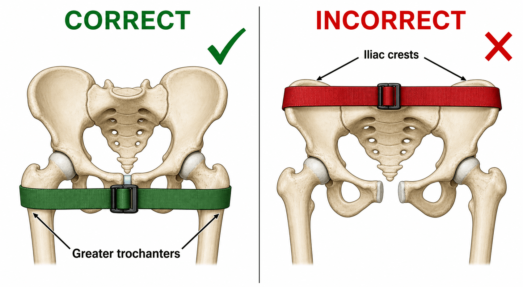
Apply at the level of the greater trochanters (NOT the iliac crests) before log-roll, before transfer and before imaging. The binder wraps the greater trochanters, reduces the open book and tamponades pelvic volume. A folded sheet is a perfectly acceptable alternative.
Run the ATLS primary survey with a FAST scan to look for free intra-abdominal fluid. A negative FAST in a haemodynamically unstable patient with an unstable ring points toward retroperitoneal pelvic bleeding and on to angioembolisation or pelvic packing.
For APC injuries, an anterior external fixator (or pelvic C-clamp) closes the open book and reduces pelvic volume. Place within the first hour if the patient is unstable. Definitive internal fixation comes later.
On-going haemorrhage after binder and ex-fix in an unstable patient is treated by angioembolisation (CT angiography first if time allows) or retroperitoneal pelvic packing in theatre. Young-Burgess APC II, APC III, VS and CM have the highest yield.
Binder · FAST · Ex-fix · EmboliseResuscitation order
Hook:Damage-control orthopaedics — do not definitively fix the pelvis on day one in a cold, coagulopathic patient.
A pelvic ring injury is rarely isolated; the marks come from a structured search for the associated injuries the ring is notorious for. Recite them by system:
- Genitourinary — the highest-yield miss. Suspect a urethral injury (more common in men, at the membranous urethra) with blood at the urethral meatus, a high-riding or boggy prostate on PR, a scrotal/perineal haematoma, or inability to void — perform a retrograde urethrogram BEFORE attempting catheterisation (a blind catheter can convert a partial to a complete urethral tear). Bladder rupture is screened with a CT cystogram and is extraperitoneal (the common type with pelvic fracture, usually managed by catheter drainage) or intraperitoneal (needs surgical repair).
- Open fracture / visceral communication. A per-rectal and per-vaginal examination is mandatory — blood signals an occult open fracture communicating with the rectum or vagina, and any perineal, buttock or groin wound makes it an open pelvic fracture. An open posterior/perineal wound needs early faecal diversion (defunctioning colostomy) and debridement; open pelvic fractures carry a markedly higher mortality.
- Morel-Lavallée lesion — a closed degloving where skin and fat shear off the deep fascia (typically over the greater trochanter or flank), filling with haematolymph. It is highly prone to infection and must be recognised before placing an incision through it.
- Lumbosacral neurology — document L5/S1 motor and sensory function and sphincter tone in every sacral/posterior-ring injury (detailed in the sacral-zone box below).
Definitive Fixation by Pattern
Definitive fixation is dictated by the Tile type and the direction of instability, not by the Young-Burgess label. The general rule is to fix the side of greatest instability first, and to fix posteriorly when there is posterior displacement or vertical instability.
- Posterior Fixation
- None
- Anterior Fixation
- Symptomatic only
- Practical Notes
- Most A1 to A3 injuries are non-operative; mobilise as tolerated
- Posterior Fixation
- Usually not required if anterior reduction is solid
- Anterior Fixation
- Symphyseal plate (open) or external fixator
- Practical Notes
- APC II — anterior fixation alone is often enough
- Posterior Fixation
- Consider iliosacral screw for crescent or sacral component
- Anterior Fixation
- Anterior plating for symphyseal or ramus component
- Practical Notes
- Beware the LC III — fix the open-book side, not just the LC side
- Posterior Fixation
- Iliosacral screw (or trans-sacral plate)
- Anterior Fixation
- Anterior symphyseal / ramus fixation
- Practical Notes
- Reduce posteriorly first; reduce vertically before transversely
- Posterior Fixation
- Bilateral iliosacral or trans-sacral fixation
- Anterior Fixation
- Anterior symphyseal / ramus fixation
- Practical Notes
- Highest biomechanical demand on the posterior construct
- Posterior Fixation
- Acetabular ORIF plus posterior pelvic fixation
- Anterior Fixation
- Anterior as needed
- Practical Notes
- Sequence the acetabulum and the ring; do not lose either reduction
Reduce vertically before transversely. A vertically displaced Tile C is reduced with longitudinal traction on the leg first; a transverse open-book is then closed with the internal rotation of the leg or a sheet / C-clamp. Out-of-sequence reduction makes the SI joint look like a C1 when it is really a C2 — and the fixation construct is wrong.
Do not place an external fixator through the iliac crest for an LC-pattern pelvis — the half-pin pulls the iliac wing laterally and opens the fracture. Use a supra-acetabular pin or a C-clamp on the posterior pelvis instead. Half-pin placement in the wrong pattern is one of the most common causes of failed resuscitation of a lateral-compression injury.
When the posterior lesion runs through the sacrum, examiners want the Denis classification, because the zone predicts the neurology and changes the operation:
- Zone I — alar (lateral to the neural foramina). The lowest neurological-injury rate (around 6% in Denis's original series); when a deficit occurs it is usually the L5 nerve root running over the ala.
- Zone II — transforaminal (through the foramina). Intermediate rate (around 28%), typically L5, S1 or S2 roots; these fractures can be vertically unstable (a shear pattern) and the foraminal comminution makes iliosacral-screw placement less forgiving.
- Zone III — central (medial to the foramina, involving the spinal canal). The highest rate (around 57%), with bowel, bladder and sexual dysfunction and cauda equina features because the sacral canal is involved — assess saddle sensation and sphincter tone specifically.
Spinopelvic dissociation: a transverse sacral fracture connecting bilateral vertical (zone II/III) fractures gives the U- or H-shaped sacral fracture — the spine is mechanically separated from the pelvis. It is classically an axial-load / fall-from-height injury, frequently has cauda equina signs, and standard iliosacral screws alone are insufficient: it needs lumbopelvic (spinopelvic) fixation — triangular osteosynthesis (iliosacral screw plus an L5/ilium construct). This is the posterior-ring nuance that distinguishes a Tile C "complete posterior disruption" worth real marks.
Limitations and How the Two Systems Fit Together
- Tile is a stability language, Young-Burgess is a mechanism language. They correlate (APC III equals Tile C, LC I equals Tile A) but answer different questions: Tile drives fixation, Young-Burgess drives resuscitation and prognosis.
- Both are radiographically based and require a CT for confident typing. A single AP pelvis is not enough — inlet and outlet views (and CT with thin cuts through the sacrum and SI joints) are required to assess posterior displacement and SI widening.
- Neither system is a stand-alone prognostic score. The Burgess group's work (Dalal 1989, Burgess 1990) linked mechanism type to transfusion need and mortality, but Starr et al. (2002) found that physiological parameters — shock on arrival, the Revised Trauma Score and age — predicted mortality, transfusion and the need for arteriography, whereas the fracture pattern (Young-Burgess class) did NOT show consistent links to outcome. The lesson: use mechanism to anticipate the injury pattern, but resuscitate to physiology.
- Paediatric and fragility fractures of the pelvis are usually Tile A or LC I and follow different fixation principles; the classification has not been validated in these groups.
- Open pelvic fractures are an emergency in their own right — classification is for planning, but haemorrhage control, debridement and diversion of the faecal stream (for open perineal wounds) come first.
- Vertical stability testing (push-pull under fluoroscopy or examination under anaesthesia) is the arbiter between a Tile B and a Tile C when imaging is equivocal.
Guidelines, Registries and Global Practice
- AO / OTA Fracture and Dislocation Classification (2018 revision) co-exists with Tile and Young-Burgess. The AO system describes the type (A / B / C) and subtype (1 / 2 / 3) of the posterior arch plus the anterior arch, and is used in registries and in AO-affiliated centres. Translation from Tile to AO / OTA is direct: Tile A becomes 61-A, Tile B becomes 61-B, Tile C becomes 61-C.
- NICE (NG39, UK) and BOA–BOAST 3 guidance emphasises the pelvic binder at the level of the greater trochanters within the first 30 minutes for a suspected unstable ring injury, with early CT and early involvement of a pelvic-reconstruction surgeon.
- AAOS and EAST (US) guidelines recommend mechanism-based triage in the resuscitation bay and early angioembolisation for arterial-phase bleeding, particularly in APC II, APC III, VS and CM.
- German Trauma Society (DGU) Pelvic Trauma Registry and the British Pelvic Trauma Registry data show that posterior fixation of Tile C injuries (iliosacral screws, trans-sacral plating, or triangular osteosynthesis) is associated with lower residual displacement and lower late posterior pain than anterior-only fixation.
- Resource-setting variation: in low-volume or low-resource centres the sheet binder and external fixator remain the workhorses, with definitive posterior fixation concentrated in tertiary pelvic-reconstruction units. Outcome gaps are largest for Tile C injuries transferred late.
- Global practice variation in arterial control: the relative use of angioembolisation versus retroperitoneal pelvic packing varies by region and by the local availability of interventional radiology. Scandinavian and North American centres favour angioembolisation; several European trauma networks use early retroperitoneal packing as a damage-control step before angiography.
Viva practice
Exam Viva
Practise clinical reasoning and management decisions out loud
“A 28-year-old motorcyclist is haemodynamically unstable after a high-speed collision. His pelvis is visibly 'open' on clinical exam and the AP pelvis shows a symphyseal diastasis of about 5 cm with bilateral sacroiliac joint widening. How do you classify this with Tile and Young-Burgess, and what do you do in the first 30 minutes?”
“A 70-year-old pedestrian is hit from the side by a car. AP pelvis shows an impacted right sacral ala fracture and an ipsilateral superior pubic ramus fracture. She is haemodynamically stable. How do you classify and manage this?”
Exam cheat sheet
Tile A / B / C
- A — stable, posterior ring intact
- B — rotationally unstable, vertically stable, posterior SI partially torn
- C — rotationally and vertically unstable, complete posterior disruption
- Posterior lesion (sacrum / SI / iliac) drives the fixation, not the anterior
Young-Burgess APC / LC / VS / CM
- APC — external rotation (open book); I (diastasis less than 2.5 cm), II, III (complete SI)
- LC — internal rotation; LC I sacral impaction, LC II crescent, LC III windswept
- VS — vertical shear, always unstable (Tile C)
- CM — combined mechanism, treat by the worst component
Resuscitation priorities
- Binder at the greater trochanters, before log-roll and before CT
- Massive transfusion, FAST, anterior ex-fix or C-clamp for open-book injuries
- CT angio and arterial embolisation, or retroperitoneal packing if bleeding continues
- Damage-control orthopaedics — definitive fixation only after physiological optimisation
Fixation by Tile type
- A — non-operative
- B1 — anterior fixation alone is usually enough (symphyseal plate or ex-fix)
- B2 / B3 — anterior plus posterior if crescent or sacral component is unstable
- C — posterior fixation (iliosacral screw, trans-sacral plate, or triangular osteosynthesis) plus anterior
Evidence
Pelvic ring fractures: should they be fixed?
- Introduced the A / B / C stability classification based on the integrity of the posterior osteoligamentous complex.
- Defined stable (A), rotationally unstable but vertically stable (B), and completely (rotationally and vertically) unstable (C) injuries.
- Linked the stability category to the biomechanical rationale for anterior versus posterior fixation.
Pelvic ring disruptions: effective classification system and treatment protocols
- 210 consecutive high-energy pelvic ring disruptions classified by mechanism (LC, APC, VS, CM) with mechanism-based treatment protocols.
- Mean blood replacement rose sharply with mechanism: LC 3.6 units, VS 9.2, CM 8.5, and APC 14.8 units.
- Mortality by mechanism was APC 20%, CM 18%, LC 7% and VS 0% in this series (cause of death was the pelvic fracture itself in under half).
According to PubMed, the A/B/C stability system is from Tile 1988 (J Bone Joint Surg Br 1988;70(1):1-12; DOI 10.1302/0301-620X.70B1.3276697). The mechanism-based transfusion/mortality data (blood: LC 3.6, VS 9.2, CM 8.5, APC 14.8 units; mortality APC 20%, CM 18%, LC 7%, VS 0%) are from Burgess et al. 1990 (PMID 2381002) and the organ-injury patterns from Dalal et al. 1989 (PMID 2746708). Starr et al. 2002 (DOI 10.1097/00005131-200209000-00003) found that physiology (shock, Revised Trauma Score, age) — not the Young-Burgess class — predicted mortality, transfusion and arteriography.