Diagnose the gap | correct the cause | use the least necessary constraint
- A balanced TKA is not simply a thick insert. It is stable in extension, flexion and mid-flexion with a centred patella and no coronal thrust.
- Distal femoral resection changes extension gap. Posterior femoral condylar resection and posterior tibial slope mainly change flexion gap.
- Flexion instability often presents with stairs, chair-rise difficulty, recurrent effusions and vague anterior/periretinacular pain.
- Mid-flexion instability is a real but difficult diagnosis. It requires careful examination between full extension and 90° rather than a single endpoint test.
- Constraint treats residual instability after the cause is corrected. It does not compensate for malrotation, malalignment, infection or unrecognised bone loss.
- “Varus-valgus stress must be tested at full extension, 30° and 90° because each position loads different stabilisers.
- “A thicker polyethylene insert tightens flexion and extension together; it cannot selectively fix a flexion gap problem without risking stiffness or overstuffing.
- “In a CR TKA, PCL incompetence is a classic cause of flexion instability; conversion to PS may be enough only if collaterals and component position are acceptable.
- “In revision, reconstruct the platform first: infection status, fixation, component position, bone loss, joint line, posterior offset and ligament competence.
Overview and Definitions
TKA balancing means creating a knee that is stable through the full arc of motion without excessive constraint, stiffness or asymmetric soft-tissue tension. It is a bone, ligament, implant and alignment problem at the same time.
The two gaps. The extension gap is the space and soft-tissue tension with the knee at full extension, and the flexion gap is the same at 90° flexion. Each is affected mainly by its own set of structures:
- Extension gap - distal femoral resection, posterior capsule, collateral tension and osteophytes
- Flexion gap - posterior condylar resection, femoral sizing, posterior offset, tibial slope and PCL status
What moves each gap. Distal femoral resection mainly changes the extension gap. Posterior condylar resection mainly changes the flexion gap and posterior offset, and more posterior tibial slope opens the flexion gap, especially in CR knees.
Rectangular and asymmetric gaps. A rectangular gap opens evenly medially and laterally under tension, which is needed for coronal stability and central tracking. In an asymmetric gap one compartment remains tighter or looser than the other, and depending on the cause it needs selective release, a recut, component correction or constraint.
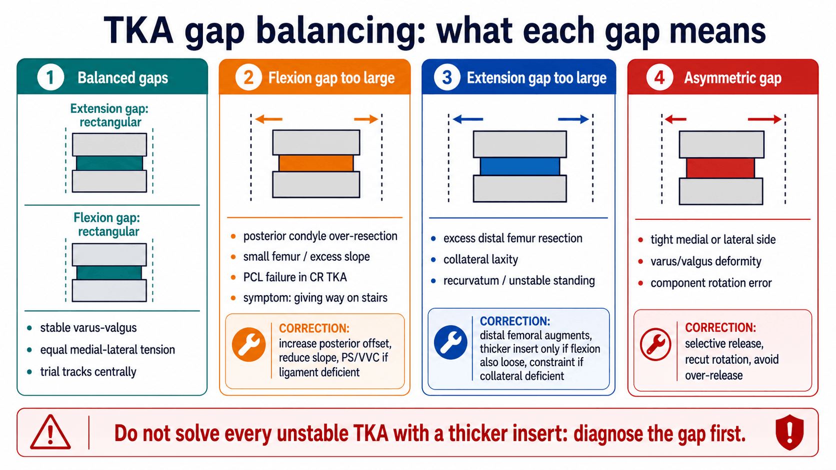
The gap diagnosis. Where in the arc the knee is loose points to the likely problem:
- Loose at 90° but stable in extension - flexion gap too large (flexion instability)
- Loose in extension but acceptable at 90° - extension gap too large (extension instability)
- Loose at 30° to 60° but stable at the endpoints - mid-flexion instability
- Varus or valgus opening at multiple flexion angles - collateral incompetence, asymmetric release, malalignment or bone loss
- Painful instability with raised inflammatory markers - possible periprosthetic joint infection
Surgically Relevant Anatomy
The knee is stabilised by bone cuts, implant geometry and soft tissues. A balanced TKA depends on respecting how each structure behaves in extension, flexion and mid-flexion.
Medial side. The superficial MCL is the primary medial restraint, especially against valgus stress, and over-releasing it in a varus TKA causes medial instability and may force higher constraint. The deep MCL and posteromedial capsule produce medial tightness near the joint line.
Lateral side. The LCL and posterolateral structures give lateral stability, especially in valgus knees, and the release sequence depends on whether the tightness is in extension, flexion or both. The IT band is associated with lateral tightness in extension and the popliteus with posterolateral tightness in flexion.
The PCL. In a CR TKA the PCL is a posterior stabiliser and a flexion-gap restraint. Incompetence creates flexion instability; excessive tension can limit flexion or cause lift-off.
Posterior osteophytes and capsule. These block extension and distort gap assessment. Remove the osteophytes before judging true extension balance.
The extensor mechanism. It provides patellar tracking and active extension. Extensor failure can mimic instability and changes the reconstruction choices.
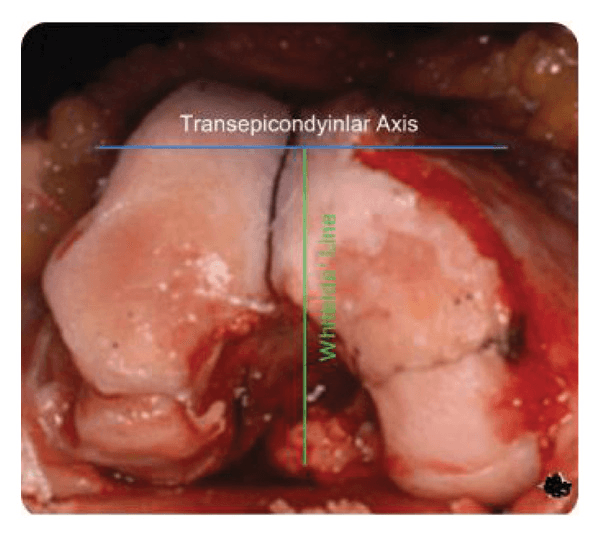
Femoral rotational landmarks. The transepicondylar axis and Whiteside line are key rotational references in the distal femur. In a valgus knee with lateral condylar hypoplasia the posterior condylar axis can be misleading, so rotation must be checked against more than one landmark.
Pathophysiology
Instability occurs when the soft-tissue envelope and implant geometry no longer control the tibia under load. The mechanism is usually mechanical, but pain and swelling from infection or loosening can make the knee feel unstable; this is why infection and fixation are checked before revising gaps.
Loss of posterior condylar offset. The flexion gap opens because the femur is effectively undersized posteriorly. The patient gives way in flexion, has difficulty on stairs and shows an AP drawer at 90°.
Excess posterior tibial slope. The tibia rests on a posteriorly sloped platform, increasing flexion laxity. The result is flexion instability, especially in a CR TKA with PCL insufficiency.
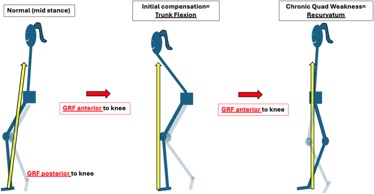
Excess distal femoral resection. The extension gap opens more than the flexion gap, giving a recurvatum tendency or instability in stance.
Malrotation. Femoral or tibial malrotation makes the flexion gap asymmetric, and patellofemoral tracking may fail. It presents with pain, stiffness, patellar symptoms or asymmetric flexion instability.
Collateral incompetence. The knee cannot resist varus-valgus load despite acceptable bone cuts. It opens coronally at 0°, 30° or 90°, and may require VVC/CCK or a hinge.
Joint-line elevation. Collateral isometry and patellofemoral mechanics change through mid-flexion. Elevation can contribute to mid-flexion instability, stiffness, patellar problems or extensor dysfunction.
Genu recurvatum. An extension gap that is too large, a distal femur that is over-resected, or a knee that hyperextends preoperatively lets the knee fall into hyperextension. Recurvatum is strongly associated with neuromuscular and connective-tissue conditions: classically poliomyelitis, where quadriceps weakness makes patients lock the knee in recurvatum to stand, and also rheumatoid, Charcot and Ehlers-Danlos knees.
Primary TKA Balancing
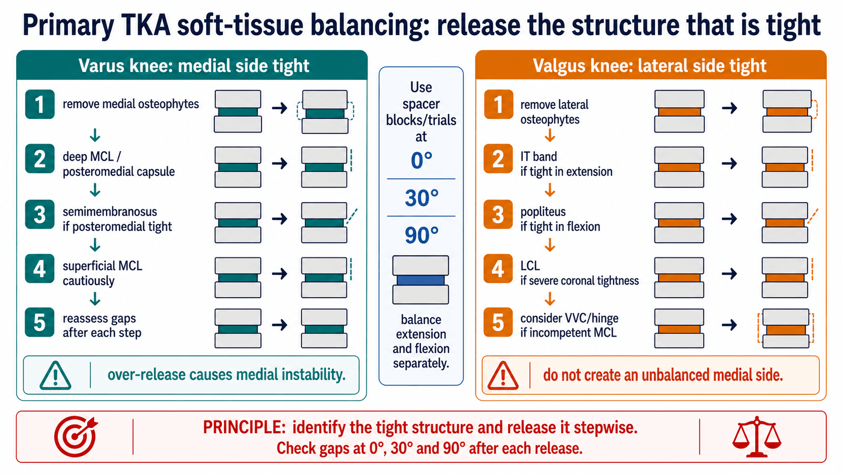
Soft-tissue balancing should be stepwise. Remove osteophytes first, release the structure that is tight, then reassess at 0°, 30° and 90° before proceeding.
Before any release. Do not release soft tissue until the basics are correct:
- Confirm the tibial cut is not in unintended varus or valgus
- Confirm distal femoral resection and femoral rotation are appropriate
- Remove posterior, medial and lateral osteophytes
- Clear meniscal remnants and loose bodies that tent the collateral ligaments
- Test with spacer blocks or trials at extension, 30° and 90°
- Correct obvious bone-cut error before sacrificing ligament
The varus knee. The medial side is tight. Release in order:
- Remove medial tibial and femoral osteophytes. If this corrects the gap, stop releasing.
- Release the deep MCL and posteromedial capsule from the proximal tibia, a good first soft-tissue step for most fixed varus knees.
- Release semimembranosus if posteromedial tightness persists; it is useful when extension and posteromedial flexion remain tight.
- Pie-crust or cautiously lengthen the superficial MCL if severe medial tightness remains. The risk is medial instability, so reassess repeatedly.
- For severe fixed deformity, consider reduction osteotomy, sliding medial epicondyle osteotomy or constraint instead of uncontrolled MCL destruction.
The valgus knee. The lateral side is tight, and which structure to release depends on the flexion angle at which it is tight:
- Tight mainly in extension - the IT band and posterolateral capsule. Release these extension-side structures first and reassess.
- Tight mainly in flexion - the popliteus or LCL, depending on the pattern. Release cautiously and avoid creating lateral flexion laxity.
- Tight in flexion and extension - the LCL and broader posterolateral complex. Release sequentially; there is a high chance of needing increased constraint.
- Medial side incompetent - an attenuated MCL in severe valgus. Do not rely on soft-tissue release alone; consider VVC/CCK or hinge.
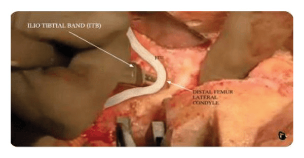
Pie-crusting the IT band. When a valgus knee remains tight laterally in extension, multiple controlled oblique punctures in the iliotibial band just proximal to the joint line can lengthen it. Reassess the extension gap after each increment to avoid lateral over-release.
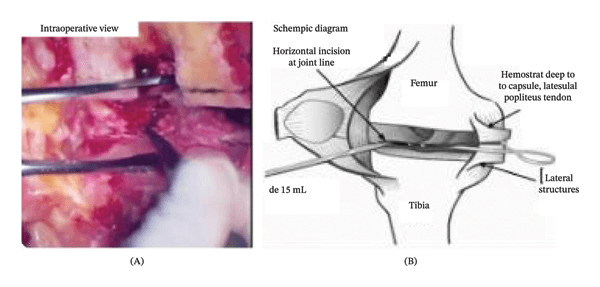
Inside-out posterolateral capsular release. The release runs from the lateral PCL margin towards the iliotibial band. A haemostat placed deep to the capsule protects the popliteus tendon and common peroneal nerve while the tight extension-side structure is released.
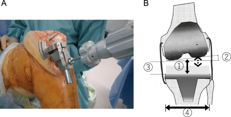
Measuring laxity. An offset tensor quantifies the joint-component gap under a defined distraction force at full extension and 90° flexion. Subtracting insert thickness from the measured lateral centre gap makes residual lateral flexion laxity explicit, instead of relying on a subjective feel.
Clinical Assessment of the Unstable TKA
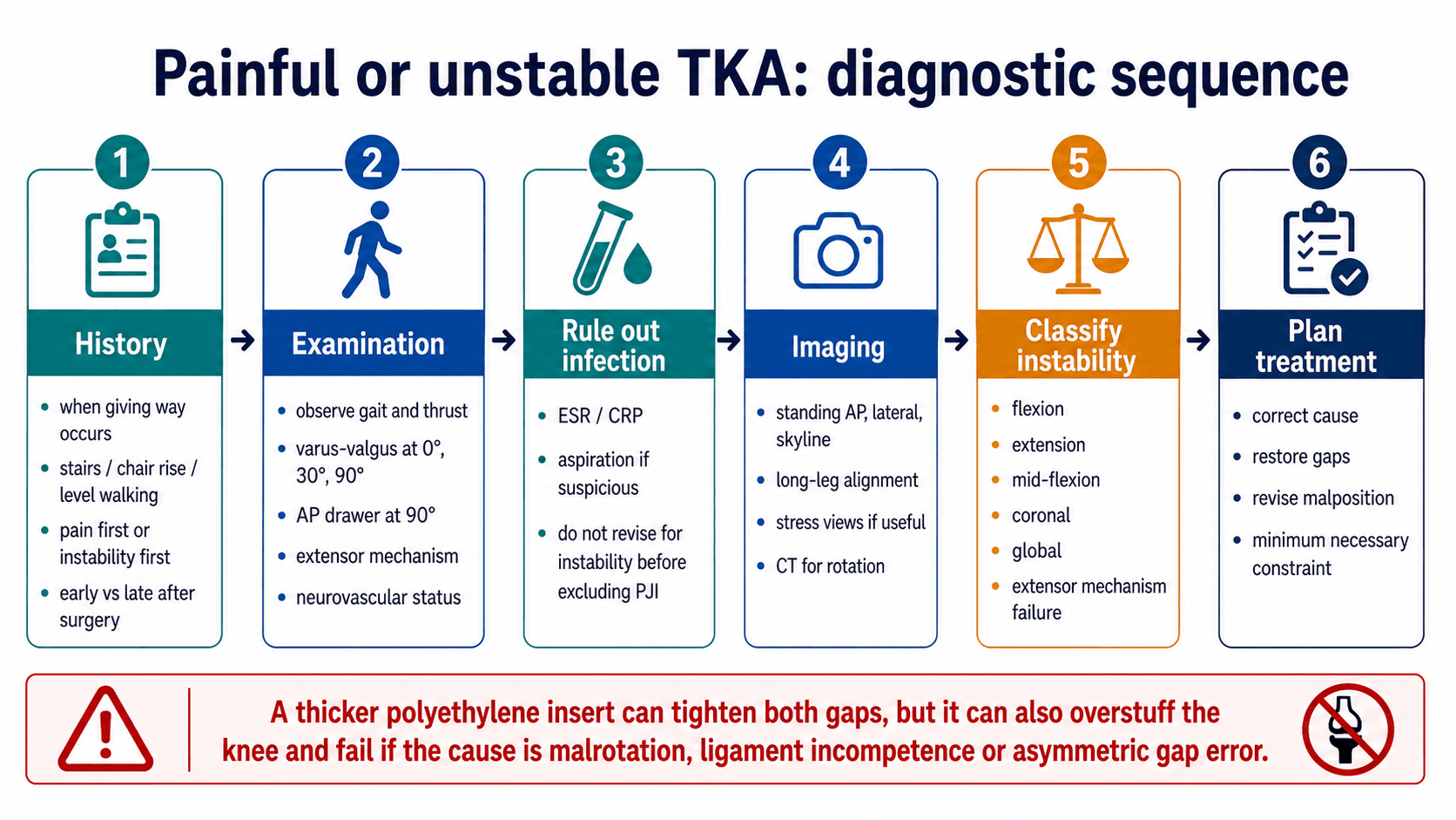
History. Ask when the knee gives way. Flexion instability is classically worse with stairs, descending slopes, rising from a chair and activities that load the knee in flexion. Extension instability or recurvatum is more obvious during stance and level walking, and mid-flexion instability may feel vague: “not trusting the knee” between full extension and deep flexion.
The other points that shape the plan:
- Timing after primary TKA: immediate, early, late or after trauma
- Pain first or instability first
- Recurrent effusions or swelling after activity
- Stairs, chair rise, pivoting, level walking and downhill walking
- Previous infection, wound issues, manipulation, stiffness or revision
- Implant type if known: CR, PS, constrained condylar, rotating hinge
- Neuromuscular disease, extensor mechanism symptoms or falls
Examination. Varus-valgus stress is tested at full extension, 30° and 90° because each position loads different stabilisers.
- How To Perform It
- Watch walking from front, side and back; look for varus/valgus thrust, recurvatum, limp and extensor lag.
- Positive Meaning
- Thrust suggests coronal instability or malalignment; recurvatum suggests extension instability or neuromuscular control problem.
- How To Perform It
- Patient supine, knee fully extended, stabilise femur and apply varus/valgus force at ankle.
- Positive Meaning
- Opening in extension suggests collateral incompetence, excessive extension gap or global instability.
- How To Perform It
- Unlock the knee slightly and repeat stress testing.
- Positive Meaning
- Isolates collateral behaviour better than full extension; useful for mid-flexion/coronal laxity.
- How To Perform It
- Knee flexed to 90°, foot supported, stress through the tibia.
- Positive Meaning
- Opening in flexion suggests flexion-gap asymmetry or collateral incompetence in flexion.
- How To Perform It
- Stabilise foot, flex knee to 90°, translate tibia anterior and posterior.
- Positive Meaning
- Excess AP translation suggests flexion instability, PCL failure in CR TKA or post-cam problems in PS TKA.
- How To Perform It
- Test coronal stability around 30° to 60° with the patient relaxed.
- Positive Meaning
- Instability here with stable endpoints supports mid-flexion instability.
- How To Perform It
- Assess active straight-leg raise, extensor lag, patellar tracking and apprehension.
- Positive Meaning
- Extensor failure or patellar instability can mimic giving way and changes reconstruction plan.
What mimics instability. The complaint of a knee that gives way is not specific. Several diagnoses present similarly and must be separated before any constraint or insert decision, because the wrong label leads to the wrong operation.
- Discriminating Features
- Objective laxity on stress at a defined flexion angle, recurrent effusions, pattern fits a specific gap.
- Key Test or Pitfall
- Stress at 0°, 30° and 90°; document the gap that opens.
- Discriminating Features
- Rest and night pain, raised CRP/ESR, effusion, sometimes warmth; instability may be a late effect of bone or soft-tissue destruction.
- Key Test or Pitfall
- ESR, CRP and aspiration with cell count, differential and culture before labelling as mechanical.
- Discriminating Features
- Start-up pain, progressive radiolucent lines, component migration or subsidence.
- Key Test or Pitfall
- Serial radiographs and comparison; loosening needs revision not insert exchange.
- Discriminating Features
- Buckling on stairs, extensor lag, quadriceps weakness, patellar maltracking; the knee gives way without true coronal laxity.
- Key Test or Pitfall
- Active straight-leg raise and extensor lag are easily missed if only collaterals are tested.
- Discriminating Features
- Anterior pain, apprehension, lateral tracking, clunk; often from component malrotation.
- Key Test or Pitfall
- Skyline view and CT rotation; do not treat as a tibiofemoral problem.
- Discriminating Features
- Pain, stiffness, patellar symptoms and asymmetric flexion gap rather than uniform laxity.
- Key Test or Pitfall
- CT rotational profile; soft-tissue release cannot fix a malrotated component.
- Discriminating Features
- Generalised giving way, falls, recurvatum, weakness out of proportion to mechanical findings.
- Key Test or Pitfall
- Neurological examination; bracing and rehabilitation rather than revision.
Investigations
What to request.
- Standing AP, lateral and skyline radiographs of the knee
- Full-length standing hip-to-ankle alignment radiograph
- Comparison with prior radiographs to assess subsidence, loosening and joint-line change
- Stress radiographs when clinical laxity needs documentation
- CT rotational profile for suspected femoral or tibial component malrotation
- ESR, CRP and aspiration when infection is possible
- Bone scan or SPECT/CT, considered selectively when loosening is unclear and infection has been addressed
What to measure. Each measurement looks for a cause of instability:
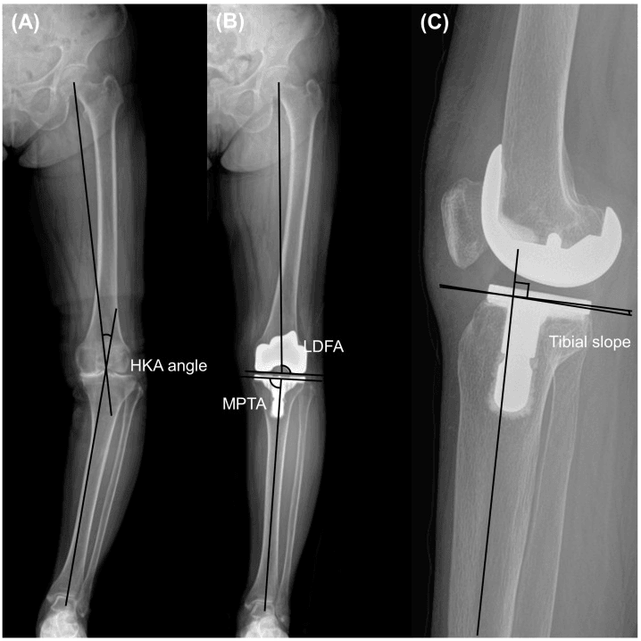
- Mechanical alignment - on the full-length standing film, femoral head centre to ankle centre through the knee. Malalignment may drive coronal thrust and demands component or osteotomy-level correction.
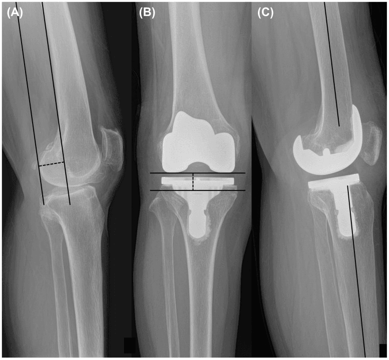
- Component coronal position - on the AP knee and long-leg films. A varus tibial or a valgus or varus femoral error can create asymmetric gaps.
- Posterior tibial slope - on a true lateral radiograph.
- Posterior condylar offset - on the lateral radiograph, compared with pre-operative or early post-operative films if available.
- Joint-line position - compared with the fibular head, adductor tubercle or prior films.
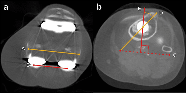
- Component rotation - on CT, using recognised femoral and tibial rotational references.
- Fixation and bone loss - radiolucent lines, migration and osteolysis, with CT if needed. Loose or deficient components need revision reconstruction, not soft-tissue-only treatment.
Management Principles
Flexion instability does not simply mean PS, nor global instability a hinge. The surgeon must decide whether the cause is infection, component malposition, PCL failure, collateral incompetence, joint-line elevation, bone loss, posterior offset loss, tibial slope, extensor mechanism failure or true global laxity.
Infection first. Pain may come from infection, loosening, malrotation, instability or extensor failure, so in a painful, loose TKA think of infection first. Aspirate when suspicion persists after blood tests, and do not start definitive instability treatment until the infection pathway is complete: missed infection makes revision fail.
Component position before gaps. Do not revise instability without assessing component position. Reconstruct the platform first: fixation, component position, bone loss, joint line, posterior offset and ligament competence.
Non-operative care. It is reasonable in the situations below, although physiotherapy does not correct a mechanical gap mismatch:
- Mild subjective symptoms, stable implant, no progressive laxity - physiotherapy for quadriceps and hip strength, gait retraining and proprioception
- Low-demand patient with mild coronal laxity and no loosening - functional brace and activity modification, a temporary or palliative option; monitor progression
- Early flexion symptoms without objective gross laxity - strengthening, swelling control and careful review. Persistent recurrent effusion or an objective drawer needs surgical planning
Revision by failure pattern. Each pattern has its correction and its trap.
- Do Not Do
- Do not simply thicken insert if extension gap is already balanced.
- Preferred Strategy
- Increase posterior offset, reduce excessive slope, correct rotation, convert CR to PS/VVC depending collateral competence.
- Do Not Do
- Do not overstuff flexion gap with insert alone.
- Preferred Strategy
- Restore distal femoral joint line with augments, revise malposition and increase constraint if collateral deficient.
- Do Not Do
- Do not diagnose only at 0° and 90°.
- Preferred Strategy
- Assess 30° to 60°, joint line, femoral design, component position, collateral isometry and malrotation.
- Do Not Do
- Do not leave malalignment uncorrected and rely on constraint alone.
- Preferred Strategy
- Revise alignment, reconstruct bone loss and use VVC/CCK if collateral support remains incomplete.
- Do Not Do
- Do not use PS when collaterals cannot control the knee.
- Preferred Strategy
- Rotating hinge if VVC cannot control instability or bone/ligament loss is severe.
Isolated insert exchange is unsafe when instability is due to malrotation, malalignment, loose components, component undersizing, bone loss, PCL failure with a CR design, collateral incompetence, extensor mechanism failure or infection. It may be considered only when components are well-fixed, well-positioned, infection is excluded and both gaps can be corrected without overstuffing.
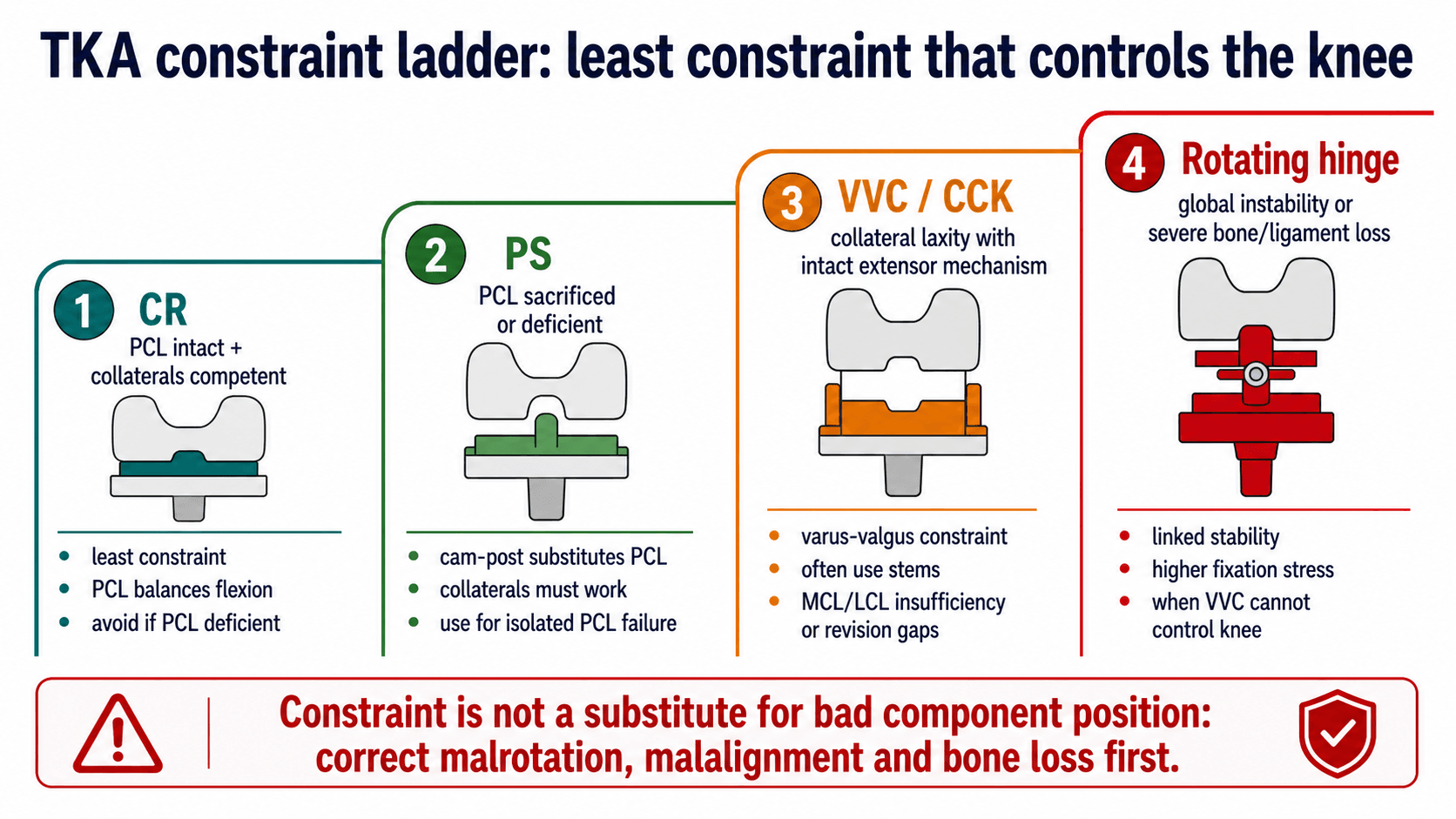
Constraint. Constraint is mechanical stability built into the implant. It is useful when the soft tissues cannot stabilise the knee, but it increases load transfer to the fixation interfaces. Constraint rises only when soft-tissue control falls: choose the least constraint that controls the reconstructed knee after infection, component position, bone loss and ligament competence have been addressed.
- CR - PCL and collaterals competent
- PS - PCL deficient but collaterals competent
- VVC/CCK - coronal laxity with a usable extensor mechanism
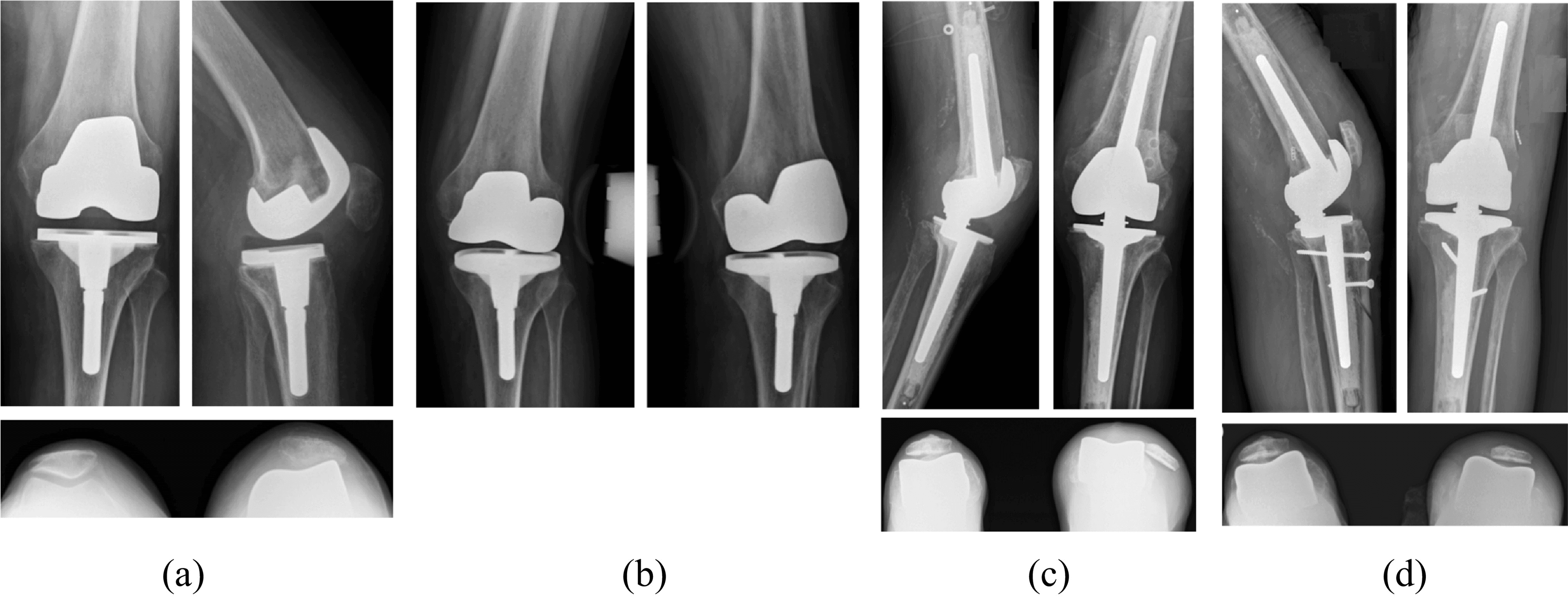
- Hinge - global instability or severe bone or ligament loss
GAPSSafe Revision Sequence
Hook:GAPS keeps the revision plan cause-based rather than insert-based.
Genu recurvatum is one of the three Cottino tibiofemoral instability patterns, and its management is distinct and examinable.
Prevention at operation. To treat or prevent recurvatum, tighten the extension gap: under-resect or build up (augment) the distal femur so the extension gap is relatively snug, and avoid excessive posterior tibial slope. This is the opposite of correcting a flexion contracture.
The neuromuscular knee. In a polio or quadriceps-deficient knee a small degree of recurvatum may be functionally necessary for the patient to stand, and over-correcting it can leave them unable to lock the knee. Preserve the protective recurvatum or use a hinge: these patients often need a constrained or rotating-hinge implant or bracing rather than relying on soft-tissue balance.
Recurrence. Recurvatum tends to recur if the underlying muscle imbalance is not addressed, so the gap cuts alone do not settle it.
Operative Technique
Revision for instability
Position. Supine, with a tourniquet available, the previous incision planned and the full limb accessible for alignment and stress testing. Poor exposure makes component removal and gap assessment unreliable.
Imaging and equipment. Have an image intensifier if needed, extraction tools, augments, stems, cones or sleeves, and PS, VVC/CCK and hinge options available. Starting without the next constraint level can force a compromised reconstruction.
Preparation. Antibiotics after cultures if there is an infection concern, an extensile exposure plan, and aspiration and cultures as indicated. Do not ignore infection because the referral says instability.
Approach. Use the prior midline incision where safe; a medial parapatellar arthrotomy is common. Plan a quadriceps snip or tibial tubercle osteotomy if exposure is difficult, because avulsing the extensor mechanism changes the entire reconstruction.
Assessment at operation. Assess polyethylene wear, PCL status if CR, collateral integrity, the extensor mechanism, component fixation and bone loss. Do not remove well-fixed components before confirming why the knee is unstable.
Reconstruction. Remove failed components when needed, rebuild the tibial platform and femoral joint line, restore posterior offset and correct slope and rotation. A thicker insert cannot correct a malpositioned femoral or tibial component.
Structures at risk. Protect the patellar tendon, MCL, LCL, popliteus, posterior capsule, popliteal vessels and peroneal nerve in valgus knees. Peroneal nerve risk rises with valgus correction and flexion contracture correction.
Fixation. Use stems for load sharing when constraint, bone loss or augments increase interface stress, and augments, cones or sleeves for bone defects. Constraint without fixation support increases loosening risk.
Closure and aftercare. Confirm stability at 0°, 30°, 60° and 90°, range of motion, patellar tracking and neurovascular status, and brace if there is soft-tissue concern. Failure to document the final stability arc leaves uncertainty after revision.
Complications and Failure Management
Instability revisions fail for identifiable reasons, and each has its prevention or salvage:
- Persistent flexion instability - posterior offset not restored, excessive slope persisting or PCL failure not addressed. Revise the femur or tibia as needed, with PS or VVC depending on collateral competence.
- New stiffness - an overstuffed knee, excessive insert thickness or joint-line error. Correct the gap cause rather than upsizing the insert indiscriminately.
- Persistent coronal laxity - collateral incompetence underestimated. Increase to VVC/CCK or hinge, and reconstruct bone loss and alignment.
- Aseptic loosening - high constraint without adequate fixation or bone support. Use stems, augments, cones or sleeves and an appropriate fixation strategy.
- Missed infection - pain and instability treated as mechanical only. PJI work-up before revision, and cultures at surgery when indicated.
- Extensor mechanism failure - exposure injury, chronic patellar maltracking or an unrecognised extensor deficit. Plan extensile exposure carefully and reconstruct the extensor mechanism when needed.
Guidelines, Registries & Global Practice
Global epidemiology
Instability is one of the leading mechanical causes of TKA revision worldwide, consistently appearing alongside infection and stiffness as a top reason for early revision and alongside infection, loosening and wear for late revision. As polyethylene quality improved, the relative share of wear-driven failure fell and technique-sensitive causes such as instability became proportionally more important. Valgus deformity accounts for roughly 10 percent of primary TKA, and these knees carry the greatest balancing and constraint challenge.
Society guidance, side by side
There is no single dedicated international guideline for "TKA instability"; practice is shaped by arthroplasty society consensus, registry data and revision-burden reporting rather than by a single algorithm.
- Emphasis Relevant to Balancing and Instability
- Evidence-based work on the painful TKA stresses systematic workup, mandatory infection exclusion (ESR, CRP, aspiration) before attributing symptoms to a mechanical cause.
- Emphasis Relevant to Balancing and Instability
- Revision activity concentrated in networked revision centres (the revision-network model), so complex instability and constraint decisions are increasingly made by specialist revision teams.
- Emphasis Relevant to Balancing and Instability
- Principles-based teaching: diagnose the loose gap, correct the cause (alignment, rotation, offset, bone loss) and use the least constraint that stabilises the reconstructed knee.
- Emphasis Relevant to Balancing and Instability
- Supports staged, cause-based revision and structured periprosthetic infection pathways before mechanical revision for instability.
Registry evidence
- Relevance to Instability and Constraint
- Tracks revision indications including instability and the use of constrained and hinged implants; informs implant and constraint selection by survivorship.
- Relevance to Instability and Constraint
- Reports revision causes and implant survivorship, with instability a recurring early-revision indication and higher-constraint designs monitored for survival.
- Relevance to Instability and Constraint
- Captures contemporary revision burden and the share attributable to instability and mechanical complications.
- Relevance to Instability and Constraint
- Long-standing survivorship data and revision-cause reporting that frame instability within total revision burden.
High- versus limited-resource practice
- Typical Reality
- Full revision implant ladder (augments, cones/sleeves, stems, VVC and hinge), CT rotation, navigation/robotics and specialist revision centres available.
- Implication
- Reconstruction can be matched precisely to the cause and to residual soft-tissue competence.
- Typical Reality
- Restricted constraint inventory, limited revision implants, less access to CT and cross-sectional imaging, fewer revision-trained surgeons.
- Implication
- Greater emphasis on correct primary balancing and alignment; instability is harder to salvage so prevention matters more.
- Typical Reality
- Infection exclusion before mechanical revision is universal and low-cost (clinical assessment, ESR, CRP, aspiration).
- Implication
- The single non-negotiable step is affordable everywhere and prevents failed revision.
Controversies and Areas of Uncertainty
Several balancing questions remain genuinely unresolved, and a strong exam answer acknowledges the uncertainty rather than overstating dogma.
- Current Position
- Mechanical alignment remains the default reference; kinematic and functional alignment aim to reproduce native soft-tissue tension.
- Why It Is Unsettled
- Registry-scale survivorship advantage for kinematic alignment is not yet proven; balance is achieved by different philosophies.
- Current Position
- Meta-analysis shows near-identical gaps, with only around 1 mm or 1 degree difference in rotation and joint line.
- Why It Is Unsettled
- Differences are small and probably clinically minor; most surgeons use a hybrid approach.
- Current Position
- There is no universally accepted laxity threshold (in millimetres or degrees) that defines pathological instability.
- Why It Is Unsettled
- Testing positions and grading systems are inconsistent, so the same knee can be labelled differently.
- Current Position
- Joint-line elevation, femoral undersizing and reduced femoral radius of curvature are proposed.
- Why It Is Unsettled
- Most evidence is computational and cadaveric; clinical studies conflict and several factors are inconclusive.
- Current Position
- Sensors, navigation and robotics improve the precision of gap and load balancing.
- Why It Is Unsettled
- Improved precision has not yet translated into proven long-term survivorship or function gains.
- Current Position
- Use the least constraint that controls the corrected knee.
- Why It Is Unsettled
- The exact point at which a surgeon should step from PS to VVC, or VVC to hinge, is judgement-based, not rule-based.
The mechanical-vs-kinematic-vs-functional alignment debate becomes concrete with the CPAK classification (Coronal Plane Alignment of the Knee, MacDessi 2021) - the modern framework for phenotyping the native knee before deciding how to balance it:
- Two variables: (1) the arithmetic HKA (aHKA = MPTA - LDFA) = the constitutional limb alignment (varus, neutral or valgus), and (2) the joint-line obliquity (JLO = MPTA + LDFA) = whether the joint line is apex-distal, neutral or apex-proximal.
- Nine types (a 3x3 grid): combining varus/neutral/valgus constitutional alignment with three joint-line-obliquity bands gives CPAK Types I-IX, with Type I (constitutional varus + apex-distal joint line) the commonest phenotype.
- Why it matters for balancing: it makes explicit that most knees are not natively neutral - a mechanically aligned TKA cuts every knee to neutral with a horizontal joint line (changing the soft-tissue envelope and requiring releases), whereas kinematic/functional alignment aims to reproduce the patient's pre-arthritic CPAK phenotype (constitutional alignment and joint-line obliquity), theoretically needing fewer releases and giving more "natural" balance.
- The unresolved question: which CPAK phenotypes are safe to restore versus which should be corrected toward neutral (e.g. how much residual varus or joint-line obliquity is acceptable) - this is the live controversy, with no proven long-term survivorship winner yet.
Exam point: CPAK = aHKA (alignment) x JLO (joint-line obliquity) giving nine phenotypes; it frames the alignment debate as "restore the native phenotype (kinematic/functional) vs cut to neutral (mechanical)" and explains why balancing strategy depends on the knee's constitutional shape.
Clinical Scenarios
Practise clinical reasoning and management decisions out loud
“A patient has recurrent effusions and giving way when descending stairs 18 months after a cruciate-retaining TKA. The knee is stable in extension but has increased anterior-posterior translation at 90°.”
“During primary TKA for a severe valgus knee, the extension gap remains laterally tight after bone cuts and osteophyte removal. The medial side is attenuated.”
“A patient is referred 3 years after a posterior-stabilised TKA with pain, swelling and a feeling that the knee is unreliable in the middle of the range. Stress testing is convincing for stability at full extension and at 90°, but the knee opens medially and laterally when tested at 30° to 45°. Inflammatory markers are normal.”
Examine
- Gait: thrust or recurvatum
- Varus-valgus at 0°, 30°, 90°
- AP drawer at 90°
- Mid-flexion stress
- Extensor mechanism
Image
- Standing AP/lateral/skyline
- Long-leg alignment
- Compare prior films
- CT for rotation
- ESR/CRP and aspirate if suspicious
Correct
- Flexion: posterior offset/slope/PCL
- Extension: distal femoral joint line
- Asymmetric: release or recut
- Global: constraint after reconstruction
- Never miss infection
“Diagnose which gap is wrong, correct the cause, then choose the least constraint that controls the knee.”
Evidence Signals
Gap balancing versus measured resection
- The goal of TKA is symmetric balanced flexion and extension gaps.
- Gap balancing aims to tension the collaterals and make the femoral component parallel to the tibial cut in flexion.
- The review argues gap balancing can produce more rectangular flexion gaps, while acknowledging controversy.
Gap balancing meta-analysis
- Eight observational comparison studies were pooled.
- Overall gap differences did not differ significantly between techniques (pooled mean difference -0.09 mm, 95% CI -0.40 to +0.21, p = 0.55).
- ONE GAP COMPARISON DID DIFFER, and the paper flags it in its own conclusion: the medial/lateral EXTENSION gap difference was 0.58 mm greater with measured resection (p = 0.008).
- Gap balancing produced greater femoral external rotation (0.77 degrees) and more joint-line elevation (1.17 mm), but differences were around 1 mm or 1 degree.
References
- Daines BK, Dennis DA. Gap balancing vs. measured resection technique in total knee arthroplasty. Clin Orthop Surg. 2014;6(1):1-8. doi:10.4055/cios.2014.6.1.1.
- Moon YW, Kim HJ, Ahn HS, Park CD, Lee DH. Comparison of soft tissue balancing, femoral component rotation, and joint line change between the gap balancing and measured resection techniques in primary total knee arthroplasty: a meta-analysis. Medicine. 2016;95(39):e5006. doi:10.1097/MD.0000000000005006.
- Siddiqi A, Smith T, McPhilemy JJ, Ranawat AS, Sculco PK, Chen AF. Soft-Tissue Balancing Technology for Total Knee Arthroplasty. JBJS Rev. 2020;8(1):e0050. doi:10.2106/JBJS.RVW.19.00050.
- Stambough JB, Edwards PK, Mannen EM, Barnes CL, Mears SC. Flexion Instability After Total Knee Arthroplasty. J Am Acad Orthop Surg. 2019;27(17):642-651. doi:10.5435/JAAOS-D-18-00347.
- Cottino U, Sculco PK, Sierra RJ, Abdel MP. Instability After Total Knee Arthroplasty. Orthop Clin North Am. 2016;47(2):311-316. doi:10.1016/j.ocl.2015.09.007.
- Chang MJ, Lim H, Lee NR, Moon YW. Diagnosis, causes and treatments of instability following total knee arthroplasty. Knee Surg Relat Res. 2014;26(2):61-67. doi:10.5792/ksrr.2014.26.2.61.
- Mehta N, Burnett RA, Kahlenberg CA, Miller R, Chalmers B, Cross MB. Mid-Flexion Instability After Total Knee Arthroplasty: Diagnosis, Implant Design, and Outcomes. Orthopedics. 2023;46(1):e13-e19. doi:10.3928/01477447-20220719-01.
- Vajapey SP, Pettit RJ, Li M, Chen AF, Spitzer AI, Glassman AH. Risk Factors for Mid-Flexion Instability After Total Knee Arthroplasty: A Systematic Review. J Arthroplasty. 2020;35(10):3046-3054. doi:10.1016/j.arth.2020.05.026.
- Rossi R, Cottino U, Bruzzone M, Dettoni F, Bonasia DE, Rosso F. Total knee arthroplasty in the varus knee: tips and tricks. Int Orthop. 2019;43(1):151-158. doi:10.1007/s00264-018-4116-3.
- Alesi D, Meena A, Fratini S, et al. Total knee arthroplasty in valgus knee deformity: is it still a challenge in 2021? Musculoskelet Surg. 2022;106(1):1-8. doi:10.1007/s12306-021-00695-x.
- Rezaei A, Moon J, Lichtig A, et al. Precision soft tissue balancing: grid-assisted pie-crusting in total knee arthroplasty. Front Surg. 2024;11:1331902. doi:10.3389/fsurg.2024.1331902.
- Le DH, Goodman SB, Maloney WJ, Huddleston JI. Current modes of failure in TKA: infection, instability, and stiffness predominate. Clin Orthop Relat Res. 2014;472(7):2197-2200. doi:10.1007/s11999-014-3540-y.