Transphyseal Distal Humerus Fracture
Overview & Epidemiology
A transphyseal fracture is a separation through the distal humeral physis, in Salter-Harris terms a type I or II. The entire distal humeral epiphysis, carrying the elbow joint and the forearm with it, shears off the metaphysis and displaces as a unit.
Who and how. It typically occurs in children younger than 3 years. The three classic mechanisms are birth trauma after a difficult delivery, non-accidental trauma, and a fall from a small height.
Why it is missed. The injury is rare, and that is itself a trap: unfamiliarity drives the misdiagnosis as an elbow dislocation or a normal elbow. In infants and toddlers the epiphysis is largely cartilaginous, so neither the fracture line nor the displaced epiphysis is directly visible on plain radiographs.
Anatomy & Biomechanics
The ossification sequence. The ossification centres of the elbow appear in the CRITOE order at roughly 1, 3, 5, 7, 9 and 11 years, the capitellum first. In the under-3 age group of this injury often only the capitellum, and sometimes the radial head, has begun to ossify, so most of the epiphysis, and the physis itself, is radiolucent cartilage.
Why that matters. That single fact explains the whole diagnostic difficulty: the separation occurs through tissue the X-ray cannot show. It has to be read indirectly, from the bony alignment and from the position of the ossified capitellum relative to the metaphysis.
CRITOE
Hook:CRITOE at 1-3-5-7-9-11 years: in a transphyseal fracture most of the epiphysis is unossified, which is exactly why it is invisible on X-ray.
Mechanism. The physis is the mechanically weakest link in the immature elbow, so a shear or rotatory force (birth canal forces, a twist or a fall) separates the epiphysis rather than dislocating the joint. The displacement is typically posteromedial and produces an apparent varus tilt, which becomes cubitus varus if it heals malreduced.
Blood supply and growth. The medial crista of the trochlea and the medial condylar ossification centre have a relatively tenuous blood supply, which is why osteonecrosis of the medial condyle is a recognised complication. Because the injury crosses the physis, partial growth arrest and progressive angular deformity are possible.
Classification
Salter-Harris. Mechanistically the injury is a type I separation, through the physis only, in the youngest infants, or a type II, with a metaphyseal Thurston-Holland fragment, in slightly older children. Either way the whole epiphysis moves with the forearm, which distinguishes it from an isolated condylar fracture.
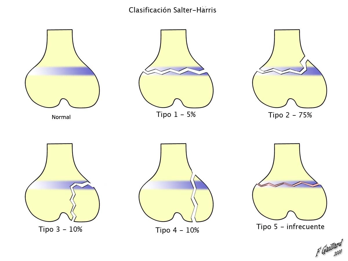
DeLee classification. DeLee groups transphyseal separations by how much of the epiphysis has ossified, which also tracks the diagnostic difficulty and the differential.
- Age and ossification
- Birth to ~1 year, before the capitellum ossifies
- Salter-Harris
- Pure type I
- The trap
- The hardest to diagnose; most easily mistaken for a congenital or elbow dislocation
- Age and ossification
- ~1 to 3 years, capitellum ossified
- Salter-Harris
- Type I or II, often with a small lateral metaphyseal flake
- The trap
- -
- Age and ossification
- Older children
- Salter-Harris
- Type II with a larger metaphyseal fragment
- The trap
- The one most easily confused with a lateral condyle fracture
Clinical Assessment
Presentation. A child under 3 with a swollen, painful elbow and pseudoparalysis of the arm. The injury is often birth-related, presenting in the first days of life, or follows a fall or abuse. On examination feel for crepitus or a muffled "bag of bones" feel, and assess the neurovascular status of the limb.
A transphyseal distal humerus fracture is a recognised abuse-associated injury in the under-3 age group. A history that does not fit the injury, an absent or inconsistent mechanism, delayed presentation, other bruises or injuries, or carer behaviour that does not fit should trigger your local safeguarding pathway alongside treatment of the fracture: a full history, examination for other injuries, a skeletal survey, and social and child-protection involvement.
The baby who will not move the arm. Pseudoparalysis is pain-driven non-use, and a transphyseal separation is only one cause of it; the examiner wants the full differential.
- Septic arthritis of the elbow or proximal humeral osteomyelitis, the must-exclude: an irritable, often febrile baby with raised CRP/ESR, in whom ultrasound-guided aspiration is mandatory before the non-use is attributed to a fracture
- Obstetric brachial plexus palsy, a neurological, flaccid "waiter's tip" posture that is relatively painless and lacks the swelling and crepitus of a fracture
- Clavicular or proximal humeral physeal birth fracture, which also presents as pseudoparalysis after a difficult delivery
Plain films, comparison of both elbows and, where infection is possible, inflammatory markers and aspiration sort these out. Never assume the cartilaginous transphyseal injury without excluding sepsis.
Investigations & Imaging
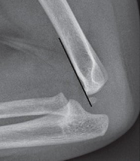
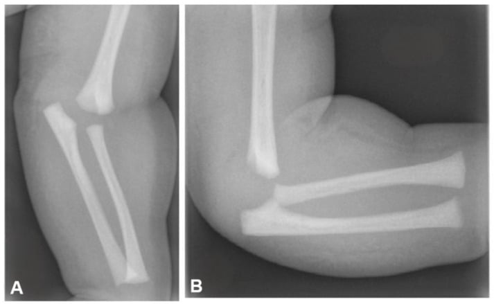
Radiographs. Obtain AP and lateral radiographs of both elbows for comparison. The epiphysis cannot be seen, so the diagnosis rests on indirect signs:
- The forearm is not aligned with the humerus, while the radius and ulna stay aligned with each other
- The radio-capitellar line is preserved, the radial shaft axis still pointing at the capitellum
- The ossified capitellum moves with the proximal radius and ulna, posteromedially, relative to the metaphysis
- A small metaphyseal (Thurston-Holland) flake may be the only bony sign in a Salter-Harris II injury
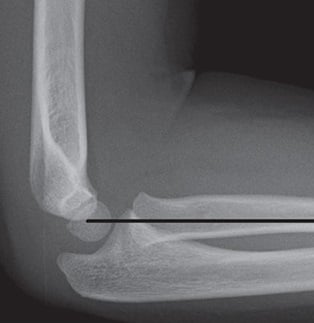
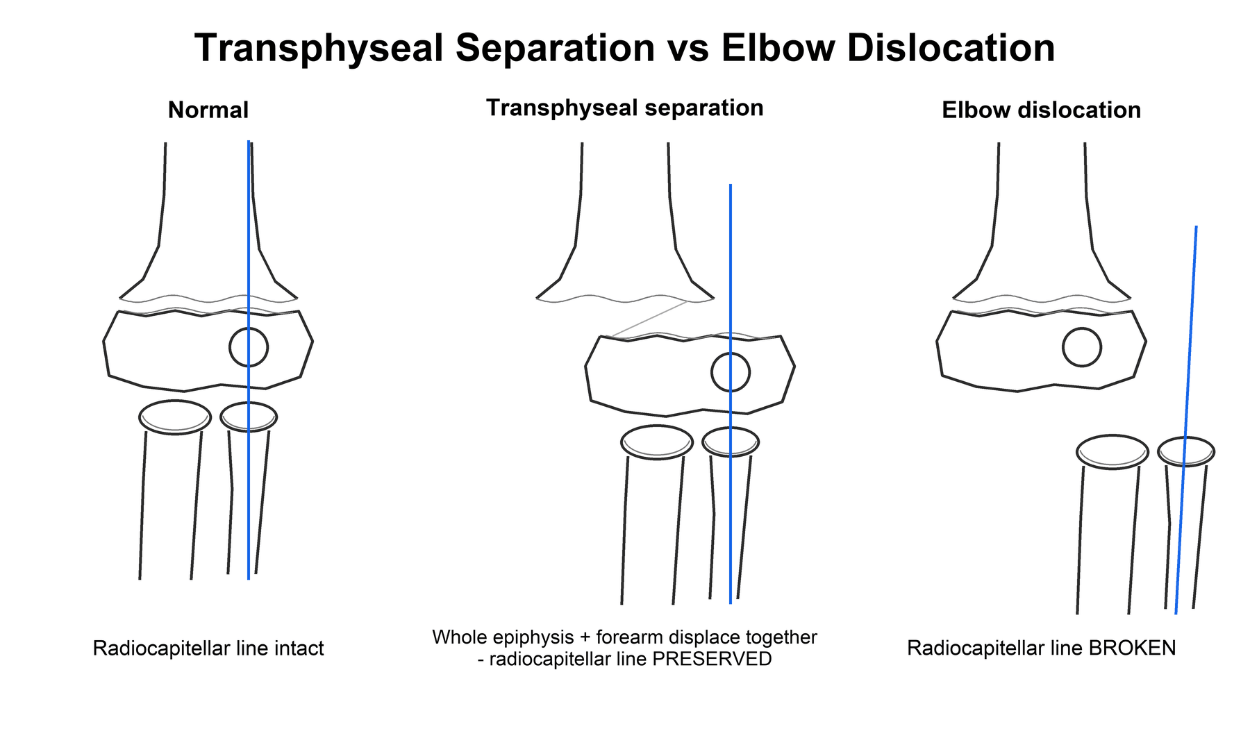
The classification is really there to stop two errors. In a transphyseal separation only the physis is disrupted: the whole distal epiphysis and the forearm displace together, and the radius and ulna keep their normal relationship to each other and to the capitellum.
In an elbow dislocation the forearm dislocates relative to the humerus and the radio-capitellar relationship is lost, the radius no longer pointing at the capitellum. True elbow dislocation is rare under 3 years, so an apparent dislocation in a toddler is usually a transphyseal separation.
A lateral condyle fracture displaces only an isolated lateral fragment, whereas a transphyseal separation moves the whole epiphysis, and the forearm, as a unit with the radio-capitellar relationship intact.
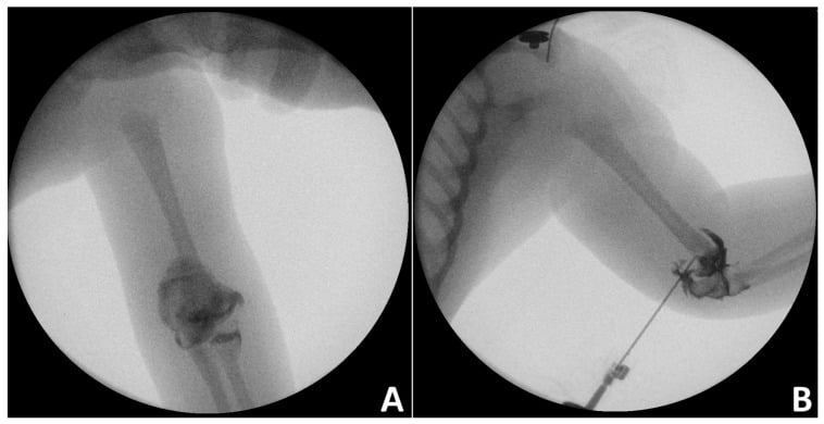
Seeing the cartilage. Three modalities demonstrate the cartilaginous epiphysis and confirm the reduction:
- Intra-operative arthrogram, the commonest adjunct: it outlines the epiphysis and confirms an accurate reduction before and after pinning
- Ultrasound, which uses no ionising radiation, shows the unossified epiphysis and its displacement, and is increasingly used at the bedside in neonates
- MRI, definitive for cartilage but rarely needed acutely, and often requiring sedation at this age
Management Algorithm
Treatment is guided by displacement, age and the soft tissues, and mirrors supracondylar fracture principles. The aim is an anatomic reduction that restores the carrying angle and protects the physis and its blood supply.
Undisplaced or minimally displaced. Immobilise in an above-elbow cast or splint with close radiographic follow-up. Infants have enormous remodelling potential, so minor residual displacement in the plane of movement is well tolerated; malalignment in the varus/valgus plane is not, so watch the carrying angle.
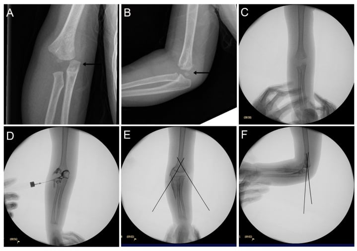
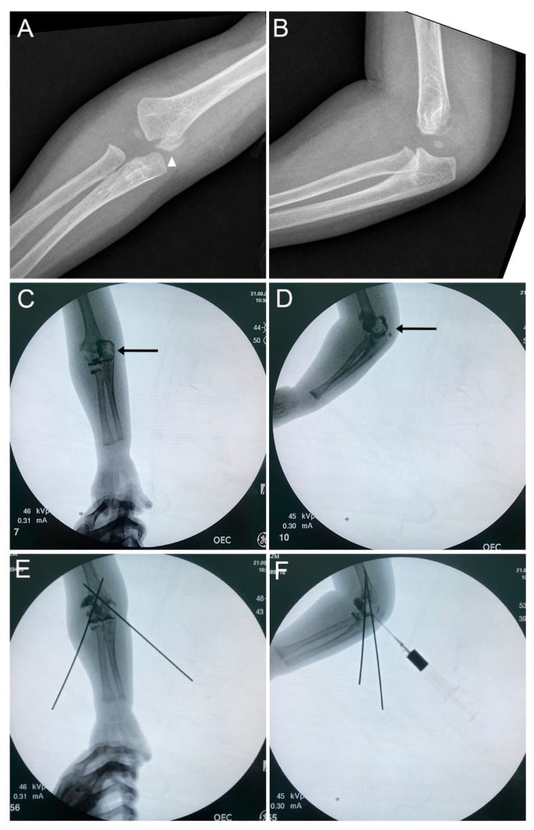
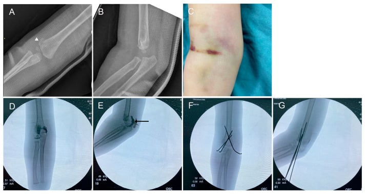
Displaced. Closed reduction and percutaneous pinning (CRPP) under anaesthesia, arthrogram-assisted, is the standard: a gentle reduction is confirmed on the arthrogram and then stabilised with smooth K-wires.
Neonates and birth injuries. Some centres accept closed reduction and casting, given the rapid healing and remodelling, while others pin. Small series report uniformly good outcomes with either when alignment is restored. Diagnosis and gentle realignment matter more than the specific fixation.
Surgical Technique
Reduction. Under general anaesthesia, with an image intensifier, apply longitudinal traction and correct the typical posteromedial displacement and any rotation. The landmarks are cartilaginous, so judge the reduction by forearm-humerus alignment and confirm it with an arthrogram, a small volume of contrast in the joint outlining the epiphysis. Pinning without one is an error: a cartilaginous reduction cannot be judged on plain screening alone.
Fixation. Stabilise with smooth K-wires in a supracondylar configuration: two divergent lateral pins, or crossed lateral and medial pins when more stability is needed. If a medial pin is used, take care to protect the ulnar nerve. Smooth wires cross the physis with minimal growth risk in this setting.
Before leaving theatre. Re-check the arthrogram after pinning to confirm the epiphysis is reduced. Do not accept varus or valgus malalignment to save operative time; it will not remodel.
Complications
Cubitus varus is the most common complication, an alignment and cosmetic deformity from malunion (malreduction). An established deformity can be corrected later with a lateral closing-wedge (supracondylar) osteotomy to restore a near-normal carrying angle, but prevention, through an accurate initial reduction confirmed on arthrogram, is far preferable.
The others:
- Osteonecrosis of the medial condyle, from a vascular insult to its tenuous blood supply
- Growth arrest, physeal injury causing partial arrest, angular deformity or a length problem
- Missed or delayed diagnosis, the cartilaginous epiphysis mistaken for a dislocation or a normal elbow
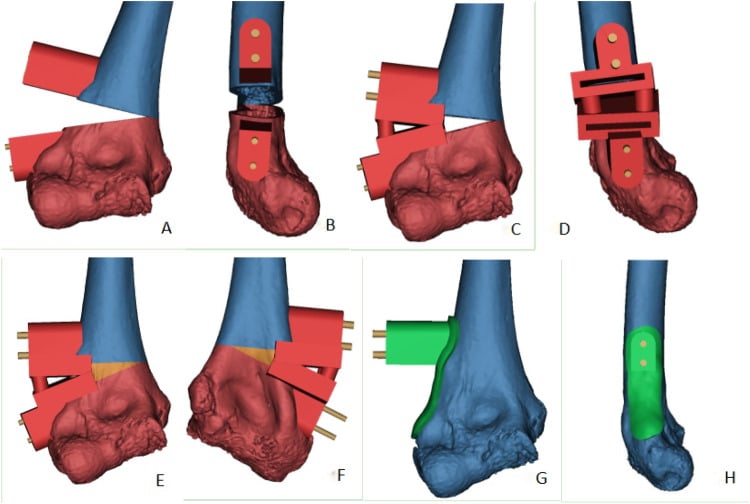
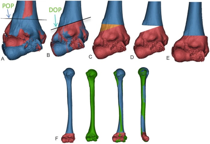
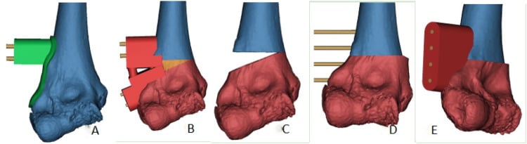
The same tenuous central blood supply that produces medial-condyle osteonecrosis can cause a fishtail deformity, a recognised late sequela of distal humeral physeal injury. Osteonecrosis or growth disturbance of the central (trochlear/medial-condylar) portion of the physis leaves a V-shaped notch in the distal humeral metaphysis, the lateral and medial pillars growing while the centre does not.
It is usually a delayed, often initially asymptomatic radiographic finding, but it can cause ulnar deviation, stiffness and pain, and may predispose to a later condylar fracture and to ulnar-nerve symptoms. The deformity declares itself only with continued growth, which is a key reason to follow these children well beyond union rather than discharging them at fracture healing.
Postoperative Care & Rehabilitation
After pinning. The elbow is protected in an above-elbow cast for around 3-4 weeks (union is rapid at this age), and the pins are removed in clinic once the fracture has united. Infants and toddlers regain elbow movement readily and rarely need formal physiotherapy. Give simple pin-site care advice and watch for pin-site irritation or infection.
Follow growth, not just union. Because the injury crosses the physis, follow-up should continue beyond union to detect progressive cubitus varus or growth disturbance. Counsel families that the carrying angle and any subtle deformity are best judged over months, and re-examine the contralateral elbow for comparison at review.
Outcomes & Prognosis
Two populations. In the neonatal birth-injury series, where the fracture was recognised within days and realigned promptly, every case united with full range of motion and no deformity requiring repeat surgery. The systematic review of children under 3 pooled 101 children from 37 studies, including those diagnosed late and those injured non-accidentally, and reported residual findings in 44.8%: most often cubitus varus (39.5% of those affected), then residual deformity and limited range of motion. It concludes that nearly half of these elbows had complications.
Reconciling them. The two answers come from different children. A neonate recognised in the first days of life and realigned promptly is not the same problem as a toddler whose injury was read as a dislocation and splinted. The fracture does well when it is recognised early and aligned, and recognition is precisely what fails: a physeal fracture was suspected on the initial radiograph in only 55.6% of cases.
What decides the result. The dominant determinants of a poor result are late or missed diagnosis (the cartilaginous epiphysis) and residual varus/valgus malalignment. The main long-term issue is cubitus varus, with medial-condyle osteonecrosis or growth arrest less often.
Guidelines, Registries & Global Practice
There is no dedicated registry for this rare injury, and management is extrapolated from paediatric supracondylar fracture principles, which national bodies address directly (e.g. BOA/BOAST standards in the UK, and AAOS/POSNA guidance in North America) - emphasising timely assessment, neurovascular documentation and appropriate-urgency surgery. The one point with strong global consensus specific to this injury is the safeguarding obligation: across health systems (e.g. NICE/RCPCH child-maltreatment guidance in the UK and equivalent mandatory-reporting frameworks elsewhere), an unexplained or inconsistent fracture in a child under 3 must trigger a non-accidental-injury assessment including a skeletal survey. Practice on fixation vs casting in neonates varies by centre and resource setting, united by the principle that restoring alignment and protecting the physis matters more than the specific construct.
MCQ Practice Points
Q: A 2-year-old has a swollen elbow after a fall; the forearm is not aligned with the humerus but the radial shaft still points at the capitellum. Most likely diagnosis? A: Transphyseal distal humeral separation - the preserved radio-capitellar line excludes a dislocation, and true dislocation is rare under 3.
Q: Why can a displaced transphyseal fracture look almost normal on a plain radiograph? A: The distal humeral epiphysis is largely cartilaginous in the under-3 age group (only the capitellum has usually ossified - CRITOE 1-3-5-7-9-11 years), so the physis and the displaced epiphysis are radiolucent; the diagnosis is inferred from forearm-humerus malalignment.
Q: What single intra-operative investigation best confirms reduction of the cartilaginous epiphysis? A: An arthrogram (ultrasound or MRI are alternatives) - it outlines the epiphysis and confirms alignment before and after pinning.
Q: What is the most common late complication, and how is an established case corrected? A: Cubitus varus from malunion; an established deformity is corrected with a lateral closing-wedge (supracondylar) osteotomy.
Q: A transphyseal distal humerus fracture in a 10-month-old with no clear mechanism - what must you not forget? A: It is a recognised non-accidental-injury pattern - undertake a safeguarding assessment including a skeletal survey.
Clinical Decision Scenarios
Practise clinical reasoning and management decisions out loud
“A 14-month-old presents with a swollen elbow and is not using the arm; the parent's account of the injury is vague. Radiographs suggest the forearm is not aligned with the humerus. What is your diagnosis, how do you confirm it, and what else must you consider?”
“How would you treat a displaced transphyseal distal humerus fracture, and what complications would you warn about?”
“The radiology report on a 2-year-old's elbow says 'lateral condyle fracture'. The whole forearm looks displaced posteromedially with the capitellum. How do you reconcile this, and how does the CRITOE sequence help your interpretation?”
Who & how
- Children under 3; birth trauma, NON-ACCIDENTAL trauma, or low fall
- Separation through the distal humeral physis (SH I/II); whole epiphysis + forearm displace (usually posteromedial)
- Recognised abuse-associated injury - screen for NAI
Diagnosis
- Epiphysis cartilaginous so not directly visible on plain films (CRITOE 1-3-5-7-9-11y)
- Clue: forearm NOT aligned with humerus, but radio-capitellar line PRESERVED (vs dislocation)
- Confirm with arthrogram (or ultrasound/MRI); compare contralateral elbow
Treatment
- Displaced: closed reduction + percutaneous pinning (like supracondylar), arthrogram-assisted
- Undisplaced/neonate: immobilisation + close follow-up (high remodelling potential)
- Aim: anatomic reduction to restore carrying angle, protect physis/blood supply
Complications
- Cubitus varus (most common, malunion) corrected by lateral closing-wedge osteotomy if established
- Medial condyle osteonecrosis; growth arrest
- Missed diagnosis from the invisible cartilaginous epiphysis
Evidence & Key Studies
Fracture-separation of the distal humeral epiphysis
- Original series of 16 patients defining the injury: ALL showed posteromedial displacement of the distal humeral epiphysis on the initial radiograph - the cardinal diagnostic sign.
- Child abuse was documented or suspected in 6 of the 16 fractures - establishing the non-accidental-injury association.
- Treated by closed reduction and immobilisation (elbow 90 degrees flexion, forearm pronated, 3 weeks); cubitus varus developed in 3 from inadequate reduction but did not progress.
Transphyseal Fracture of the Distal Humerus
- Typically occurs in children younger than 3 years from birth trauma, non-accidental trauma, or a fall from a small height; prompt accurate diagnosis is crucial.
- Recognising that the forearm is not aligned with the humerus on plain radiographs aids diagnosis; surgery is most commonly performed with the aid of an arthrogram (closed reduction and percutaneous pinning, as for supracondylar fractures).
- The most common complication is cubitus varus from malunion (also medial-condyle osteonecrosis and growth arrest); a corrective lateral closing-wedge osteotomy can restore the carrying angle.
The original description - posteromedial displacement as the cardinal sign, the abuse association (6 of 16) and cubitus varus from inadequate reduction - is the DeLee et al. series, which also underpins the DeLee classification (J Bone Joint Surg Am 1980;62(1):46-51, PMID 7351415). The age group, mechanisms (including non-accidental trauma), the forearm-humerus malalignment clue, arthrogram-assisted CRPP and the complication profile come from the Abzug et al. review (DOI); the neonatal diagnostic-and-outcome data from the Galeotti et al. case series (DOI); the quantitative imaging/misdiagnosis and complication figures from the Nguyen et al. systematic review (DOI); and the reduction-quality/outcome comparison from the Chen et al. series (DOI). The CRITOE ossification sequence and the Salter-Harris and DeLee classifications are standard, long-established paediatric concepts. (See also our Supracondylar Humerus Fracture, Lateral Condyle Fractures and Physis topics.)