Distal Humeral Physeal Separation
- A transphyseal distal humerus fracture is a separation through the (largely CARTILAGINOUS) DISTAL HUMERAL PHYSIS, typically in children YOUNGER THAN 3 years, caused by BIRTH TRAUMA, NON-ACCIDENTAL TRAUMA, or a fall from a small height.
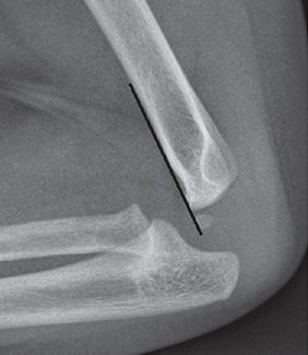
- It is easy to miss because in infants the distal humeral epiphysis is mostly UNOSSIFIED, so the fracture line and the displaced epiphysis are not directly visible on plain films - you must infer it from the MALALIGNMENT of the forearm with the humerus.
- The whole distal epiphysis (and with it the forearm) displaces as a UNIT, usually POSTEROMEDIALLY; crucially the proximal radius and ulna keep their NORMAL relationship to EACH OTHER and to the capitellum (radio-capitellar line maintained) - which DISTINGUISHES it from an elbow DISLOCATION (rare in this age) and from a LATERAL CONDYLE fracture.
- Because the cartilage is invisible on X-ray, an intra-operative ARTHROGRAM (or ultrasound/MRI) is commonly used to define the epiphysis and confirm reduction; treatment is CLOSED REDUCTION and PERCUTANEOUS PINNING, using techniques similar to supracondylar fractures.
- The most common complication is CUBITUS VARUS from malunion; others include OSTEONECROSIS of the medial condyle and GROWTH ARREST; an established cubitus varus can be corrected later with a lateral closing-wedge osteotomy.
- Given the age group and mechanism, you must actively consider and screen for NON-ACCIDENTAL INJURY (this fracture is a recognised abuse-associated injury), especially when the history is inconsistent.
- “In a child under 3 with a swollen elbow, if the forearm is not aligned with the humerus but the radius and ulna are aligned with each other (and the radio-capitellar line is intact), think transphyseal separation - not an elbow dislocation.
- “The distal humeral epiphysis is cartilaginous, so use an ARTHROGRAM (or ultrasound/MRI) to see it and confirm reduction.
- “Always consider NON-ACCIDENTAL INJURY in this age group - and remember cubitus varus is the commonest late complication.
The whole distal epiphysis + forearm displace together (usually posteromedially). The radius and ulna keep their normal relationship to each other and to the capitellum (radio-capitellar line maintained) - only the physis is disrupted. Common in infants/toddlers.
The forearm dislocates relative to the humerus with loss of the radio-capitellar relationship (the radius no longer points at the capitellum). True elbow dislocation is rare under 3 years - so an apparent 'dislocation' in a toddler is usually a transphyseal separation.
Who, How & Why It's Missed
Transphyseal fractures of the distal humerus typically occur in children younger than 3 years. The three classic mechanisms are birth trauma (a difficult delivery), non-accidental trauma, and a fall from a small height. The injury is a physeal separation - in Salter-Harris terms a type I or II - so the entire distal humeral epiphysis (carrying the elbow joint and the forearm) shears off the metaphysis and displaces, most often posteromedially.
In infants and toddlers the distal humeral epiphysis is largely cartilaginous - the capitellum ossifies around 1-2 years and the other ossification centres later - so the fracture line and the displaced epiphysis are not directly visible on plain radiographs. The clue is MALALIGNMENT of the forearm with the humerus: the forearm axis no longer lines up with the humeral shaft, even though the radius and ulna remain aligned with each other. Comparison with the contralateral elbow, and recognising that the radio-capitellar line is preserved (excluding dislocation), helps make the diagnosis.
Diagnosis
- A young child (under 3) with a swollen, painful elbow and pseudoparalysis of the arm
- Often birth-related (presents in the first days of life) or after a fall/abuse
- A history that does not fit the injury should raise non-accidental injury concern
- Plain radiographs (both elbows): look for forearm-humerus malalignment with a preserved radio-capitellar relationship; the epiphysis itself is invisible if cartilaginous
- Arthrogram (intra-operative), ultrasound, or MRI demonstrate the cartilaginous epiphysis and confirm the diagnosis and reduction
A transphyseal distal humerus fracture is a recognised abuse-associated injury in the under-3 age group. Where the mechanism is unclear or inconsistent, where there are other injuries, or where presentation is delayed, follow your local safeguarding pathway (full history, examination for other injuries, skeletal survey, social/child-protection involvement) alongside treating the fracture.
Management
Displaced transphyseal fractures are treated with closed reduction and percutaneous pinning (CRPP), using techniques similar to supracondylar humerus fractures. Because the epiphysis is cartilaginous, an intra-operative arthrogram is commonly used to visualise the epiphysis and confirm an accurate reduction before and after pin placement. Smooth K-wires stabilise the reduction and are removed once healed (which is rapid in this age group). Truly undisplaced injuries may be managed with immobilisation and close follow-up. The aims are an anatomic reduction to restore the carrying angle and to protect the physis and blood supply.
Complications
| 0 | 1 |
|---|---|
| Cubitus varus | MOST common; from malunion (malreduction) - a cosmetic/alignment deformity |
| Osteonecrosis of the medial condyle | Vascular insult to the medial condylar ossification centre |
| Growth arrest | Physeal injury -> partial arrest, angular deformity or length issue |
| Missed diagnosis | Cartilaginous epiphysis -> mistaken for dislocation/normal; delayed treatment |
If a cubitus varus deformity becomes established from malunion, it can be corrected later with a lateral closing-wedge (supracondylar) osteotomy to restore a near-normal carrying angle. Prevention - through accurate initial reduction confirmed on arthrogram - is far preferable.
Evidence & Key Studies
Transphyseal fracture of the distal humerus
- Typically occurs in children younger than 3 years from birth trauma, non-accidental trauma, or a fall from a small height; prompt accurate diagnosis is crucial.
- Recognising that the forearm is not aligned with the humerus on plain radiographs aids diagnosis; surgery is most commonly performed with the aid of an arthrogram (closed reduction and percutaneous pinning, as for supracondylar fractures).
- The most common complication is cubitus varus from malunion (also medial-condyle osteonecrosis and growth arrest); a corrective lateral closing-wedge osteotomy can restore the carrying angle.
Transphyseal distal humerus fracture (Instructional Course Lecture)
- Reinforces the under-3 age group and the birth/non-accidental/low-fall mechanisms, and the importance of recognising forearm-humerus malalignment.
- Describes arthrogram-assisted closed reduction and percutaneous pinning as the standard surgical management.
- Lists cubitus varus (malunion), medial-condyle osteonecrosis and growth arrest as the principal complications, correctable by lateral closing-wedge osteotomy when cubitus varus is established.
According to PubMed, the age group, mechanisms (including non-accidental trauma), the forearm-humerus malalignment diagnostic clue, arthrogram-assisted CRPP, and the complication profile come from the cited Abzug et al. review and the matching instructional course lecture. (See also our Supracondylar Humerus Fracture, Lateral Condyle Fractures and Physis topics.)
Clinical Decision Scenarios
Practise clinical reasoning and management decisions out loud
“A 14-month-old presents with a swollen elbow and is not using the arm; the parent's account of the injury is vague. Radiographs suggest the forearm is not aligned with the humerus. What is your diagnosis, how do you confirm it, and what else must you consider?”
“How would you treat a displaced transphyseal distal humerus fracture, and what complications would you warn about?”
Mnemonics & Memory Aids
INFANT
Hook:An INFANT's elbow: malaligned forearm, think NAI, arthrogram, not a dislocation, pin it.
VANG
Hook:VANG: varus, AVN, malalignment sign, growth arrest - the transphyseal fracture essentials.
Who & how
- Children under 3; birth trauma, NON-ACCIDENTAL trauma, or low fall
- Separation through the distal humeral physis (SH I/II); whole epiphysis + forearm displace (usually posteromedial)
- Recognised abuse-associated injury - screen for NAI
Diagnosis
- Epiphysis cartilaginous -> not directly visible on plain films
- Clue: forearm NOT aligned with humerus, but radio-capitellar line PRESERVED (vs dislocation)
- Confirm with arthrogram (or ultrasound/MRI); compare contralateral elbow
Treatment
- Displaced: closed reduction + percutaneous pinning (like supracondylar), arthrogram-assisted
- Undisplaced: immobilisation + close follow-up
- Aim: anatomic reduction to restore carrying angle, protect physis/blood supply
Complications
- Cubitus varus (most common, malunion) -> lateral closing-wedge osteotomy if established
- Medial condyle osteonecrosis; growth arrest
- Missed diagnosis from the invisible cartilaginous epiphysis