Oculo-Auriculo-Vertebral Spectrum - the Orthopaedic Burden of a Craniofacial Diagnosis
- Goldenhar syndrome is the severe end of the OCULO-AURICULO-VERTEBRAL SPECTRUM (OAVS), a disorder of first and second branchial arch development. It is usually SPORADIC, has a male predominance, and is RIGHT-SIDED more often than left, with bilateral involvement in a substantial minority.
- The orthopaedic burden is spinal: CERVICAL ANOMALIES including congenital fusion (with KLIPPEL-FEIL overlap), hemivertebrae, occipitalisation of the atlas, os odontoideum and atlantoaxial instability, plus CONGENITAL SCOLIOSIS which is progressive and unresponsive to bracing.
- EVERY child needs WHOLE-SPINE imaging at diagnosis - plain radiographs of the whole spine plus dedicated cervical views including flexion-extension laterals, with CT for bony detail and MRI of the whole spine to look for intraspinal anomalies (tethered cord, diastematomyelia, syringomyelia, Chiari malformation) BEFORE any deformity surgery.
- Limb anomalies occur in a minority and include RADIAL DEFICIENCY (radial longitudinal deficiency and thumb hypoplasia), Sprengel deformity, club foot and hip dysplasia.
- The ANAESTHETIC AIRWAY is the single most dangerous aspect for any operation on any part of the body: mandibular hypoplasia, restricted mouth opening from temporomandibular and ramus deficiency, and a stiff or unstable cervical spine that must not be extended. Plan the airway before you plan the operation.
- “Say the full name in the viva: oculo-auriculo-vertebral spectrum, or hemifacial microsomia when the eye findings are absent - the vertebral component is what makes it an orthopaedic problem.
- “Klippel-Feil is not a separate diagnosis here but an overlapping description: congenital cervical fusion, a short neck, a low posterior hairline and restricted cervical motion.
- “Congenital scoliosis in this population behaves like congenital scoliosis anywhere - the growth imbalance determines progression, bracing does not control the congenital curve, and a fully segmented unilateral hemivertebra is a high-risk pattern.
- “Look for what travels with it: cardiac anomalies, renal anomalies (agenesis or ectopia), and hearing loss - each of which changes the anaesthetic and perioperative plan.
Every anaesthetic, for every operation.
- Mandibular hypoplasia with a receding chin, restricted mouth opening from temporomandibular joint and ramus deficiency, and a small pharyngeal space make intubation genuinely difficult.
- A fused or unstable cervical spine means the neck must not be extended to help - and extension may itself risk the cord.
- Plan for videolaryngoscopy, a fibreoptic technique or an awake or spontaneously breathing approach, with an experienced paediatric anaesthetist, a difficult-airway trolley and a surgical airway plan available.
- Do not discover this in the anaesthetic room.
Not just the level that is symptomatic.
- Congenital vertebral anomalies are frequently multiple and non-contiguous, so the whole spine must be imaged.
- Include dedicated cervical views with flexion-extension laterals for instability, and CT for bony detail of the upper cervical spine.
- MRI of the whole spine is mandatory before any deformity surgery - intraspinal anomalies are common and change the plan.
A common and damaging misconception.
- A congenital curve arising from a structural vertebral anomaly does not respond to a brace, because the deforming force is asymmetric growth in the anomalous segment.
- Bracing may have a role in controlling a flexible compensatory curve above or below the congenital segment.
- Serial radiographic surveillance identifies the progressive curve; surgery is the treatment for progression.
They change perioperative management.
- Cardiac anomalies occur in a substantial minority - obtain an echocardiogram.
- Renal anomalies (unilateral agenesis, ectopia, hydronephrosis) mandate a renal ultrasound and affect drug and fluid management.
- Hearing loss is very common and affects consent, cooperation and rehabilitation - involve audiology early.
- There is meaningful overlap with the VACTERL association, so look for the whole pattern.
GOLDENHARThe Goldenhar Triad and Its Extensions
Hook:The first three letters give you the classic triad; the rest give you everything that will actually determine how you manage the child.
Bar Beats HemiCongenital Scoliosis Progression Risk
Hook:Ask two questions of every anomaly: is growth ASYMMETRIC, and is the anomalous segment SEGMENTED? Asymmetric growth potential equals progression.
Overview
Goldenhar syndrome is the name traditionally given to the severe, classically triadic end of the oculo-auriculo-vertebral spectrum (OAVS) - a heterogeneous disorder of first and second branchial arch development. The historical triad is hemifacial microsomia, epibulbar dermoid and vertebral anomalies, described by Gorlin and colleagues as oculoauriculovertebral dysplasia, building on Goldenhar's earlier account.
Modern practice treats it as a spectrum rather than a discrete syndrome. At the mild end sits isolated hemifacial microsomia or an isolated preauricular tag; at the severe end sits the full triad with extracranial anomalies. The term hemifacial microsomia is often used where the ocular findings are absent, and craniofacial microsomia as an umbrella term.
For the orthopaedic surgeon the essential point is simple: the diagnosis is made by the face and the ear, but the orthopaedic problems are in the spine. A child referred with a craniofacial diagnosis must have the spine imaged, screened and followed, because congenital cervical and thoracic anomalies in this population are common, frequently multiple, and clinically silent until they are not.
The framing to give in a viva, because it demonstrates you understand your own role in a multidisciplinary condition.
- The face is managed by others. Craniofacial surgery, ENT and audiology, ophthalmology and orthodontics own the mandible, the ear and the eye.
- The spine is yours, and it carries the two risks that can be catastrophic and are both preventable:
- An unrecognised unstable or stenotic cervical spine, which can produce myelopathy or a devastating cord injury during positioning or intubation for a craniofacial or dental procedure performed by someone else.
- A progressive congenital scoliosis that is not identified early, is inappropriately braced, and presents as a severe fixed deformity with thoracic insufficiency.
- The corollary: your single most valuable contribution is often not an operation but a complete set of screening images at diagnosis and a surveillance plan through growth, communicated to the wider team - together with a clear written statement about cervical stability and safe neck positioning that the anaesthetist and every other surgeon can rely on.
- The second corollary: because the airway is difficult and the neck may be unstable, every anaesthetic in this child is a shared risk, and the orthopaedic assessment of the cervical spine is what makes that anaesthetic safe.
A point worth making because it shows you understand the evidence rather than a list.
- Looking from the face to the spine: children with the oculo-auriculo-vertebral spectrum have a high frequency of vertebral anomalies, which is why screening is justified. Reported frequencies vary widely between series, driven largely by how completely each cohort was imaged and by the diagnostic criteria used, so quoting a single percentage is unwise.
- Looking from the spine to the face: in a large retrospective review of 668 consecutive patients with congenital spine deformity, Tsirikos and McMaster identified 14 with Goldenhar conditions. Among those 14, thoracic scoliosis was the commonest deformity, in 10 patients - eight from an isolated hemivertebra and two from a unilateral unsegmented bar with a contralateral hemivertebra at the same level. Thoracolumbar kyphosis occurred in four.
- The mechanistic link they emphasised is elegant and worth quoting: the association is non-random, representing unilateral errors in the morphogenesis of the spine and of the first and second branchial arches occurring in the same developmental window.
- Consistent with that, the side of the vertebral anomaly correlated with the side of the hemifacial microsomia in 5 of 11 patients with scoliosis or kyphoscoliosis in that series - a correlation, but far from an absolute rule, so never predict the side of the spinal anomaly from the face. Image both sides and the whole spine.
Aetiology and Epidemiology
Incidence and Demographics
- Incidence is commonly quoted at approximately 1 in 3,500 to 1 in 5,600 live births, making the spectrum one of the commoner craniofacial malformations after cleft lip and palate. Precise figures vary widely because the diagnostic criteria applied across the spectrum differ between studies.
- Sex: a male predominance. In the largest single craniofacial clinic series described, Rollnick and colleagues reported 294 patients with a male to female ratio of 191 to 103.
- Laterality: right-sided involvement is more common than left, and bilateral involvement occurs in a substantial minority. In the same 294-patient series, microtia was unilateral in 193 and bilateral in 98, and in only 34 of the 98 bilateral cases was the microtia symmetric - a useful reminder that bilateral does not mean equal.
- Inheritance: the great majority of cases are sporadic. Familial cases with autosomal dominant and, less commonly, autosomal recessive patterns are described with variable expressivity, and a proportion have identifiable chromosomal abnormalities or copy number variants. Recurrence risk in a sporadic case is low but not zero, and formal genetic counselling should be offered.
- Associated exposures reported in some series include maternal diabetes, assisted reproduction, and vasoactive drug exposure in early pregnancy - associations rather than established causes.
Pattern of Orthopaedic Involvement
The honest position on frequencies: reported figures vary widely between series because they depend on whether the whole spine was imaged and on which end of the spectrum the cohort came from. What is consistent across series is the pattern rather than the percentage.
- What the literature consistently shows
- A high frequency across series, with the reported proportion depending heavily on how completely the spine was imaged
- Practical consequence
- Image the WHOLE spine in every child - do not rely on symptoms or on a percentage
- What the literature consistently shows
- Cervical anomalies are prominent enough that anomalies of the cervical spine feature among the minimal diagnostic criteria used to define the spectrum
- Practical consequence
- Dedicated cervical views with flexion-extension laterals in every child
- What the literature consistently shows
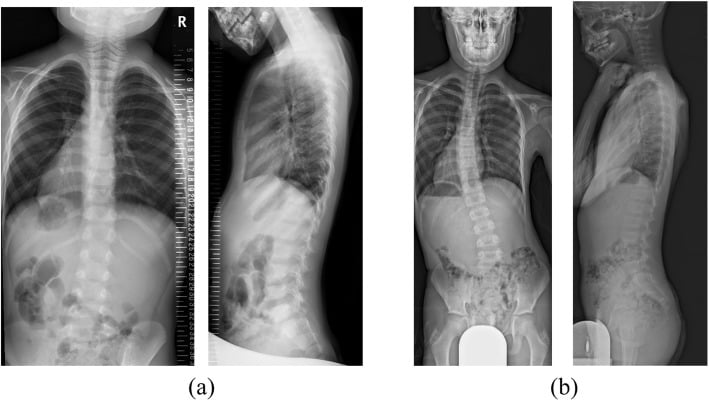
- Thoracic scoliosis is the commonest spinal deformity; in the Tsirikos and McMaster subgroup it affected 10 of 14 patients, most from an isolated hemivertebra
- Practical consequence
- Distinguish a structural congenital curve from a flexible compensatory one, and surveil through growth
- What the literature consistently shows
- Thoracolumbar kyphosis from posterior hemivertebrae, wedge vertebrae or an anterior unsegmented bar is described
- Practical consequence
- Assess the sagittal profile, not just the coronal curve
- What the literature consistently shows
- Occur alongside thoracic vertebral anomalies
- Practical consequence
- Assess the thorax and respiratory function - thoracic insufficiency is the outcome to fear
- What the literature consistently shows
- Avon and Shively, reviewing 23 patients, reported spinal anomalies, club foot, congenital dislocation of the hip, Sprengel deformity and radial limb defects as the main orthopaedic findings
- Practical consequence
- Examine the hips, feet, shoulder girdle and upper limbs in every child
- What the literature consistently shows
- Reported in a substantial minority across series
- Practical consequence
- Echocardiogram at diagnosis - it changes perioperative management
- What the literature consistently shows
- Unilateral agenesis, ectopia, hydronephrosis and duplex systems are described
- Practical consequence
- Renal ultrasound at diagnosis; a solitary kidney changes drug and fluid management
- What the literature consistently shows
- Very common, predominantly conductive from external and middle ear anomalies
- Practical consequence
- Audiology from the newborn period; it affects speech, schooling and cooperation
- What the literature consistently shows
- Tethered cord, diastematomyelia, syringomyelia and Chiari malformation occur in children with congenital vertebral anomalies and are frequently silent
- Practical consequence
- MRI of the WHOLE spine before any deformity surgery
Clinical Presentation
Craniofacial and Sensory Features
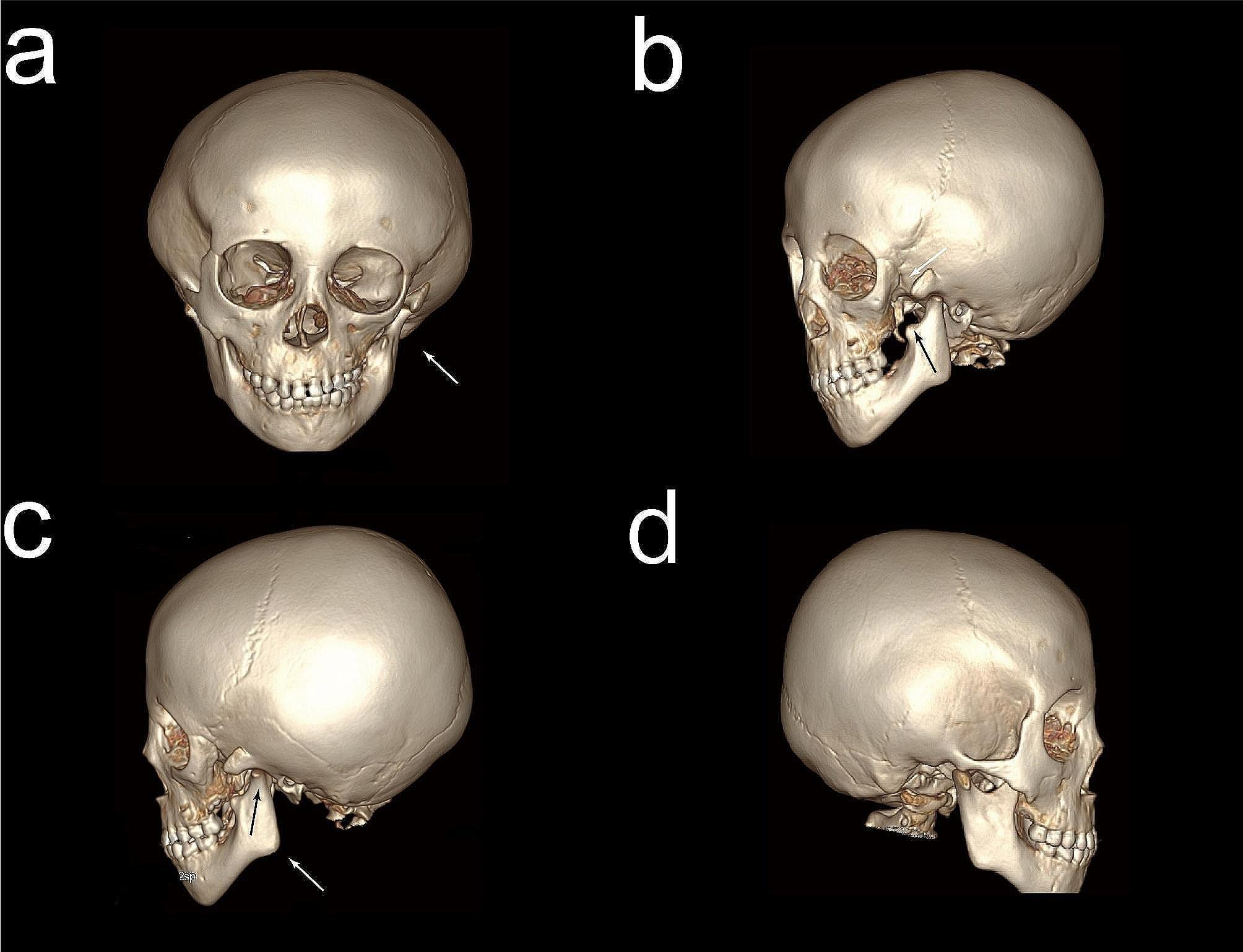
- Hemifacial microsomia - asymmetric hypoplasia of the mandible (ramus, condyle and temporomandibular joint) and to a lesser degree the maxilla and zygoma, producing facial asymmetry, chin deviation to the affected side, an occlusal cant and restricted mouth opening.
- Ear anomalies - microtia or anotia, preauricular tags and pits, external auditory canal stenosis or atresia, middle ear ossicular anomalies, and conductive hearing loss. Ear anomalies are the most consistently present feature across the spectrum, and microtia is one of the minimal diagnostic criteria used to define it.
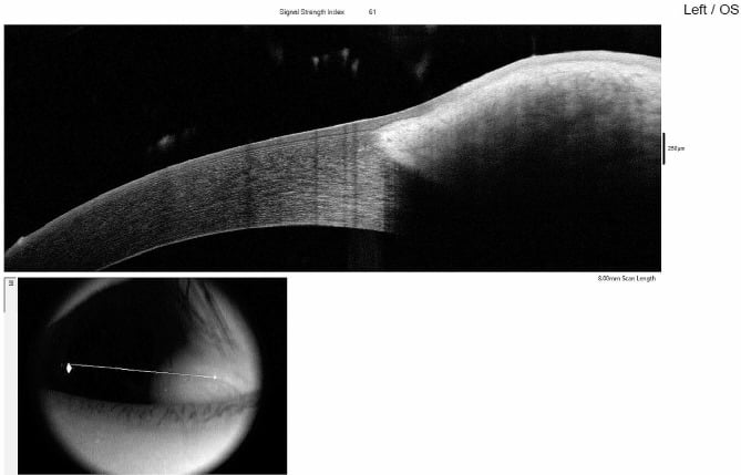
- Ocular anomalies - epibulbar dermoid or lipodermoid (the classic finding, typically at the inferolateral limbus), coloboma of the upper eyelid, microphthalmia, anophthalmia, strabismus and Duane retraction syndrome.
- Other first and second arch features - macrostomia or lateral facial cleft, cleft lip and palate, facial nerve weakness, parotid hypoplasia, and velopharyngeal insufficiency.
- Airway and feeding - a small mandible and pharynx may cause obstructive sleep apnoea, feeding difficulty and failure to thrive in infancy.
Orthopaedic Features
What to Look For
- Short neck, low posterior hairline and restricted cervical motion - the classic Klippel-Feil clinical triad, present in only a minority of children with congenital cervical fusion, so its absence does not exclude the anomaly.
- Torticollis or a fixed head tilt, which may be structural (from a hemivertebra or unilateral fusion) or compensatory (for hearing or visual asymmetry) - and the two must be distinguished.
- Congenital cervical fusion at one or multiple non-contiguous levels.
- Occipitocervical anomalies - occipitalisation of the atlas, basilar invagination, posterior element deficiency.
- Odontoid anomalies - os odontoideum, odontoid hypoplasia or aplasia - the key substrate for atlantoaxial instability.
- Neurological assessment is mandatory and must be documented at every review: tone, power, reflexes, clonus, plantar responses, gait, hand function and dexterity, and bladder function. Subtle myelopathy in a child presents as deteriorating handwriting, clumsiness, a change in gait or new toe-walking rather than as a complaint of neck pain.
- Symptoms of instability or stenosis: neck pain, torticollis, headache, paraesthesiae, weakness, loss of dexterity, deteriorating gait, and in the worst case transient quadriparesis after a minor injury.
The Klippel-Feil Overlap
- Klippel-Feil syndrome is a description, not a competing diagnosis - congenital fusion of two or more cervical vertebrae, with or without the clinical triad.
- What matters clinically is not the number of fused levels but the consequences:
- Adjacent segment hypermobility. Fused segments transfer motion to the mobile levels above and below, which become hypermobile and degenerate early. This is the mechanism of late instability and myelopathy.
- Occipitocervical involvement carries the highest risk, particularly occipitalisation of the atlas combined with a mobile fusion below it, which concentrates all motion at the atlantoaxial joint.
- Associated anomalies to look for: Sprengel deformity, scoliosis, rib anomalies, renal anomalies, congenital heart disease, hearing loss and, occasionally, mirror movements from synkinesis.
- Activity counselling is a genuine and often neglected part of management: children with multiple fused levels, occipitocervical anomalies or documented instability should be advised against collision and contact sports and against activities involving axial neck loading.
Assessment and Screening Imaging
A child diagnosed anywhere on the oculo-auriculo-vertebral spectrum requires a defined screening set, not an ad hoc investigation of whatever is symptomatic. Congenital vertebral anomalies are frequently multiple and non-contiguous and are usually clinically silent.
Spine imaging
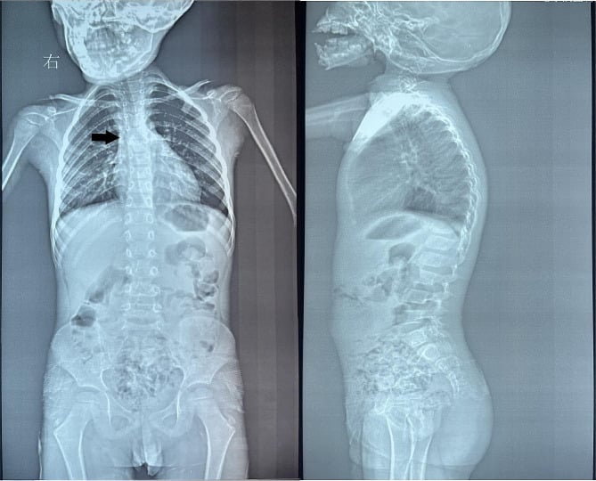
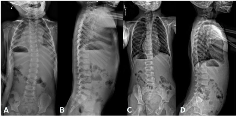
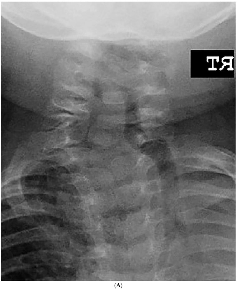
- Whole-spine plain radiographs, anteroposterior and lateral, standing where age permits - to identify every vertebral anomaly, count and characterise them, measure the curves and assess coronal and sagittal balance.
- Dedicated cervical spine radiographs - anteroposterior, lateral, open-mouth (odontoid) view where achievable, and flexion-extension laterals to assess instability. Measure the atlanto-dens interval and the space available for the cord.
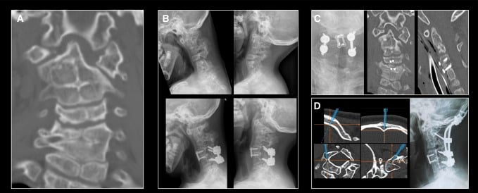
- CT of the cervical spine where the plain films show or suspect an occipitocervical or odontoid anomaly - it defines the bony anatomy, the pedicle and lateral mass dimensions, and the vertebral artery course for surgical planning.
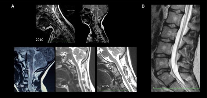
- MRI of the WHOLE spine - mandatory before any deformity surgery and indicated at diagnosis in most children, to identify intraspinal anomalies: tethered cord, diastematomyelia, syringomyelia, Chiari malformation, low-lying conus and intraspinal lipoma. These are common enough in congenital spinal deformity that operating without an MRI is indefensible.
Systemic screening 5. Echocardiogram - cardiac anomalies occur in a substantial minority. 6. Renal ultrasound - unilateral agenesis, ectopia and hydronephrosis are recognised, and a solitary kidney changes drug and fluid management. 7. Formal audiology assessment - hearing loss is very common and affects speech, development, consent and cooperation. 8. Ophthalmology assessment - epibulbar dermoid, lid coloboma, refractive error and amblyopia risk. 9. Genetics referral - for diagnostic clarification, chromosomal and copy number analysis where indicated, and family counselling. 10. Chromosomal breakage testing and a full blood count if there is radial deficiency - to exclude Fanconi anaemia and TAR syndrome. This is not optional.
Clinical Assessment
- What to do
- Assess mouth opening, thyromental distance, mandibular size and retrusion, cervical range of motion, and history of snoring or apnoea; review previous anaesthetic records
- What you are looking for
- A predictably difficult airway with a neck that cannot or must not be extended
- Pitfalls
- Assuming a previous uneventful anaesthetic in infancy predicts an easy airway later - the mandible becomes relatively more deficient with growth
- What to do
- Inspect for a short neck, low hairline, torticollis and webbing; measure range of motion in all planes; document a full neurological examination including gait, dexterity, reflexes, clonus and plantar responses
- What you are looking for
- Restricted motion, fixed tilt, and any sign of myelopathy
- Pitfalls
- Recording a normal examination without testing gait, tandem walking and hand dexterity - the earliest myelopathic signs in a child
- What to do
- Adams forward bend test with a scoliometer, assess shoulder and pelvic balance, leg length, sagittal profile, and flexibility on side-bending; measure and plot height and sitting height
- What you are looking for
- A structural curve at the level of a vertebral anomaly, versus a flexible compensatory curve; kyphosis; growth velocity
- Pitfalls
- Failing to distinguish a compensatory curve from a congenital one and bracing the wrong thing; assessing only the coronal plane
- What to do
- Inspect the whole back for a hairy patch, dimple, sinus, naevus, haemangioma or subcutaneous lipoma
- What you are looking for
- Cutaneous stigmata of an underlying intraspinal anomaly or dysraphism
- Pitfalls
- Examining only the region of the curve; subtle midline stigmata are easily missed
- What to do
- Assess forearm length and bowing, wrist stability and deviation, thumb size, stability and thenar function, first web space, and grip and pinch pattern; assess ELBOW motion and hand-to-mouth function
- What you are looking for
- Radial longitudinal deficiency (Bayne and Klug) and thumb hypoplasia (Blauth), particularly the IIIA versus IIIB distinction
- Pitfalls
- Missing an unstable carpometacarpal joint and planning a reconstruction that will fail; ignoring elbow stiffness before planning wrist correction
- What to do
- Inspect and palpate the scapular position and assess shoulder abduction; look for an omovertebral bar clinically and radiographically
- What you are looking for
- Sprengel deformity, which frequently accompanies cervical anomalies
- Pitfalls
- Attributing limited abduction to the glenohumeral joint when the scapula is undescended and tethered
- What to do
- Ortolani and Barlow in the infant, hip range and leg lengths thereafter; examine the feet for talipes and other deformity
- What you are looking for
- Developmental dysplasia or congenital dislocation of the hip, and club foot
- Pitfalls
- Deferring hip examination because the craniofacial problem dominates the consultation
- What to do
- Plot height, sitting height and weight; assess chest wall symmetry and expansion; formal pulmonary function testing where age permits
- What you are looking for
- Thoracic insufficiency where there are rib anomalies or a hypoplastic hemithorax
- Pitfalls
- Assessing the spine radiographically but never assessing the thorax functionally
- What to do
- Confirm that craniofacial, ENT and audiology, ophthalmology, cardiology, nephrology, genetics, anaesthesia and, where relevant, hand surgery are all engaged; write a clear statement about cervical stability and safe neck positioning into the record
- What you are looking for
- A coordinated plan and a documented cervical spine status available to every other team
- Pitfalls
- Each specialty managing its own organ with nobody owning the overall plan or the neck
Grading Systems to Know
Management
State the team structure explicitly in an exam; it demonstrates that you understand this is a coordinated condition, not a series of isolated problems.
- Craniofacial and maxillofacial surgery - mandibular distraction, orthognathic surgery, soft-tissue augmentation, macrostomia repair, timed against facial growth.
- ENT and audiology - hearing assessment from the newborn period, bone-conduction or conventional hearing aids, external ear reconstruction or a prosthesis, airway assessment and management of obstructive sleep apnoea.
- Ophthalmology - epibulbar dermoid management, lid coloboma repair, refraction and amblyopia therapy.
- Cardiology and nephrology - screening and management of associated anomalies.
- Genetics - diagnosis, testing and family counselling.
- Paediatric anaesthesia - airway planning for every procedure, in the light of the documented cervical spine status.
- Speech and language therapy, dietetics and psychology - feeding, communication and the very substantial psychosocial burden of a visible facial difference.
- Orthopaedics and spinal surgery - the cervical spine and its stability, spinal deformity surveillance and correction, the shoulder girdle, and the limbs.
- Hand surgery - radial deficiency and thumb reconstruction.
- A named coordinator. Someone must own the overall plan; in practice this is usually the craniofacial team, but the orthopaedic contribution must be explicit and written down.
Principles
- Most congenital cervical anomalies are managed non-operatively with surveillance, activity modification and clear documentation.
- Surveillance: clinical review with a documented neurological examination, and repeat imaging including flexion-extension laterals at intervals determined by the anatomy and by growth. Increase frequency during growth spurts and if symptoms change.
- Activity counselling: advise against collision and contact sports and activities involving axial neck loading where there are multiple fused levels, occipitocervical anomalies, or any documented instability. Put this in writing.
- Communicate the cervical status to everyone. Every anaesthetist and every other surgeon needs to know whether the neck is stable and how far it may safely be moved. A written statement in the record and a note for the parents is one of the most valuable things you will do.
When to Fuse
Indications for surgical stabilisation:
- Any objective neurological deficit or documented myelopathy attributable to the cervical spine.
- Demonstrated instability on flexion-extension imaging - an increased or dynamically changing atlanto-dens interval, or a space available for the cord that is reduced below the safe threshold (a space available for the cord of less than about 13 mm is widely used as the level at which the risk of cord compromise becomes significant).
- Progressive deformity producing an unacceptable fixed head position or compromising function or the airway.
- Symptomatic adjacent segment degeneration with instability above or below a congenital fusion.
- Transient neurological events after minor trauma - an ominous sign that should prompt definitive stabilisation.
What the Operation Involves
- Level selection is dictated by the anatomy: an occipitocervical fusion where the occipitoatlantal junction is involved or the atlas is occipitalised, or an atlantoaxial fusion for isolated instability with a competent occipitoatlantal junction.
- Preoperative CT with vascular assessment is essential. The vertebral artery course is frequently anomalous in congenital cervical anomalies - high-riding, medially placed or aberrant - and lateral mass, pedicle and transarticular screw trajectories must be planned individually.
- Autograft, meticulous decortication and rigid instrumentation where the bone stock permits; in a small child, instrumentation options are limited by anatomy and onlay grafting with halo or rigid orthosis immobilisation may be required.
- Neuromonitoring and careful positioning - the neck must not be extended to obtain the view.
- Counsel about the cost of fusion: loss of motion, loading of adjacent levels, and in a growing child the effect on subsequent cervical growth and alignment.
The Positioning and Intubation Risk
- The most likely moment of catastrophe in this condition is not during spinal surgery but during an anaesthetic for something else - a dental procedure, a mandibular distraction, an ear reconstruction - when the neck is extended to secure a difficult airway in a child with unrecognised instability.
- Preventing that is an orthopaedic responsibility: screen the cervical spine, document the status, and state explicitly what neck movement is safe.
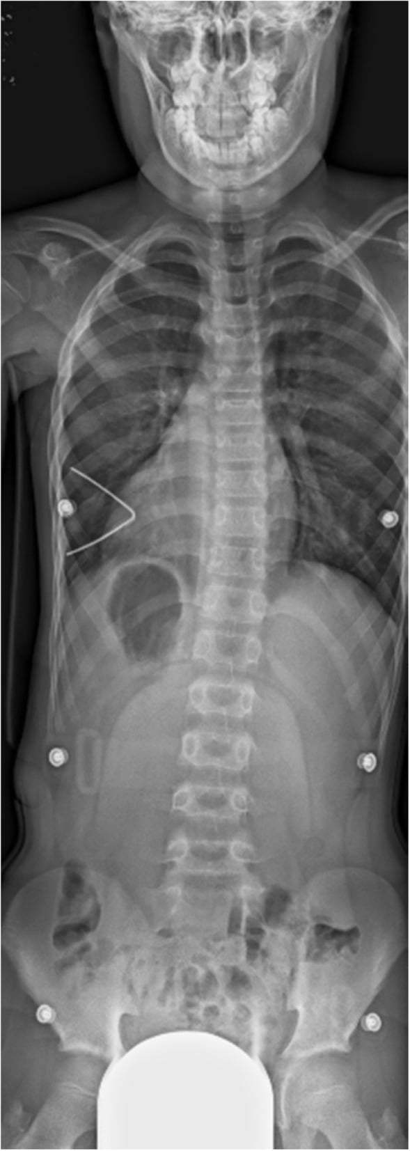
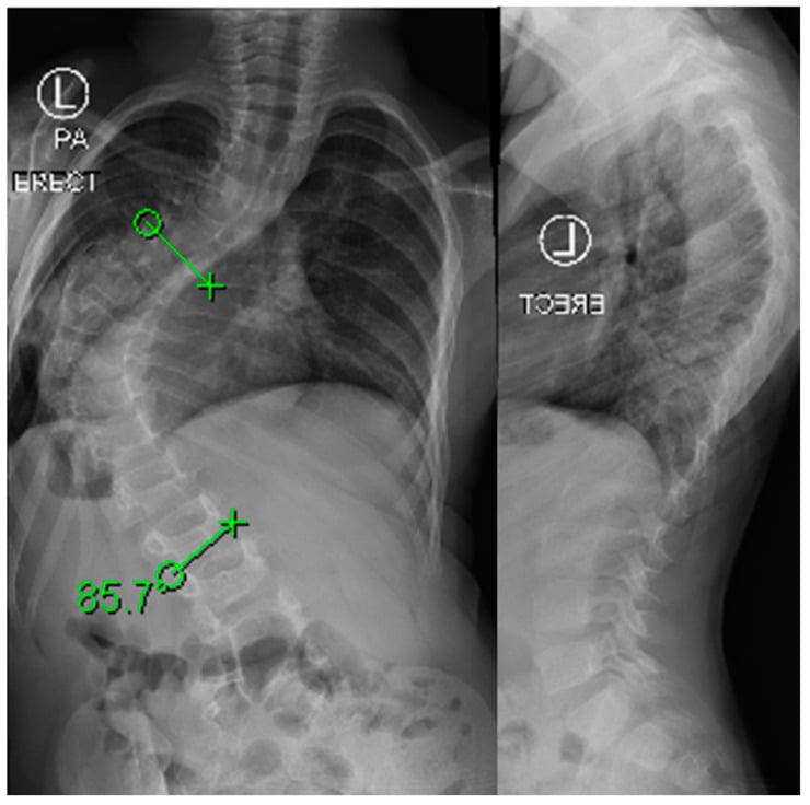
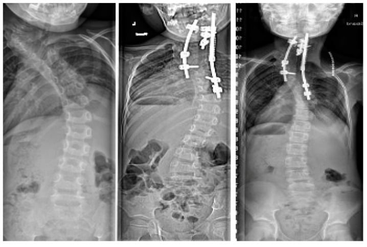
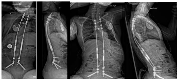
Complications
- Mechanism
- Neck extension to secure a difficult airway in a child with unrecognised cervical instability or a narrow space available for the cord
- Avoidance
- Screen the cervical spine at diagnosis; document stability and safe range in writing; manual in-line stabilisation; videolaryngoscopic or fibreoptic technique
- Mechanism
- Mandibular hypoplasia, restricted mouth opening and a small pharynx
- Avoidance
- Senior paediatric anaesthetist, planned difficult-airway technique, spontaneous ventilation preserved, surgical airway plan available
- Mechanism
- Failure to surveil, or inappropriate reliance on bracing for a congenital curve
- Avoidance
- Whole-spine imaging at diagnosis, stratify progression risk by anomaly type, serial radiographs with increased frequency during growth spurts, operate on progression
- Mechanism
- Multiple rib anomalies and a hypoplastic hemithorax limiting lung growth and respiratory mechanics
- Avoidance
- Assess the thorax and respiratory function, not just the curve; consider growth-friendly instrumentation rather than early definitive fusion
- Mechanism
- Correction stretching a cord that is tethered or has an unrecognised intraspinal anomaly
- Avoidance
- MRI of the WHOLE spine before surgery; address intraspinal pathology first; mandatory neuromonitoring
- Mechanism
- Anomalous vertebral artery course, which is common in congenital cervical anomalies
- Avoidance
- Preoperative CT with vascular assessment; individualised screw trajectories; consider alternative fixation or onlay grafting
- Mechanism
- Motion transferred from fused to mobile segments, which become hypermobile and degenerate early
- Avoidance
- Long-term surveillance with documented neurological examination; activity counselling; stabilise symptomatic instability
- Mechanism
- Failure to perform chromosomal breakage testing
- Avoidance
- Test every child with radial longitudinal deficiency - it is a life-threatening, treatable diagnosis
- Mechanism
- Straightening the wrist in a child with a stiff elbow who relied on radial deviation to reach the mouth
- Avoidance
- Assess elbow motion and hand-to-mouth function before planning wrist correction
- Mechanism
- Excessive caudal translation of the scapula
- Avoidance
- Limit translation; consider clavicular osteotomy; intraoperative monitoring
- Mechanism
- Cumulative burden of repeated lengthening procedures in a young child
- Avoidance
- Counsel families explicitly about the number of procedures and the high cumulative complication rate
- Mechanism
- Unrecognised or undertreated conductive hearing loss
- Avoidance
- Audiology from the newborn period, early amplification, educational support
- Mechanism
- Visible facial difference, repeated hospital admissions, multiple operations
- Avoidance
- Early and continuing psychology input, peer support, and involvement of the child in decisions as they mature
Outcomes and Follow-Up
Prognosis
- Life expectancy is usually normal in the absence of a severe cardiac anomaly or a significant airway problem. The burden is one of function, appearance, hearing and multiple operations rather than mortality.
- Craniofacial outcomes have improved considerably with distraction osteogenesis and staged reconstruction, and facial symmetry can be substantially improved - though facial growth means results must be reassessed through adolescence.
- Hearing outcomes depend on early identification and amplification; conductive loss is generally amenable to bone-conduction devices or reconstruction.
- Spinal outcomes depend almost entirely on whether progression is identified early. A congenital curve identified in infancy and treated when short and flexible has a far better outcome than the same curve identified as a severe rigid deformity in adolescence.
- Cervical spine outcomes are usually good with surveillance alone; the poor outcomes are the ones caused by an unrecognised anomaly during someone else's anaesthetic.
- Upper limb function after pollicisation for a Blauth IIIB or higher thumb is generally good, with a sensate opposable thumb; results after wrist centralisation are more variable and recurrence of radial deviation is common.
Follow-Up Strategy
Lifelong Surveillance in Oculo-Auriculo-Vertebral Spectrum
Whole-spine radiographs, dedicated cervical views including flexion-extension laterals, CT of the cervical spine if an occipitocervical or odontoid anomaly is suspected, MRI of the whole spine, echocardiogram, renal ultrasound, audiology, ophthalmology and genetics. Chromosomal breakage testing if there is radial deficiency. Write a clear statement of cervical stability and safe neck positioning into the record.
Six-monthly to annual orthopaedic review with documented neurological examination and spinal radiographs, more often if a high-risk anomaly is present. Hip and foot examination. Serial splinting for radial deficiency. Audiology and speech surveillance. Confirm the airway plan is in the notes before every anaesthetic.
Annual clinical and radiographic review of the spine, with growth plotted. Reassess cervical range and neurology. Hand surgery interventions are commonly timed in this window. Reinforce activity counselling as the child takes up sport.
Four- to six-monthly review with radiographs during peak height velocity. This is when a previously stable congenital curve can progress rapidly, and when adjacent segment problems above a cervical fusion begin to declare themselves.
Establish the final spinal and limb status, complete any staged reconstruction, and plan transition to adult services with a full written summary - especially the cervical spine status, which the patient will need for every future anaesthetic.
Continued access for new neck or neurological symptoms and for adjacent segment problems. Provide the patient with a written record of their cervical anatomy and safe positioning to carry - it may prevent a catastrophe during an unrelated operation decades later.
What to Tell the Family
- This is a spectrum of conditions affecting the development of the face, ear, eye and spine, almost always occurring by chance and not caused by anything that happened in pregnancy.
- Several teams will be involved, and one of them will coordinate the overall plan.
- We will image the whole spine now, even though the spine looks and feels normal, because the changes are common, often silent, and much easier to manage if we know about them.
- The neck matters for every future anaesthetic. We will give you a written record of it, and you should show it to any doctor before any operation, however minor.
- If there is a curve in the spine, we will follow it closely, especially during growth spurts. A brace will not straighten a curve caused by an abnormally shaped vertebra, but that does not mean nothing can be done - it means we watch carefully and act if it progresses.
- Hearing is very often affected, and treating it early makes a real difference to speech and school.
- Ask for genetic counselling. The chance of it happening again is low, but the question deserves a proper answer.
Guidelines, Registries & Global Practice
Global Framing
- The oculo-auriculo-vertebral spectrum occurs worldwide, and there is no established ethnic predilection. Reported frequencies of individual features differ substantially between series, driven largely by how completely each cohort was imaged and how the diagnostic criteria were applied rather than by real biological variation. This is why a single quoted percentage for spinal involvement is unreliable, and why the practical instruction is simply to image every child.
- Because the diagnosis spans several specialties, care is delivered through craniofacial or cleft and craniofacial networks in most high-income systems, with orthopaedics as a contributing specialty rather than the coordinating one. Where such networks do not exist, the spine is the component most likely to be neglected - which makes the orthopaedic screening contribution disproportionately valuable.
- No dedicated registry exists for the spectrum. Data come from congenital anomaly registries (which capture the craniofacial and limb features), craniofacial network databases, and paediatric spinal deformity registries and study groups that capture the congenital scoliosis component.
Side-by-Side Guidance
- Position relevant to orthopaedic management
- Multidisciplinary team management from diagnosis with defined screening for associated anomalies, staged reconstruction timed to facial growth, and a documented airway plan for every anaesthetic.
- Position relevant to orthopaedic management
- Whole-spine imaging in any child with a congenital vertebral anomaly, stratification of progression risk by anomaly type, MRI of the whole spine before any corrective surgery, mandatory neuromonitoring, and preference for growth-friendly techniques where the thorax is at risk.
- Position relevant to orthopaedic management
- Early identification and excision of a progressive fully segmented hemivertebra while the curve is short; explicit acknowledgement that bracing does not control a congenital curve; long-term surveillance through growth.
- Position relevant to orthopaedic management
- Similar principles with strong emphasis on network-based multidisciplinary care, structured surveillance protocols and clear written communication of cervical spine status between specialties.
- Position relevant to orthopaedic management
- A predicted difficult paediatric airway requires a senior anaesthetist, a stepwise plan with videolaryngoscopy and fibreoptic options, preservation of spontaneous ventilation, and an explicit surgical airway plan - with cervical spine status established beforehand.
- Position relevant to orthopaedic management
- Serial splinting from infancy, mandatory exclusion of Fanconi anaemia and TAR syndrome, preservation of elbow motion and hand-to-mouth function, and pollicisation for Blauth IIIB and above.
Areas of Genuine Uncertainty
- The true frequency of vertebral and cervical anomalies across the spectrum, because it depends entirely on how completely cohorts are imaged and on where on the spectrum the cohort sits. Reported figures vary widely and should be quoted with that caveat.
- Optimal cervical surveillance interval and imaging modality in an asymptomatic child with congenital fusion. No comparative data exist, and the balance between detecting silent instability and cumulative radiation exposure is unresolved.
- Whether asymptomatic radiographic instability should be stabilised prophylactically or surveilled - a genuine and unresolved debate in congenital cervical anomalies generally.
- Optimal timing of hemivertebra excision. Early surgery gives a shorter fusion and better correction but carries a higher technical and neurological risk in a small child.
- Growth-friendly instrumentation versus early definitive fusion where the thorax is compromised - the trade-off between preserving thoracic growth and the very substantial cumulative complication burden of repeated procedures.
- Genetic aetiology. No single gene explains the majority of cases, and the yield of genomic testing in a sporadic case with a classic phenotype is modest, so the role of routine genomic testing is still evolving.
- Whether activity restriction genuinely prevents neurological events in congenital cervical fusion - it is universally advised on mechanistic grounds and never studied.
- Why the side of the vertebral anomaly correlates only partially with the side of the facial deformity - the correlation was present in only 5 of 11 patients with scoliosis in the Tsirikos and McMaster series, so the developmental mechanism is clearly not simply unilateral.
High- versus Limited-Resource Practice
- Well-resourced settings: established craniofacial networks, whole-spine MRI and CT, three-dimensional planning, intraoperative neuromonitoring, growth-friendly spinal instrumentation, mandibular distraction, bone-conduction hearing devices, genomic testing, and full multidisciplinary support including psychology.
- Limited-resource settings: the highest-value interventions are cheap. Plain radiographs of the whole spine including flexion-extension cervical laterals identify almost all the anomalies that matter and cost very little. A documented neurological examination at every visit detects myelopathy. A written statement of cervical spine status carried by the family may prevent a cord injury during an unrelated anaesthetic anywhere in the world. Newborn hearing screening and early amplification transform developmental outcomes. Where MRI is unavailable, the honest position is that corrective spinal deformity surgery should not be undertaken, because the risk of operating on an unrecognised tethered cord is unacceptable - and referral to a centre with MRI is the correct decision.
- Everywhere: the two interventions that most change outcomes are screening the whole spine at diagnosis and communicating the cervical spine status in writing to everyone who will ever anaesthetise this child. Neither requires advanced technology, and both prevent catastrophes.
MCQ Practice Points
Q: What is the full name of the condition of which Goldenhar syndrome is the severe end? A: The OCULO-AURICULO-VERTEBRAL SPECTRUM (OAVS) - a disorder of first and second branchial arch development. Hemifacial or craniofacial microsomia is used where the ocular features are absent.
Q: Which four features were used by Rollnick and colleagues as the minimal diagnostic criteria for the spectrum? A: Microtia, mandibular hypoplasia, anomalies of the CERVICAL SPINE, and epibulbar dermoid or lipodermoid. Note that a spinal feature is one of the defining criteria.
Q: What spinal imaging does every child with this diagnosis need? A: WHOLE-SPINE radiographs plus dedicated cervical views including FLEXION-EXTENSION laterals, with CT for an occipitocervical or odontoid anomaly and MRI of the whole spine - mandatory before any deformity surgery.
Q: What is the commonest spinal deformity, and the commonest underlying anomaly? A: Thoracic scoliosis, most often caused by an isolated hemivertebra (10 of 14 patients in the Tsirikos and McMaster subgroup, 8 of them from an isolated hemivertebra).
Q: What is the Klippel-Feil clinical triad, and how often is it present? A: A short neck, low posterior hairline and restricted cervical motion - present in only a minority of children with congenital cervical fusion, so its absence does not exclude the anomaly.
Q: Does bracing control a congenital scoliosis? A: No. The deforming force is asymmetric growth within the anomalous segment. Bracing may control a flexible compensatory curve above or below it.
Q: Which congenital vertebral anomaly carries the highest progression risk? A: A unilateral unsegmented bar with a contralateral hemivertebra, followed by a unilateral unsegmented bar alone, then a fully segmented hemivertebra. Block vertebrae carry the lowest risk.
Q: What canal dimension is used as the threshold for significant risk of cord compromise? A: A space available for the cord of less than about 13 mm. Measure it alongside the atlanto-dens interval on flexion-extension views.
Q: Why is the airway so dangerous in this condition? A: Mandibular hypoplasia, restricted mouth opening from temporomandibular and ramus deficiency, and a small pharynx create a difficult airway in a child whose cervical spine may be fused or unstable - so the usual rescue manoeuvre, neck extension, is unavailable or unsafe.
Q: A child with this spectrum has radial longitudinal deficiency. What test must you not omit? A: Chromosomal breakage testing to exclude FANCONI ANAEMIA, plus a platelet count for TAR syndrome, an echocardiogram (Holt-Oram) and renal and vertebral assessment (VACTERL).
Exam Viva Scenarios
Practise clinical reasoning and management decisions out loud
“A 3-year-old girl with a confirmed diagnosis of Goldenhar syndrome is referred to you from the craniofacial clinic before planned mandibular surgery, with a request to 'clear the spine'. She has no neck or back symptoms and her parents report normal development apart from hearing difficulty. What do you do?”
“A 6-year-old boy with Goldenhar syndrome has a right thoracic curve that has increased from 28 to 45 degrees over 18 months. Radiographs show a fully segmented hemivertebra at T7 on the convexity, with a partially fused rib pair adjacent to it. He is otherwise well. His parents ask about a brace. Discuss your management.”
“A 12-year-old with Goldenhar syndrome is listed by another team for a dental procedure under general anaesthesia at short notice. The anaesthetist telephones you the evening before because the anaesthetic record from a procedure at age 2 says 'grade 1 view, uneventful'. There are no recent cervical spine images. What is your advice?”
Identity
- Severe end of the OCULO-AURICULO-VERTEBRAL SPECTRUM (OAVS)
- First and second branchial arch developmental disorder
- Triad: hemifacial microsomia, epibulbar dermoid, vertebral anomalies
- Male predominance; RIGHT more often than left; bilateral usually asymmetric
- Mostly sporadic; VACTERL overlap
Craniofacial and Sensory
- Mandibular hypoplasia - Pruzansky and Kaban grades I to III
- Microtia, anotia, preauricular tags and pits, canal atresia
- Conductive hearing loss - very common, screen from birth
- Epibulbar dermoid, upper lid coloboma, microphthalmia
- Facial nerve weakness, macrostomia, cleft lip and palate
Orthopaedic Burden
- Vertebral anomalies common - often multiple, non-contiguous and SILENT
- Cervical fusion (Klippel-Feil overlap), occipitalisation, os odontoideum
- Atlantoaxial instability and adjacent segment hypermobility
- THORACIC scoliosis is commonest, usually from an isolated hemivertebra
- Thoracolumbar kyphosis; rib anomalies; thoracic insufficiency risk
- Radial deficiency, Sprengel, club foot, congenital hip dislocation
Screening at Diagnosis
- WHOLE-SPINE radiographs AP and lateral
- Cervical views including open-mouth and FLEXION-EXTENSION laterals
- Measure atlanto-dens interval and space available for the cord (under 13 mm is significant)
- CT for occipitocervical or odontoid anomaly; MRI of the WHOLE spine
- Echocardiogram, renal ultrasound, audiology, ophthalmology, genetics
- Chromosomal breakage testing if radial deficiency (Fanconi anaemia)
Spine Management
- Bracing does NOT control a congenital curve
- Progression risk: unilateral bar plus contralateral hemivertebra worst; block vertebra best
- MRI before any correction - tethered cord, diastematomyelia, syrinx
- Progressive segmented hemivertebra: EARLY excision plus short fusion
- Fuse the cervical spine for deficit, documented instability or progressive deformity
- Preoperative CT for the anomalous VERTEBRAL ARTERY course
The Airway
- Small mandible, restricted mouth opening, small pharynx
- Gets HARDER with growth - an easy infant anaesthetic means nothing
- Neck may be fused or UNSTABLE - extension unavailable or unsafe
- Senior paediatric anaesthetist; videolaryngoscopy or fibreoptic
- Preserve spontaneous ventilation; surgical airway plan available
- Manual in-line stabilisation; give the family a PORTABLE written record
Evidence Base
Oculoauriculovertebral Dysplasia
- Defined oculoauriculovertebral dysplasia as a recognisable pattern of anomalies of the eye, ear and vertebral column
- Established the association between the craniofacial features and vertebral anomalies
- Described the variability of expression between affected individuals
- Provided the terminology that underpins the modern concept of a spectrum rather than a discrete syndrome
Oculoauriculovertebral Dysplasia and Variants - Phenotypic Characteristics of 294 Patients
- The largest single craniofacial clinic series described at the time - 294 individuals with oculoauriculovertebral dysplasia and variants
- Male to female ratio 191 to 103, confirming a male predominance
- The population was subdivided by minimal diagnostic criteria, namely microtia, mandibular hypoplasia, anomalies of the cervical spine, and epibulbar dermoid or lipodermoid
- Microtia was unilateral in 193 patients and bilateral in 98, and was symmetric in only 34 of the 98 bilateral cases
- Demonstrated the wide phenotypic variability that justifies the spectrum concept
Orthopaedic Manifestations of Goldenhar Syndrome
- Reviewed 23 patients with Goldenhar syndrome alongside a review of the literature
- Noted that the craniofacial manifestations had been studied extensively while the orthopaedic findings had received little attention
- Identified spinal anomalies as the main orthopaedic association
- Also reported club foot, congenital dislocation of the hip, Sprengel deformity and radial limb defects
Goldenhar-Associated Conditions (Hemifacial Microsomia) and Congenital Deformities of the Spine
- Retrospective review of 668 consecutive patients with congenital spine deformity, of whom 14 had Goldenhar conditions
- Thoracic scoliosis was the commonest deformity, occurring in 10 of the 14 patients - 8 from an isolated hemivertebra and 2 from a unilateral unsegmented bar with a contralateral hemivertebra at the same level
- Thoracolumbar kyphosis occurred in 4 patients, from posterior hemivertebrae, wedge vertebrae or an anterior unsegmented bar
- The side of the vertebral anomaly correlated with the side of the hemifacial microsomia in only 5 of 11 patients with scoliosis or kyphoscoliosis
- Characterised the association as non-random, representing unilateral errors in the morphogenesis of the spine and of the first and second branchial arches
- THE PAPER'S STATED CONCLUSION, which is the level-specific rule: failures of SEGMENTATION were the most frequent abnormality in the CERVICAL spine, whereas failures of FORMATION most commonly occurred in the THORACIC or THORACOLUMBAR spine
- Klippel-Feil syndrome was present in 6 of the 14, and 8 of 14 (57%) needed surgery at a mean age of 9.8 years (range 2.9-19), half by combined anterior-posterior and half by posterior-only arthrodesis
Oculo-Auriculo-Vertebral Spectrum - a Review of the Literature and Genetic Update
- Comprehensive review of the clinical spectrum and its genetic basis
- Confirmed marked genetic heterogeneity, with no single gene accounting for the majority of cases
- Reviewed reported copy number variants, chromosomal rearrangements and candidate genes
- Summarised the frequency of extracranial anomalies including vertebral, cardiac and renal involvement
- Supported a developmental field defect of the first and second branchial arches as the unifying mechanism
The Natural History of Congenital Scoliosis - A Study of Two Hundred and Fifty-One Patients
- Analysed the natural history of congenital scoliosis in 251 patients stratified by the type of vertebral anomaly
- A unilateral unsegmented bar with a contralateral hemivertebra carried the worst prognosis, with rapid severe progression
- A unilateral unsegmented bar alone and a fully segmented hemivertebra were also associated with significant progression
- Block vertebrae and incarcerated or unsegmented hemivertebrae showed little progression
- Progression was greatest during the periods of rapid growth in infancy and adolescence
Hemifacial Microsomia - Anatomical Prediction of Difficult Intubation
- Retrospective review of 102 children with hemifacial microsomia over ten years, testing whether the RADIOGRAPHIC severity of mandibular hypoplasia predicts intubation difficulty
- Mandible classified as type I ('mini-mandible'), type II (abnormal condylar size and shape) or type III (absent ramus, condyle and temporomandibular joint); intubation graded A easy, B difficult, C very difficult
- In the 82 UNILATERAL patients: 70% grade A, 21% grade B, 9% grade C. NO type I mandible produced a grade C airway, whereas 25% of type III did - the correlation with mandibular type was significant (p = 0.001)
- IN THE 20 BILATERAL PATIENTS THE DISTRIBUTION INVERTED: 30% grade A, 35% grade B and 35% grade C - more than a third were very difficult
- The authors limit their conclusion to UNILATERAL disease: radiographic classification of the mandible is a useful preoperative adjunct there
Congenital Scoliosis
- INTRASPINAL ABNORMALITIES ARE PRESENT IN APPROXIMATELY ONE THIRD of patients with congenital spine deformities - the number behind routine whole-spine MRI
- Congenital spine anomalies are associated with anomalies of OTHER ORGAN SYSTEMS in more than half of patients, so cardiac, auditory, genitourinary and renal screening is part of the workup rather than an optional extra
- MRI is warranted when curve progression is established or when surgical intervention is planned
- Curve progression is best documented by measuring IDENTICAL landmarks on sequential radiographs - in a congenitally malformed spine the landmarks are not obvious, and inconsistent choices manufacture apparent progression
- Progressive deformity is generally managed by early in situ fusion, because orthotic treatment is rarely appropriate in a congenital curve
Airway Management in Children with Hemifacial Microsomia - a Retrospective Study of 311 Cases
- 136 children with hemifacial microsomia aged 5 to 17, across 311 anaesthetic procedures at a single centre
- A DIFFICULT LARYNGOSCOPIC VIEW occurred in 95 procedures (38.9%) and correlated strongly with failed direct laryngoscopy (p less than 0.001)
- Intubation succeeded in 100% of attempts with video laryngoscopy and with fibrescopy, against only 79.5% with DIRECT laryngoscopy
- Face-mask ventilation was possible in every child except one - who had BILATERAL involvement and needed two practitioners; airway-visualising equipment was the primary technique in 3 of the 4 bilateral children
- Of 60 children who underwent mandibular distraction osteogenesis, the laryngoscopic view improved in 26 (43%)