Greater Trochanteric Pain Syndrome | Lateral Hip Pain | Gluteal Tendinopathy vs Bursitis
- Greater trochanteric pain syndrome (GTPS) is the preferred umbrella term encompassing trochanteric bursitis, gluteal tendinopathy, and ITB pathology
- Up to two-thirds of lateral hip pain is due to gluteal tendinopathy, not true bursitis
- Finkelstein test is NOT for trochanteric bursitis — use palpation over greater trochanter and resisted external rotation
- Conservative management (physiotherapy, NSAIDs, activity modification) is first-line for at least 6-8 weeks
- Corticosteroid injection provides short-term relief but may worsen gluteal tendon pathology if tendinopathy is the primary driver
- “GTPS is the third most common musculoskeletal presentation in primary care after back and knee pain
- “Trendelenburg sign and weak hip abduction suggest gluteal tendinopathy rather than isolated bursitis
- “MRI differentiates bursitis (fluid in bursa) from tendinopathy (tendon thickening, partial tear)
- “Do NOT inject corticosteroid near gluteal tendon insertions without imaging guidance
Overview and Epidemiology
Greater trochanteric pain syndrome (GTPS) is the modern term for lateral hip pain arising around the greater trochanter. Trochanteric bursitis, gluteal tendinopathy and iliotibial band (ITB) friction syndrome are sub-diagnoses under this umbrella. "Trochanteric bursitis" is historically entrenched but anatomically imprecise, so do not use it as a catch-all in the exam: say GTPS, then specify the underlying pathology.
Why it matters. GTPS is one of the most common musculoskeletal complaints, with an estimated incidence of 1.8 per 1000 per year, and the third most frequent reason for a primary care musculoskeletal consultation after low back pain and knee pain. It is frequently misdiagnosed as hip osteoarthritis or lumbar radiculopathy, which delays treatment or sends it the wrong way. Distinguishing GTPS from lumbar spine referral and hip OA is a classic exam differential.
Who. Between 10 and 25 percent of adults will experience lateral hip pain. The female-to-male ratio is approximately 4:1, the peak age is 40-60 years, and perimenopausal women are at highest risk. It is associated with obesity, lumbar spine pathology, hip OA and total hip replacement, and up to one-third of cases are bilateral.
Anatomy and Pathophysiology
The footprint. The "rotator cuff of the hip" analogy is only useful if you know the footprint, which is examinable anatomy: it explains where tears occur and where injections should, and should not, go. The greater trochanter has four facets (Pfirrmann/Robertson anatomy):
- Anterior facet - insertion of gluteus minimus
- Lateral facet - the main insertion of gluteus medius
- Superoposterior facet - the second insertion of gluteus medius
- Posterior facet - the "bald" facet, with no tendon insertion, covered by the trochanteric (subgluteus maximus) bursa
The bursae. Three main bursae lie around the greater trochanter. The subgluteus maximus bursa, the trochanteric bursa proper, is the largest and the one most commonly inflamed in clinical "trochanteric bursitis"; it overlies the trochanter, between gluteus maximus and the greater trochanter and gluteus medius tendon. The subgluteus medius bursa lies deep to the gluteus medius tendon at its insertion, and the subgluteus minimus bursa deep to the gluteus minimus tendon. The ITB overlies the greater trochanter, and friction there can contribute to pain.
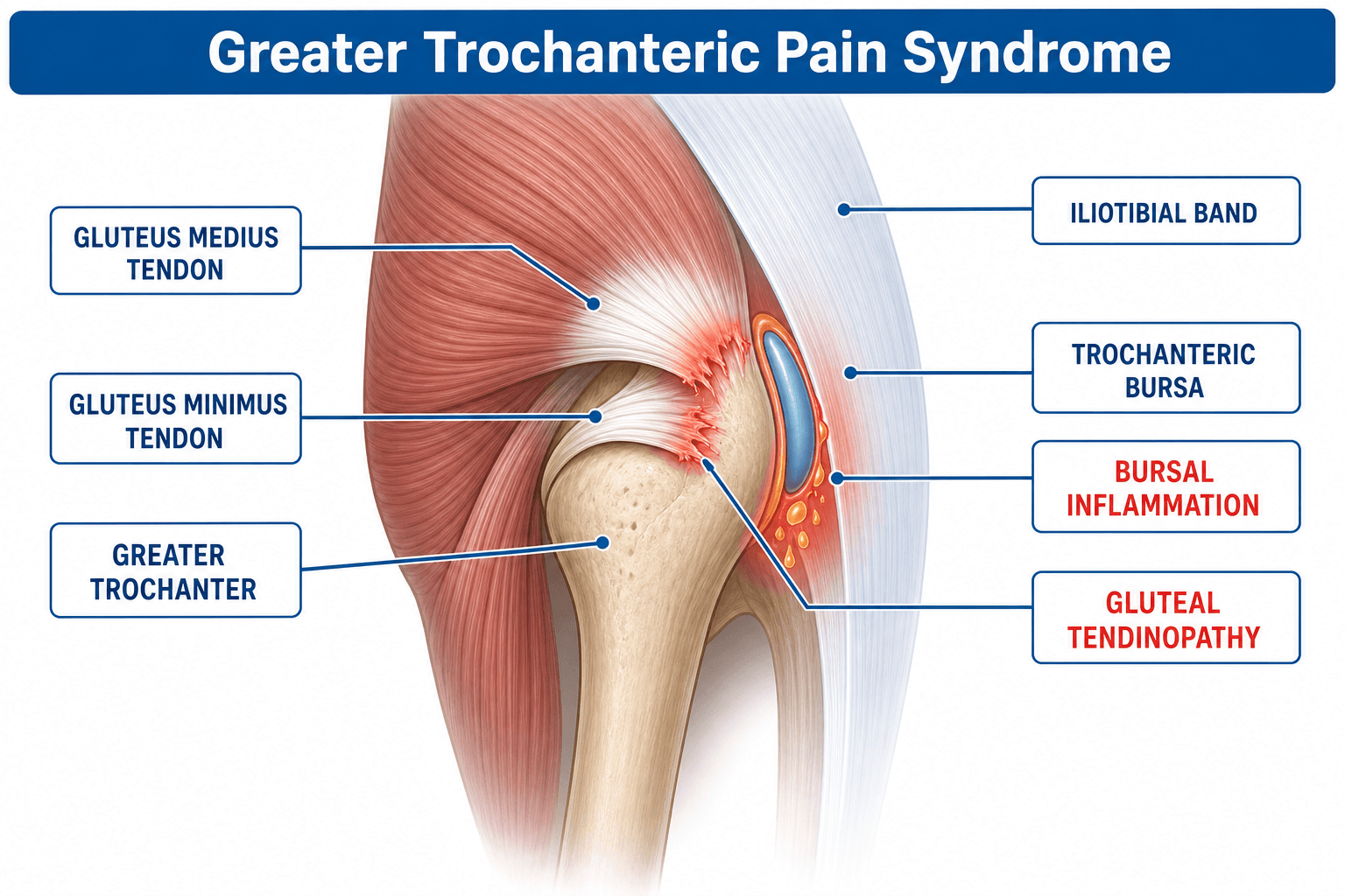
The hip's rotator cuff. Gluteus medius is the primary hip abductor and pelvic stabiliser during gait, and its insertion on the greater trochanter is analogous to the supraspinatus insertion on the greater tuberosity. Like rotator cuff disease, an overloaded tendinopathy progresses from reactive to degenerative, and partial-thickness tears may progress to full-thickness tears. The tear sequence mirrors the cuff as well: anterior gluteus minimus and lateral gluteus medius fail first, analogous to supraspinatus.
Where the tears are. Gluteus minimus tears are anterior. Gluteus medius tears, the commonest, are at the lateral and superoposterior facets, directly under the point of maximal tenderness.
Tendon more often than bursa. Modern imaging with MRI and ultrasound shows that true bursitis accounts for only a minority of cases. The majority of patients have gluteus medius or minimus tendinopathy, with or without bursal inflammation, and the bursae may be secondarily inflamed by adjacent tendon pathology, mechanical friction or direct pressure. The distinction changes management: tendinopathy requires progressive loading, the evidence-based treatment, not just anti-inflammatory measures, and corticosteroid may give temporary relief but risks further tendon degradation.
Classification and Types
GTPS is divided by the structure at fault, and the subtype guides management: tendinopathy needs loading, bursitis needs anti-inflammatory measures, and ITB syndrome needs stretching and biomechanical correction.
- Trochanteric Bursitis (isolated)
- Inflammation of subgluteal bursae without tendon pathology
- Gluteal Tendinopathy
- Degeneration / micro-tears of gluteus medius/minimus tendons
- ITB Syndrome
- Friction or tightness of ITB over greater trochanter
- Trochanteric Bursitis (isolated)
- Acute onset, overuse, direct pressure
- Gluteal Tendinopathy
- Female 40-60, insidious onset
- ITB Syndrome
- Runners, cyclists, repetitive flexion/extension
- Trochanteric Bursitis (isolated)
- Point tenderness, warm bursa, swelling, pain on direct pressure
- Gluteal Tendinopathy
- Weak abduction, Trendelenburg positive, pain on resisted abduction
- ITB Syndrome
- Pain on flexion-extension, pain at 30 degrees of flexion (Noble test), tight ITB
- Trochanteric Bursitis (isolated)
- Fluid in bursa, normal tendon
- Gluteal Tendinopathy
- Tendon thickening, increased signal, partial or full tear
- ITB Syndrome
- ITB thickening, fluid deep to ITB
- Trochanteric Bursitis (isolated)
- Approximately 15-20 percent
- Gluteal Tendinopathy
- Approximately 50-66 percent
- ITB Syndrome
- Approximately 10-15 percent
Mixed disease. Approximately 15-25 percent have tendinopathy with secondary bursitis, and show features of both tendon and bursal pathology.
Severity. Grading guides escalation. Mild cases may resolve with self-management, while severe cases need imaging and specialist input to rule out tendon tears.
- Symptoms
- Pain after activity only, no night pain
- Functional Impact
- No gait abnormality, full function
- Treatment Approach
- Self-management: ice, NSAIDs, stretching, activity modification
- Symptoms
- Pain during activity, occasional night pain
- Functional Impact
- Modified gait, limited side-lying, stairs difficult
- Treatment Approach
- Physiotherapy referral, structured rehab programme
- Symptoms
- Constant pain, significant night pain, cannot lie on affected side
- Functional Impact
- Antalgic gait, unable to climb stairs, functional limitation
- Treatment Approach
- Imaging (MRI or ultrasound), image-guided injection, specialist referral
- External (coxa saltans externa) - the iliotibial band or gluteus maximus tendon snaps over the greater trochanter. This is the type within the GTPS spectrum: a palpable or visible lateral snap, often reproducible by the patient, worse as the hip moves from extension into flexion. It is managed with ITB and abductor stretching and load management; surgical ITB lengthening (e.g. a Z-plasty or cross/diamond release) is only for refractory painful snapping.
- Internal (coxa saltans interna) - the iliopsoas tendon snaps over the iliopectineal eminence or femoral head. The snap is anterior, in the groin, reproduced moving from flexion-abduction-external rotation into extension. It is managed with stretching, ultrasound-guided iliopsoas injection, and arthroscopic or open psoas lengthening if refractory.
- Intra-articular - a loose body, labral tear or chondral flap causes a true mechanical catch or lock. This is not a tendon snap, and it needs joint imaging (MR arthrogram) and arthroscopy.
Do not lump them together as "snapping hip": a lateral snap is external, an anterior or groin snap is internal, and a true painful catch with locking is intra-articular.
Clinical Assessment
History. The pain is over the lateral aspect of the hip and may radiate to the lateral thigh, but not below the knee. Night pain is common, with difficulty sleeping on the affected side. Onset is insidious in tendinopathy and acute in bursitis. The aggravating activities are characteristic:
- Lying on the affected side
- Climbing stairs
- Rising from a chair
- Crossing the legs
Function suffers on stairs, with prolonged walking and in side-sleeping. Record any previous treatment (NSAIDs, physiotherapy, injections) with its response and how long it lasted.
Examination. Watch the gait for a Trendelenburg pattern and pelvic tilt, and look for gluteal wasting. Point tenderness over the greater trochanter, most commonly its posterolateral aspect, is the hallmark but is non-specific. Hip range of motion is usually full, unlike hip OA, which restricts internal rotation. The provocative tests:
- Single-leg stance for 30 seconds - the best provocative test; pain reproduced within 30 seconds is highly sensitive for GTPS
- Resisted abduction in side-lying at 30 degrees - provokes gluteal tendon pain at the trochanter, pointing to gluteal tendinopathy
- Trendelenburg sign - the pelvis drops on the contralateral side during single-leg stance, abductor weakness that points to tendinopathy
- Neurological screen - lumbar spine, femoral nerve stretch and sensation over the lateral thigh, to exclude radiculopathy
Differential diagnosis. The cause of lateral hip pain is not always the trochanter. L4-L5 radiculopathy or facet pain may refer to the lateral hip, and radiculopathy also brings reflex changes, positive neural tension signs and tenderness over the lumbar paraspinals rather than the trochanter. Hip OA may present with lateral hip pain, not just groin pain, femoroacetabular impingement or labral tears may refer laterally, and lateral knee OA or ITB syndrome may present similarly.
- Pain Location
- Lateral hip, may radiate to lateral thigh
- Key Examination Finding
- Point tenderness over greater trochanter
- Discriminating Test
- Single-leg stance pain, resisted abduction pain
- Pain Location
- Groin (primary), may refer laterally
- Key Examination Finding
- Restricted internal rotation, FADIR positive
- Discriminating Test
- Radiographs: joint space narrowing, osteophytes
- Pain Location
- Radiating pain below knee, dermatomal
- Key Examination Finding
- Neurological signs, positive SLR or femoral stretch
- Discriminating Test
- MRI lumbar spine, neurological examination
- Pain Location
- Groin, may refer to lateral hip
- Key Examination Finding
- Pain on FADIR, positive impingement test
- Discriminating Test
- MRI arthrogram, radiographs (cam/pincer lesions)
- Pain Location
- Anterolateral thigh, burning, tingling
- Key Examination Finding
- Sensory changes in lateral femoral cutaneous nerve distribution
- Discriminating Test
- Tinel over ASIS region, nerve conduction studies
- Pain Location
- Groin or lateral hip, weight-bearing pain
- Key Examination Finding
- Pain on hopping or fulcrum test, progressive pain
- Discriminating Test
- MRI (urgent), risk factors: female athlete triad
GTPS has lateral hip tenderness, full hip range of motion (especially internal rotation), pain on trochanteric palpation and a normal neurological examination. Do not assume lateral hip pain is trochanteric bursitis without examining the lumbar spine and hip joint.
In a female runner or military recruit with lateral hip or groin pain that is progressive and worse with weight-bearing, always consider a femoral neck stress fracture, a potentially catastrophic diagnosis to miss. A positive fulcrum test, pain when the femur is stressed over the examiner's hand as a fulcrum, should raise concern. Request an MRI if plain radiographs are normal but clinical suspicion exists.
Investigations
The diagnosis of GTPS is primarily clinical, and imaging is not required for typical presentations. When it is used, radiographs exclude other pathology and MRI defines the lesion.
Radiographs. An AP pelvis and a lateral hip (cross-table lateral or frog lateral). They are often normal in isolated GTPS, and their primary role is excluding other pathology such as hip OA, fracture and tumour. Look for:
- Greater trochanteric enthesophytes and calcific tendinopathy
- Hip OA (joint space narrowing)
- Femoral neck stress fracture, which may be normal early
- Pelvic pathology
MRI. The gold standard for differentiating bursitis from tendinopathy and for grading tendon tears. Bursitis shows fluid distension of the subgluteal bursae (high T2 signal) and peritrochanteric oedema. Tendinopathy shows increased T2 signal within the gluteus medius and minimus tendons, tendon thickening, partial or full-thickness tears and tendon retraction. The same scan assesses the hip joint for concurrent labral tears, chondral damage and femoroacetabular impingement. Request MRI when:
- Symptoms are refractory to conservative management, after a minimum of 6-8 weeks (the conservative protocol's formal review falls at 12 weeks)
- There is significant abductor weakness or a Trendelenburg sign, suggesting a gluteal tendon tear
- There is diagnostic uncertainty: possible hip OA, stress fracture or lumbar pathology
- Surgical planning is being considered
Ultrasound. The alternative, and the dynamic one: it assesses the peritrochanteric structures in motion. It shows bursal fluid, tendon thickening and calcifications, assesses ITB snapping in real time, and guides injection, so diagnostic and therapeutic injection can be done at one sitting under direct visualisation. It is operator-dependent and cannot assess deep structures or the hip joint as well as MRI.
Management Algorithm
Management escalates from conservative care to image-guided injection to surgery.
- Likely Diagnosis
- Trochanteric bursitis
- First-line Treatment
- NSAIDs, physiotherapy, ice, activity modification
- Key Pearl
- Often self-limiting with conservative care
- Likely Diagnosis
- Gluteal tendinopathy
- First-line Treatment
- Progressive abductor loading programme (12 weeks)
- Key Pearl
- Mimics bursitis but needs tendon-specific rehab
- Likely Diagnosis
- Chronic GTPS with possible partial tear
- First-line Treatment
- Image-guided injection, consider PRP if tendinopathy
- Key Pearl
- MRI before injection to confirm target pathology
Conservative Management (First-Line for All Patients)
Conservative care is first-line for every patient, for a minimum of 6-8 weeks before escalating. The aims are to reduce pain and inflammation, restore hip abductor function and address biomechanical factors.
Match the rehabilitation to the pathology. Gluteal tendinopathy follows tendinopathy principles: isometric loading first for pain reduction, then isotonic loading for strength, then functional loading. Isolated bursitis management focuses instead on anti-inflammatory measures, so always assess which pathology predominates.
Conservative Protocol
Relative rest from aggravating activities: cross-legged sitting, lying on the affected side and excessive stair climbing. Ice massage over the greater trochanter for 15 minutes, 3-4 times daily, and oral NSAIDs, if not contraindicated, for 2-4 weeks for pain relief and anti-inflammatory effect.
Sleep with a pillow between the knees and avoid sleeping on the affected side; trochanteric padding is an alternative for side-sleepers. Reassure the patient about the benign nature of the condition, and explain it and the expected timeline.
Physiotherapy referral for a structured hip abductor strengthening programme, starting with pain-free isometric hip abduction in side-lying and graduating from isometric to isotonic to functional exercise. Stretching of the ITB, hip flexors and piriformis is adjunctive, not the primary treatment.
Manual therapy (trochanteric soft tissue mobilisation, myofascial release) and biomechanical correction (footwear, gait retraining, core stability) support the loading programme.
Progressive resistance with theraband, weight-bearing exercises and single-leg balance, then functional retraining: stair climbing, sit-to-stand and return to sport-specific activities. The patient continues on an independent home exercise programme for long-term management.
Review at 12 weeks. If still symptomatic, consider imaging and injection.
Complications
The most clinically significant "complication" of GTPS is incorrect diagnosis. Patients with lumbar radiculopathy, hip OA, femoral neck stress fractures or femoroacetabular impingement may present with lateral hip pain and be inappropriately treated for bursitis.
- Incidence
- 15-30 percent develop chronic symptoms
- Risk Factors
- Delayed diagnosis, inadequate rehab, obesity
- Management
- Multidisciplinary pain management, re-evaluate diagnosis
- Incidence
- Partial tears may progress to full-thickness
- Risk Factors
- Repeated corticosteroid injections, continued overload
- Management
- Surgical repair if full-thickness with functional deficit
- Incidence
- Reported with multiple injections into tendon
- Risk Factors
- More than 3 injections, blind technique, tendinopathy
- Management
- Avoid steroid into tendon, use image guidance, limit injections
- Incidence
- Common — delayed diagnosis frequent
- Risk Factors
- Not examining lumbar spine or hip joint, reliance on X-rays
- Management
- Always perform full lumbar and hip examination
- Incidence
- Variable depending on procedure
- Risk Factors
- Obesity, smoking, diabetes, poor tissue quality
- Management
- Appropriate patient selection, rehabilitation adherence
Outcomes and Prognosis
- Treatment
- NSAIDs + physiotherapy +/- one injection
- Expected Outcome
- 80-90 percent resolution within 3-6 months
- Long-term Prognosis
- Excellent, low recurrence with activity modification
- Treatment
- Progressive loading programme (12 weeks)
- Expected Outcome
- 60-80 percent significant improvement with structured rehab
- Long-term Prognosis
- Good with ongoing maintenance exercises
- Treatment
- Rehabilitation first, consider PRP, surgery if refractory
- Expected Outcome
- 50-70 percent improve with non-operative management
- Long-term Prognosis
- Variable — may progress, monitor with MRI
- Treatment
- Surgical repair (open or endoscopic)
- Expected Outcome
- 70-85 percent good-to-excellent in case series
- Long-term Prognosis
- Functional improvement but rarely full strength return
Prognostic factors. The best prognosis belongs to isolated bursitis, an acute presentation, compliance with physiotherapy, a normal BMI and no associated lumbar or hip pathology. Chronic symptoms (over 6 months), gluteal tendon tears, obesity, concurrent lumbar spine pathology, previous hip surgery (especially lateral approach THR) and poor compliance with rehabilitation carry a poor prognosis. Early and accurate diagnosis with appropriate rehabilitation gives the best outcomes.
Guidelines, Registries & Global Practice
- GTPS incidence: Approximately 1.8 per 1000 per year in primary care populations
- Female predominance: 4:1 ratio, peak age 40-60 years, perimenopausal women at highest risk
- Associations: Obesity (BMI over 30), low back pain, hip OA, total hip replacement (lateral approach)
- Post-THR: Up to 20 percent of patients develop GTPS after total hip replacement via lateral approach due to abductor disruption
- Bilateral involvement: Reported in up to one-third of cases
- High-resource settings: MRI readily available for refractory cases, image-guided injections, specialised hip physiotherapy, endoscopic surgical options
- Limited-resource settings: Clinical diagnosis with trial of conservative management, blind injection if available, plain radiographs to exclude other pathology
- Universal principle: Diagnosis is clinical, first-line treatment is conservative (physiotherapy, NSAIDs, activity modification), and outcomes depend more on rehabilitation compliance than on access to advanced imaging or injections
- Global consensus: Conservative management before injection; imaging before surgery; GTPS as umbrella term
- Diagnosis Emphasis
- Clinical diagnosis; radiographs to exclude OA and fracture; MRI for refractory cases
- First-line Treatment
- Physiotherapy (abductor strengthening), NSAIDs, activity modification for minimum 6-8 weeks
- Injection Guidance
- Image-guided corticosteroid for true bursitis; PRP as emerging option for tendinopathy
- Diagnosis Emphasis
- GTPS as umbrella term; differentiate from hip OA and lumbar referral
- First-line Treatment
- Education plus progressive exercise program; NICE recommends physiotherapy as first-line
- Injection Guidance
- Corticosteroid injection if conservative measures fail; ultrasound guidance preferred
- Diagnosis Emphasis
- Clinical assessment; imaging not routinely needed for diagnosis
- First-line Treatment
- Self-management advice, exercise referral, consider topical or oral NSAIDs
- Injection Guidance
- Corticosteroid injection for persistent symptoms; limit to 3 injections per site
- Diagnosis Emphasis
- Clinical diagnosis with emphasis on differentiating tendinopathy from bursitis
- First-line Treatment
- Load management and progressive strengthening; avoid corticosteroid as first-line
- Injection Guidance
- PRP or image-guided injection only after structured rehabilitation trial
There is no dedicated arthroplasty or implant registry for GTPS, as most cases are managed non-operatively. However, national joint replacement registries (NJR UK, AOANJRR Australia) track post-THR abductor complications including GTPS after lateral approach. The evidence base is evolving from small case series and expert opinion to higher-quality RCTs such as the LEAP trial, which demonstrated the superiority of exercise-based management over corticosteroid injection.
Record in every GTPS presentation:
- Examination of hip joint (ROM, impingement tests) to exclude hip OA
- Lumbar spine assessment to exclude radiculopathy
- Trochanteric palpation, single-leg stance test, Trendelenburg sign, and resisted abduction
- Duration and adequacy of conservative management before injection
- Injection: image-guided vs blind, corticosteroid vs PRP, response documented
- If MRI performed: document bursitis vs tendinopathy vs tear — guides ongoing management
A missed hip OA diagnosis or femoral neck stress fracture in a patient labelled as 'trochanteric bursitis' is a recurring source of medicolegal claims. Always examine the hip joint and lumbar spine before attributing lateral hip pain to GTPS.
Controversies & Areas of Uncertainty
Corticosteroid provides faster short-term relief but worse long-term outcomes compared to exercise. PRP shows promising medium-term results for tendinopathy in limited studies, but high-quality RCTs are lacking. The choice remains resource-dependent and patient-specific. Neither injection should replace rehabilitation.
Tendinopathy-based loading protocols (isometric to isotonic to functional) are recommended, but the optimal exercise type, dose, and duration are not precisely defined. Most protocols recommend 8-12 weeks of progressive loading, with maintenance exercises ongoing. Individual tailoring based on pain response and functional progress is key.
Endoscopic gluteus medius repair is increasingly performed but has no RCT evidence of superiority over open repair. Theoretical advantages include smaller incisions and lower morbidity, but outcomes are comparable in case series. Surgeon experience and tear characteristics determine approach.
Extracorporeal shockwave therapy (ESWT) has shown benefit in some case series for refractory GTPS, particularly calcific tendinopathy. However, evidence quality is low and it remains an adjunctive option rather than a standard treatment. Not universally available.
MCQ Practice Points
Q: What is the preferred term for lateral hip pain over the greater trochanter? A: Greater trochanteric pain syndrome (GTPS). This umbrella term encompasses trochanteric bursitis, gluteal tendinopathy, and ITB pathology. 'Trochanteric bursitis' is a specific sub-diagnosis, not the overall condition. The exam rewards candidates who use the correct terminology and understand the spectrum.
Q: What is the most common underlying pathology in GTPS? A: Gluteal tendinopathy (gluteus medius or minimus). MRI studies show that approximately 50-66 percent of GTPS cases have gluteal tendon pathology as the primary driver, with isolated bursitis being less common. This distinction is important because tendinopathy requires progressive loading, while bursitis is managed with anti-inflammatory measures.
Q: What is the most sensitive clinical test for GTPS? A: The single-leg stance test. Standing on the affected leg for 30 seconds reproduces lateral hip pain in the vast majority of GTPS patients. Point tenderness over the greater trochanter is common but non-specific. Resisted abduction in side-lying at 30 degrees is more specific for gluteal tendinopathy.
Q: How do you differentiate GTPS from hip osteoarthritis clinically? A: Hip ROM. GTPS has full hip range of motion (especially internal rotation), while hip OA restricts internal rotation and flexion. GTPS pain is reproduced by trochanteric palpation and single-leg stance, while hip OA pain is reproduced by FADIR (flexion-adduction-internal rotation) and is typically groin-predominant. Radiographs show OA changes; GTPS radiographs are typically normal.
Q: What is the evidence-based first-line management for GTPS? A: Structured physiotherapy with progressive hip abductor strengthening. The LEAP trial demonstrated that education plus exercise was superior to corticosteroid injection at 52 weeks. Conservative management should be trialled for a minimum of 6-8 weeks before considering injection, and 3-6 months before considering surgery.
Q: What are the risks of corticosteroid injection in GTPS? A: Tendon degradation if injected into the gluteal tendons (rather than the bursa). Image guidance (ultrasound) is recommended to ensure accurate placement. Corticosteroid provides short-term relief (4-12 weeks) but does not offer long-term benefit and may worsen underlying tendinopathy. The LEAP trial showed exercise was superior to injection at one year.
Exam Viva Scenarios
Practise clinical reasoning and management decisions out loud
“A 52-year-old woman presents with a 3-month history of right lateral hip pain. It is worse when she lies on her right side at night and when climbing stairs. She has tried over-the-counter ibuprofen with partial relief. On examination, there is point tenderness over the right greater trochanter. Hip range of motion is full and pain-free except for pain on resisted abduction in side-lying. Trendelenburg sign is negative. Lumbar spine examination is normal. What is the diagnosis and how would you manage this?”
“A 58-year-old woman has had left lateral hip pain for 9 months despite physiotherapy and two corticosteroid injections (the last one 3 months ago). She now has a positive Trendelenburg sign on the left and reports weakness when trying to abduct the leg against gravity. MRI shows a full-thickness tear of the gluteus medius tendon at its insertion on the greater trochanter with 2 cm of retraction and associated bursal fluid. What is your diagnosis and management plan?”
Key Terminology and Anatomy
- GTPS = umbrella term for lateral hip pain (bursitis, tendinopathy, ITB syndrome)
- Gluteal tendinopathy is the most common cause (50-66 percent of GTPS), not bursitis
- Subgluteus maximus bursa is the most commonly inflamed bursa
- Gluteus medius inserts on superoposterior facet of greater trochanter (like supraspinatus analogy)
Diagnosis
- Clinical diagnosis: point tenderness over trochanter, full hip ROM, pain on single-leg stance
- Trendelenburg and weak abduction suggest tendinopathy rather than isolated bursitis
- Always examine lumbar spine and hip joint to exclude referred pain and OA
- MRI gold standard for differentiating bursitis from tendinopathy and grading tears
Differential Diagnosis
- Hip OA: groin pain, restricted internal rotation, radiographic changes
- Lumbar radiculopathy L4-L5: pain below knee, neurological signs, SLR positive
- Femoral neck stress fracture: progressive weight-bearing pain, urgent MRI needed
- Meralgia paraesthetica: burning anterolateral thigh, sensory changes
Management Algorithm
- First-line: structured physiotherapy (progressive abductor loading) for minimum 6-8 weeks
- Second-line: image-guided injection (corticosteroid for bursitis, PRP for tendinopathy)
- Third-line: surgical referral for documented tendon tears with functional deficit after 6 months
- Activity modification throughout: avoid side-sleeping, crossing legs, excessive stairs
Evidence-Based Pearls
- LEAP trial: exercise superior to corticosteroid injection at 52 weeks
- Corticosteroid injection risks tendon damage if placed into gluteal tendons
- Image-guided injection accuracy approximately 90 percent vs 50 percent blind
- Gluteal tendon tears are the hip equivalent of rotator cuff tears
Evidence Base and Key Trials
Exercise compared to a control condition or other conservative treatment options in patients with Greater Trochanteric Pain Syndrome: a systematic review and meta-analysis of randomized controlled trials
- Six randomised trials including 733 patients, appraised with ROB2 and graded with GRADE
- USE THE AUTHORS' OWN QUALIFIER: in the long term exercise SLIGHTLY reduces hip pain and disease severity and SLIGHTLY improves patient-reported function and global rating of change against a control condition. The direction is consistent and the effect is small
- Against corticosteroid injection, the one outcome exercise improved in the long term was GLOBAL RATING OF CHANGE - which is the same outcome, and the same limitation, as in the Mellor trial below
- Only two of the six trials compared exercise with corticosteroid injection, two compared it with shockwave therapy and one with a different exercise programme
- No serious adverse events were reported in any trial
Gluteal Tendinopathy: Integrating Pathomechanics and Clinical Features in Its Management
- Gluteal tendinopathy is the primary pathology in the majority of GTPS cases
- The condition is analogous to rotator cuff disease of the hip — progressive tendon degeneration
- Corticosteroid injection may provide temporary pain relief but risks further tendon degeneration
- Progressive loading (isometric to isotonic to functional) is the recommended rehabilitation approach