Principles, Transducers, Doppler & Artefacts
- Piezoelectric transducer and the pulse-echo principle: echoes from acoustic-impedance interfaces, timed to localise each structure
- Frequency trade-off: high-frequency linear probes for superficial structures, lower-frequency curvilinear probes for deep ones
- Anisotropy makes a normal tendon look hypoechoic when the beam is off-perpendicular, the classic source of false-positive diagnoses
- Shadowing behind calcification and enhancement behind fluid help the diagnosis; anisotropy and mirror image mislead
- Real-time, dynamic, cheap and radiation-free, but highly operator-dependent
- For rotator cuff tears ultrasound matches MRI in experienced hands (65 studies, surgical reference standard); MR arthrography beats both but is invasive
- “Higher frequency = better resolution, less penetration; lower frequency = deeper, poorer resolution
- “B-mode = brightness, the 2D grey-scale image; Doppler (colour, power, spectral) = flow
- “Anisotropy: keep the probe perpendicular, and heel-toe to confirm
- “Cuff tears, area under summary ROC: MR arthrography 0.935, ultrasound 0.889, MRI 0.878
Principles and Transducers
The transducer. A piezoelectric transducer converts electrical energy into high-frequency sound, and converts the returning echoes back into electrical signals. It both makes the sound and receives it.
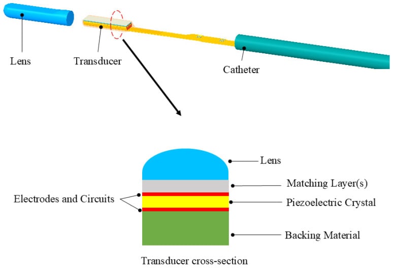
The pulse-echo principle. Sound is reflected at interfaces between tissues of differing acoustic impedance. The machine times each returning echo, its time-of-flight, to localise the structure and place the pixel, and from these echoes builds a real-time image with no ionising radiation.
Acoustic impedance. Impedance is tissue density multiplied by the speed of sound, Z = ρ × c, and the proportion of sound reflected at an interface depends on the mismatch in Z. A large mismatch, soft tissue against bone or gas, reflects almost all the sound: a strong echo, with shadowing beyond. A small mismatch, soft tissue against soft tissue, reflects a little and transmits the rest, which is what allows imaging.
The assumed speed of sound. To convert time-of-flight into depth, the machine assumes a fixed soft-tissue speed of about 1540 m/s. Real tissues differ, fat slower and bone far faster, and the difference is a source of some artefacts.
Frequency, Attenuation and Resolution
Attenuation. Sound weakens with depth through absorption, scatter and reflection, by roughly 0.5-1 dB/cm per MHz, so higher frequencies attenuate more; that is the physics behind their poorer penetration. Time-gain compensation (TGC) corrects for this depth-dependent loss.
The trade-off. This is the fundamental rule of the modality. Higher frequency gives better axial resolution but attenuates faster and so penetrates less; lower frequency penetrates deeper at the cost of resolution. Choose the probe to match the depth:
- High-frequency linear probes for superficial musculoskeletal structures
- Lower-frequency curvilinear probes for deeper structures, such as the hip
The two resolutions. Axial resolution, along the beam, improves with higher frequency, because the pulse is shorter. Lateral resolution, across the beam, improves with focusing and is best at the focal zone.
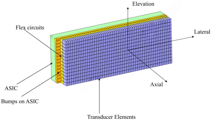
Image Modes, Doppler and Safety
Image modes. The echoes can be displayed in three modes:
- A-mode - amplitude, displayed as a graph
- B-mode - brightness, the standard 2D grey-scale image
- M-mode - motion over time
Doppler. Colour, power and spectral Doppler detect and characterise blood flow, and are useful for showing hyperaemia in inflammation or infection and the vascularity of lesions.
- Colour Doppler shows mean velocity and direction; it is angle-dependent and aliases
- Power Doppler shows signal amplitude. It is more sensitive to slow flow, non-directional and less angle-dependent, and does not alias, which makes it the best for detecting hyperaemia
- Spectral (pulsed-wave) Doppler gives a velocity waveform for quantification
The Doppler equation. The frequency shift is proportional to blood velocity × cos(angle) between the beam and the flow. The shift is therefore maximal when the beam is parallel to flow and zero at 90 degrees, so for an accurate velocity keep the insonation angle low, ideally under 60 degrees.
Aliasing. When the Doppler shift exceeds the Nyquist limit, half the pulse-repetition frequency (PRF), the signal wraps around: colour reversal, or spectral wrap on the waveform. To correct it:
- Raise the velocity scale (PRF)
- Shift the baseline
- Use a lower frequency
- Use a smaller angle
Safety and bio-effects. Ultrasound is non-ionising, but it deposits energy. The bio-effects are thermal (heating, displayed as the thermal index, TI) and mechanical (cavitation, the mechanical index, MI). The ALARA principle applies, especially in obstetric and ophthalmic use; diagnostic musculoskeletal ultrasound is very safe.
Artefacts - Pitfalls and Helpers
Artefacts divide into those that help the diagnosis and those that cause misdiagnosis. Shadowing and posterior enhancement are used deliberately, anisotropy and mirror image mislead, and reverberation is recognised so that it is not mistaken for pathology.
- What it is
- Echogenicity changes with beam angle (tendon falsely hypoechoic off-perpendicular)
- Significance
- PITFALL - mimics tear/tendinopathy; keep probe perpendicular
- What it is
- Dark band behind bone/calcification/gas (sound blocked)
- Significance
- Helpful - confirms calcification/bone
- What it is
- Increased brightness behind a fluid-filled structure
- Significance
- Helpful - confirms a cyst/fluid
- What it is
- Repeating echoes between two reflectors
- Significance
- Indicates metal/gas/foreign body
- What it is
- Duplicated structure across a strong reflector
- Significance
- Pitfall - false duplicate
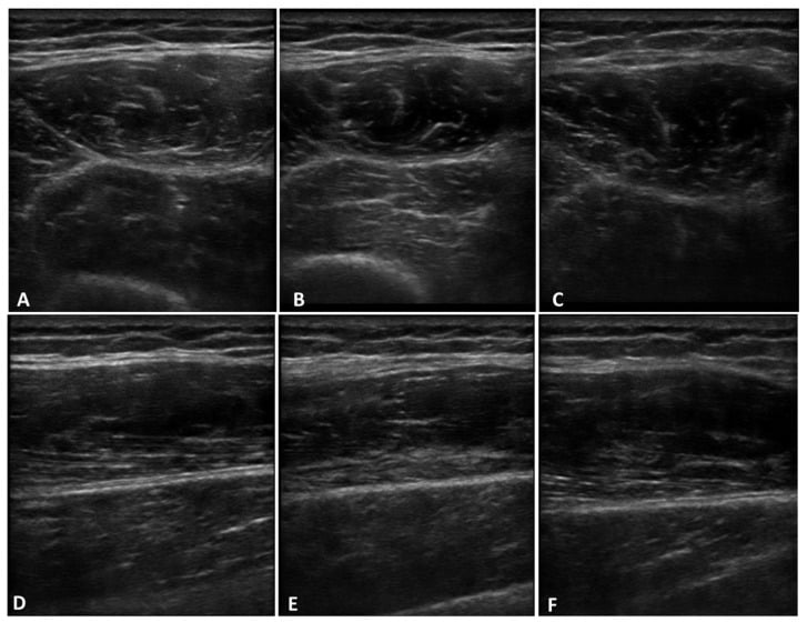
Anisotropy. This is the most important practical pitfall in musculoskeletal ultrasound. Tendons and other fibrillar structures are highly reflective only when the beam strikes them perpendicularly; angled even a few degrees off, the structure returns fewer echoes and appears artefactually hypoechoic, exactly mimicking a tear or tendinopathy. It is the classic source of false-positive diagnoses.
Avoiding it. Keep the transducer perpendicular to the structure being examined. Then tilt it, the heel-toe manoeuvre, to confirm that an apparent hypoechoic area fills in when the angle is corrected.
The helpful artefacts. Use them deliberately. Acoustic shadowing behind a focus confirms calcification, as in calcific tendinopathy; posterior enhancement behind a lesion confirms that it is fluid-filled or cystic, as in a ganglion.
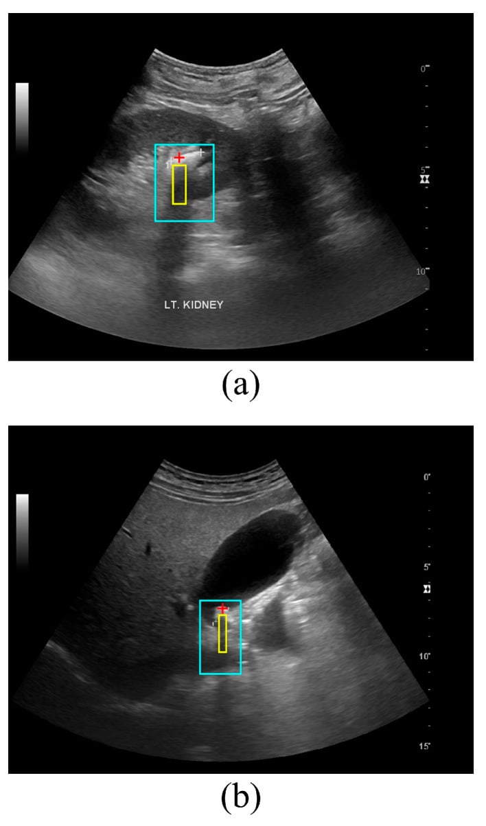
Reverberation and mirror image. These are recognised so that they are not mistaken for pathology. A comet-tail indicates metal, gas or a foreign body, and a mirror image is a false duplicate.
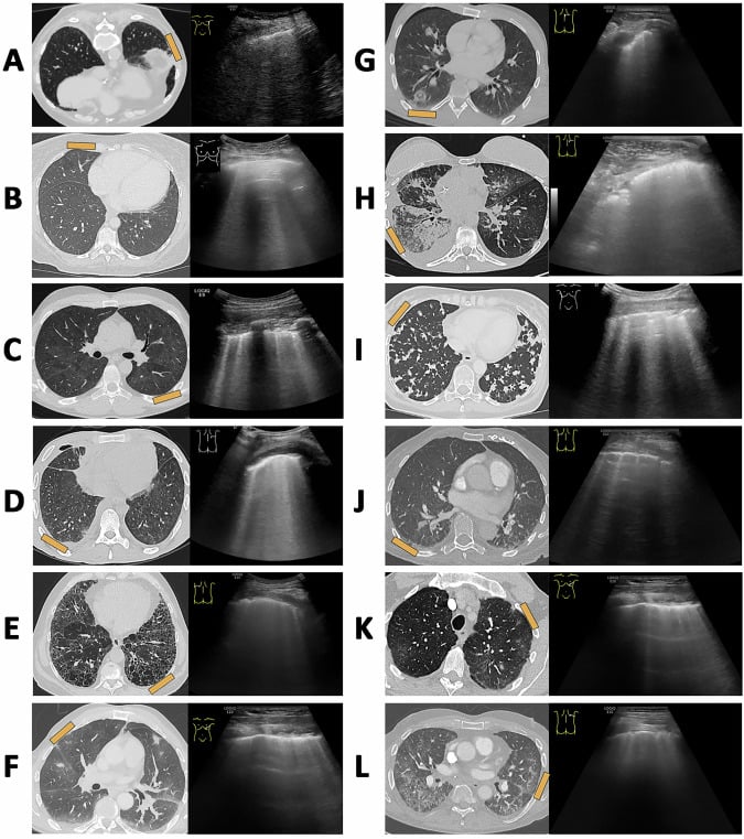
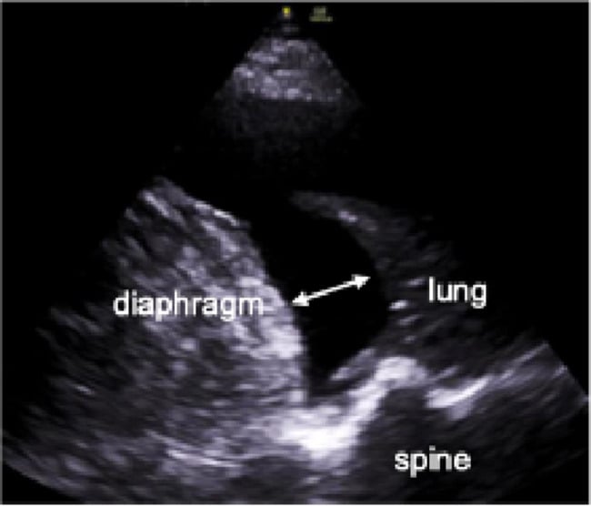
Technique. Good musculoskeletal ultrasound depends on the right probe and frequency, machine settings (depth, focus, gain and TGC), positioning and standoff pads, all of which limit artefacts. Limiting and correctly interpreting artefacts, especially anisotropy, is what avoids diagnosing pathology where none exists. Ultrasound is real-time, dynamic, cheap and radiation-free, but highly operator-dependent, and correct technique is what separates a reliable study from a misleading one.
Does the Operator Dependence Actually Cost Accuracy?
"Operator-dependent" is the stock criticism of ultrasound, and it is repeated so often that candidates assume the modality is second-rate. The accuracy data say otherwise for the commonest musculoskeletal indication.
Ultrasound against MRI for cuff tears. A meta-analysis of 65 studies, all requiring an open or arthroscopic surgical reference standard, found no significant difference in either sensitivity or specificity between MRI and ultrasound for partial-thickness or full-thickness tears (P greater than 0.05). The area under the summary ROC curve was in fact numerically higher for ultrasound (0.889) than for MRI (0.878), though that pairwise difference was not significant.
MR arthrography beat both. It was significantly more sensitive and specific than either (P less than 0.05), with the highest area under the curve at 0.935. The price is that it is invasive.
In the viva. Do not concede that ultrasound is inferior. For cuff tears it performs comparably to MRI at lower cost, in real time, with dynamic assessment and no ionising radiation, and MR arthrography is the most accurate if the added invasiveness is justified.
Where operator dependence bites. These are pooled published studies, largely from centres with a declared interest in musculoskeletal ultrasound, so the figures describe the modality in experienced hands. That is precisely what operator dependence means: the technique can match MRI, but only if the person holding the probe knows the anatomy, keeps the beam perpendicular and recognises the artefacts. The physics does not transfer with the machine.
What is not established. The figures are for the rotator cuff and should not be assumed to hold for every tendon, nerve or joint. No comparable pooled accuracy figure exists for most other musculoskeletal sites, and no published study measures how often anisotropy specifically produces a false-positive report.
Mnemonics & Memory Aids
ECHO
Hook:ECHO: Echo (pulse-echo/piezoelectric), Compromise (frequency trade-off), Hypoechoic = anisotropy, Other artefacts (shadow/enhancement/mirror).
Clinical Decision Scenarios
Practise clinical reasoning and management decisions out loud
“Explain the frequency trade-off in ultrasound and the anisotropy artefact.”
Principles
- Piezoelectric transducer (sound out and echoes in)
- Pulse-echo: reflection at acoustic-impedance interfaces; time-of-flight localises
- Real-time, dynamic, no ionising radiation; operator-dependent
Frequency & probes
- Higher frequency = better resolution, less penetration (linear, superficial)
- Lower frequency = deeper, poorer resolution (curvilinear, deep)
- Modes: A/B/M-mode (B = 2D grey-scale image)
Doppler
- Colour, power, spectral Doppler detect/characterise flow
- Shows hyperaemia (inflammation/infection) and vascularity
- Power Doppler more sensitive to low flow
Accuracy (rotator cuff)
- Ultrasound and MRI equivalent: no significant difference in sensitivity or specificity (65 studies)
- Area under summary ROC: MR arthrography 0.935, ultrasound 0.889, MRI 0.878
- MR arthrography significantly better than both, but invasive
- Figures reflect experienced operators and apply to the cuff, not every site
Artefacts
- Anisotropy (classic MSK pitfall - tendon falsely hypoechoic; keep perpendicular)
- Acoustic shadowing (calcification - helpful); posterior enhancement (fluid/cyst - helpful)
- Reverberation/comet-tail (metal/gas); mirror image (false duplicate)
Evidence & Key Studies
Diagnostic errors in musculoskeletal ultrasound and how to avoid them
- A NARRATIVE REVIEW of technique and artefacts. It reports NO error rate, no denominator and no measure of how often any artefact causes a misdiagnosis - so 'anisotropy frequently leads to diagnosing pathology where none exists' is expert description, not a measured false-positive rate. No such rate has been published.
- Correct musculoskeletal ultrasound technique - equipment settings, image-software innovations, standoff pads and correct transducer positioning - is fundamental to producing good-quality images and limiting artefacts.
- Artefacts divide into diagnostically HELPFUL ones (posterior echo enhancement deep to a fluid-filled structure; acoustic shadowing behind calcification) and MISLEADING ones - notably anisotropy-related artefacts and the mirror-reflection artefact.
- The review also covers correct differentiation of hypoechoic versus anechoic foci, and the atypical appearances that lead to specific conditions being misdiagnosed - the practical skill that separates a reliable study from a misleading one.
Accuracy of MRI, MR arthrography, and ultrasound in the diagnosis of rotator cuff tears: a meta-analysis
- Meta-analysis of 65 articles comparing MRI, MR arthrography and ultrasound, with an OPEN OR ARTHROSCOPIC SURGICAL REFERENCE STANDARD as an inclusion criterion - so the accuracy figures are anchored to what was actually found at operation.
- KEY RESULT FOR ORTHOPAEDIC PRACTICE: there was NO significant difference in either sensitivity or specificity between MRI and ultrasound for partial-thickness or full-thickness rotator cuff tears (P greater than 0.05). Operator dependence does not translate into inferior accuracy at this site.
- MR arthrography was significantly more sensitive and specific than either MRI or ultrasound (P less than 0.05) for both tear types.
- Areas under the summary ROC curve: MR arthrography 0.935, ULTRASOUND 0.889, MRI 0.878 - ultrasound numerically ahead of MRI, though pairwise comparison showed no significant difference between them.
- Limitations: pooled published studies, predominantly from centres with a declared musculoskeletal ultrasound interest, so these describe performance IN EXPERIENCED HANDS - which is the honest meaning of operator dependence. The findings are specific to the rotator cuff and should not be generalised to other tendons, nerves or joints. Published 2009, so it predates the most recent equipment generations.
The technique requirements and the division of artefacts into helpful (posterior enhancement behind fluid, shadowing behind calcification) and misleading (anisotropy, mirror image) come from the Serafin-Krol review (DOI) - a narrative review that reports no error rates. The comparative accuracy figures - no significant difference between ultrasound and MRI for partial- or full-thickness rotator cuff tears, MR arthrography superior to both, and the summary ROC areas of 0.935, 0.889 and 0.878 - come from de Jesus (DOI), a meta-analysis of 65 studies with a surgical reference standard. The piezoelectric pulse-echo principle, acoustic impedance, the assumed 1540 m/s soft-tissue speed, attenuation of roughly 0.5 to 1 dB/cm/MHz, the frequency-resolution-penetration trade-off, axial versus lateral resolution, the Doppler equation and Nyquist limit, and the thermal and mechanical indices with ALARA are standard, well-established physics.
What is not established: no published figure quantifies how often anisotropy actually produces a false-positive report - it is described as frequent by expert consensus, not measured. There is no pooled accuracy figure quoted here for sites other than the rotator cuff, no inter-observer reliability figure for musculoskeletal ultrasound generally, and no evidence defining how much training is needed to reach the accuracy reported in specialist series. The accuracy meta-analysis dates from 2009 and predates the most recent equipment.