Imaging Physics for the Orthopaedic Surgeon
- COMPUTED TOMOGRAPHY forms cross-sectional images by rotating an X-RAY tube and DETECTORS around the patient to measure X-ray ATTENUATION from many angles, then RECONSTRUCTING the data (filtered back-projection or iterative reconstruction) into a volume; modern helical multidetector CT acquires large volumes rapidly and allows multiplanar and 3D reconstruction (the basis of orthopaedic 3D planning).
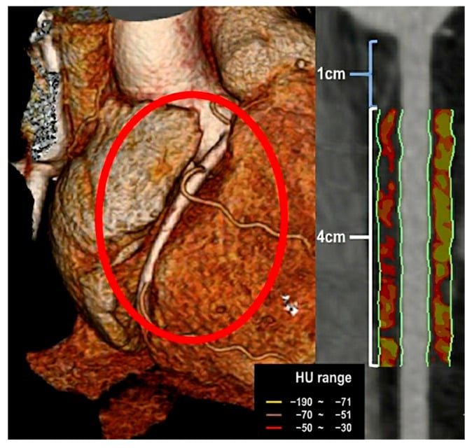
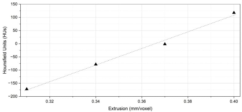
- Each voxel is assigned a HOUNSFIELD UNIT (CT number) on a scale referenced to WATER = 0 HU and AIR = -1000 HU: fat is around -100 HU, soft tissue roughly +30 to +60 HU, and dense cortical BONE up to about +1000 HU (and higher) - so knowing where water, air and bone sit lets you interpret the scale.
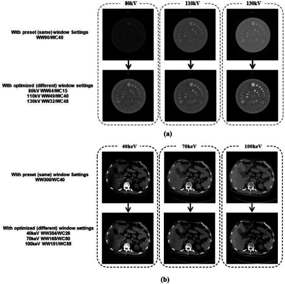
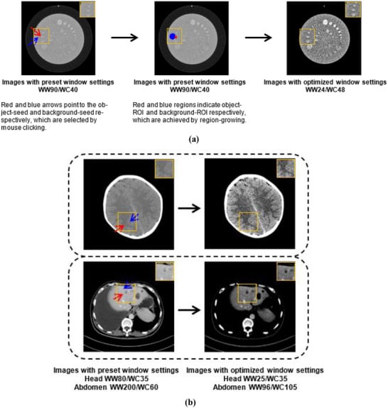
- Because the full HU range cannot be displayed simultaneously, WINDOWING is used: the window LEVEL sets the central HU and the window WIDTH the range of HU mapped across the grey scale - a narrow soft-tissue window and a wide bone window display different tissues from the same data (and explain why a fracture is best seen on a bone window).
- The key ACQUISITION PARAMETERS determine both image quality and dose: kVp (tube voltage) sets beam energy/penetration; mAs (tube current x time) sets the number of photons and hence image NOISE; PITCH (table movement per rotation) and SLICE THICKNESS affect coverage, resolution and noise - increasing dose (higher mAs, thinner slices) reduces noise/improves resolution but RAISES RADIATION DOSE, so they must be balanced.
- RADIATION DOSE is quantified by CTDIvol (volume CT dose index, mGy), DLP (dose-length product, mGy.cm) and the EFFECTIVE dose (mSv, allowing comparison of stochastic risk); CT delivers substantially more dose than plain radiographs, and the ALARA principle (As Low As Reasonably Achievable) mandates using the lowest dose that answers the clinical question - automatic exposure control and iterative reconstruction help.
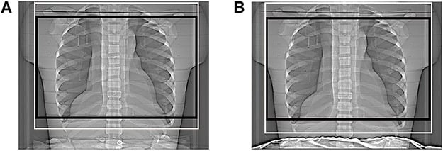
- The PRACTICAL/ORTHOPAEDIC message is to use LOW-DOSE, HARMONISED protocols optimised for both field of view and acquisition parameters: across 17 published CT protocols for total hip arthroplasty planning the recommended kV (100-150) and mAs (100-250) varied widely and most protocols were incomplete, so a consistent low-dose protocol is needed that balances image quality against dose.
- PUT A NUMBER ON THE RISK IN THE YOUNG rather than only warning about it. In a retrospective cohort of about 178,000 people first scanned under the age of 22, a cumulative red-marrow dose of roughly 50 mGy roughly TRIPLED the relative risk of leukaemia and about 60 mGy to the brain roughly tripled the risk of brain tumour - but because both cancers are rare, the ABSOLUTE excess was approximately ONE extra leukaemia and ONE extra brain tumour per 10,000 head CTs in the 10 years after a first scan in a child under 10. The correct conclusion is the authors' own: a justified CT should be done, and the dose kept as low as possible - not that CT should be withheld from children.
- “CT image = reconstructed from tissue X-ray ATTENUATION (rotating tube + detectors). Hounsfield scale: WATER = 0 HU, AIR = -1000 HU, fat ~-100, soft tissue +30-60, dense bone ~+1000.
- “WINDOWING: level = central HU, width = range displayed (bone window vs soft-tissue window). kVp = beam energy; mAs = photon number/noise; pitch + slice thickness affect coverage/resolution/noise - all trade quality vs DOSE.
- “Dose metrics: CTDIvol (mGy), DLP (mGy.cm), effective dose (mSv). ALARA + low-dose protocols; harmonise protocols.
- “Quantify the paediatric risk: ~50 mGy to marrow roughly triples relative leukaemia risk and ~60 mGy to brain roughly triples brain-tumour risk, but the ABSOLUTE excess is about 1 of each per 10,000 head CTs in a child under 10 - justify the scan, then minimise the dose.
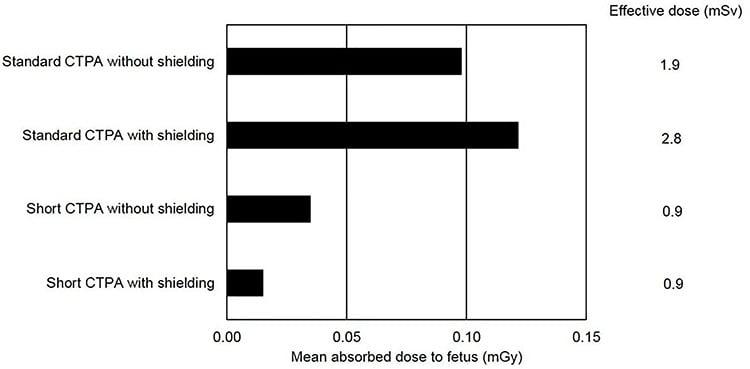
- “In pregnancy the counter-intuitive point: measured fetal dose from a standard CT pulmonary angiogram was ~0.1 mGy, and an abdominal lead SHIELD raised maternal effective dose by 47% and did not lower fetal dose, because the shield enters the automatic-exposure-control field. SHORTENING the scan cut fetal dose by 70%.
Water = 0 HU, air = -1000 HU, fat ~-100, soft tissue +30-60, dense bone ~+1000. Windowing (level = centre, width = range) picks the tissue to display (bone vs soft-tissue window).
kVp (energy), mAs (photon number/noise), pitch, slice thickness - more dose lowers noise but raises radiation dose. ALARA; low-dose protocols; protect the young.
How CT Works & the Hounsfield Scale
CT rotates an X-ray tube and detectors around the patient, measures X-ray attenuation from many angles, and reconstructs cross-sectional images (filtered back-projection/iterative); helical multidetector CT acquires volumes rapidly for multiplanar/3D reconstruction. Each voxel is given a Hounsfield unit on a scale where water = 0 and air = -1000 (fat ~-100, soft tissue +30-60, dense bone ~+1000+). Because the whole range cannot be shown at once, windowing maps a chosen HU range to the grey scale - a window level (centre) and window width (range) - so a bone window and a soft-tissue window display different structures from the same data (a fracture is seen best on a bone window).
- Approximate Hounsfield units
- -1000 HU
- Approximate Hounsfield units
- around -100 HU
- Approximate Hounsfield units
- 0 HU (reference)
- Approximate Hounsfield units
- +30 to +60 HU
- Approximate Hounsfield units
- +50 to +90 HU
- Approximate Hounsfield units
- around +1000 HU
- Approximate Hounsfield units
- about +3000 HU - it saturates the scale, which is why it streaks
Parameters, Dose & ALARA
- kVp (tube voltage): beam energy/penetration. mAs (tube current x time): photon number → image noise. Pitch and slice thickness: coverage, resolution, noise.
- Trade-off: higher mAs/thinner slices reduce noise and improve resolution but increase radiation dose - balance to answer the clinical question.
- Dose metrics: CTDIvol (mGy), DLP (mGy.cm), effective dose (mSv). CT dose far exceeds that of radiographs.
- ALARA: use the lowest dose that answers the question; automatic exposure control and iterative reconstruction lower dose; use low-dose, harmonised protocols (optimise field of view + parameters).
- Protect the vulnerable: young patients and women of childbearing age (higher radiation-induced cancer risk).
The central safety principle of CT physics is that image quality is bought with radiation dose: increasing tube current (mAs) or using thinner slices reduces image noise and improves resolution, but at the cost of a higher dose, and CT already delivers far more dose than plain radiography. The ALARA principle therefore requires using the lowest dose that still answers the clinical question, supported by automatic exposure control, iterative reconstruction, appropriate kVp selection and a field of view limited to the region of interest. This matters most in young patients and women of childbearing age, in whom the lifetime risk of radiation-induced malignancy is greater. As shown for total hip arthroplasty planning, CT protocols vary widely and are often incomplete, leading to inconsistency and unnecessary dose, so harmonised low-dose protocols that are optimised for both the field of view and the acquisition parameters are needed to balance diagnostic quality against patient dose.
The Dose Metrics, Quantified
- What each metric is. CTDIvol (mGy) is the average dose within the scanned volume, measured in a standard phantom (16 cm head/32 cm body) - it reflects the dose intensity (per rotation), independent of scan length, and is a scanner-output index rather than the patient's true dose. DLP (mGy·cm) = CTDIvol × scan length - it captures the total energy imparted over the whole scan. Effective dose (mSv) = DLP × a region-specific conversion factor (k) - it weights for tissue radiosensitivity so stochastic (cancer) risk can be compared across body regions and modalities.
- The magnitudes (for justification). A chest radiograph is around 0.02 mSv; a head CT about 2 mSv; an abdomen/pelvis CT roughly 5-10 mSv; and natural background radiation is about 2-3 mSv per year - so an abdominopelvic CT is equivalent to several years of background, the concrete basis for justification and ALARA.
Q: What do CTDIvol, DLP and effective dose measure, and how are they related?
A: CTDIvol (mGy) = the average dose in a standard phantom per rotation - the dose intensity, independent of length (a scanner index, not the patient's true dose). DLP (mGy·cm) = CTDIvol × scan length - the total energy imparted. Effective dose (mSv) = DLP × a region-specific k-factor - weighting for tissue radiosensitivity so stochastic/cancer risk can be compared. Magnitudes: chest radiograph ~0.02 mSv, head CT ~2 mSv, abdomen/pelvis CT ~5-10 mSv, annual background ~2-3 mSv.
CT Artefacts and How Dose Is Reduced
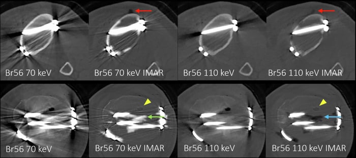
- The key CT artefacts. Beam hardening (the polychromatic beam loses low-energy photons passing through tissue → cupping/dark streaks between dense structures); metal/streak (dense implants cause beam hardening, photon starvation and scatter → bright/dark streaks obscuring peri-prosthetic bone - the orthopaedic problem); partial volume (a voxel spanning two tissues is averaged - misleading at boundaries, worse with thick slices); motion (blur/ghosting); and ring (a faulty detector element).
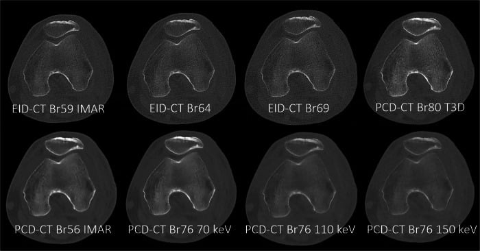
- How dose (and artefact) is reduced. Iterative (and deep-learning) reconstruction models the acquisition physics/statistics to give lower noise at a given dose - allowing roughly a third-to-half dose reduction versus filtered back-projection. Automatic tube-current modulation varies mAs by patient attenuation so dose is delivered only where needed. Lower kVp suits smaller patients and iodine-based angiography. Dual-energy CT enables material decomposition and high-keV monoenergetic / metal-artefact-reduction reconstructions that specifically reduce the orthopaedic metal artefact.
Q: What are the key CT artefacts, and how do modern techniques reduce dose and artefact?
A: Artefacts: beam hardening (beam loses low-energy photons → cupping/dark streaks), metal/streak (implants → beam hardening + photon starvation + scatter, obscuring peri-prosthetic bone), partial volume (voxel-averaging at boundaries), motion, ring (faulty detector). Reduction: iterative/deep-learning reconstruction (lower noise at a given dose → ~third-to-half dose cut vs filtered back-projection); automatic tube-current modulation (mAs by attenuation); lower kVp (small patients/angiography); dual-energy CT with high-keV monoenergetic / metal-artefact-reduction images to cut the orthopaedic metal artefact.
How Big Is the Risk, Actually - and What Shielding Does Not Do
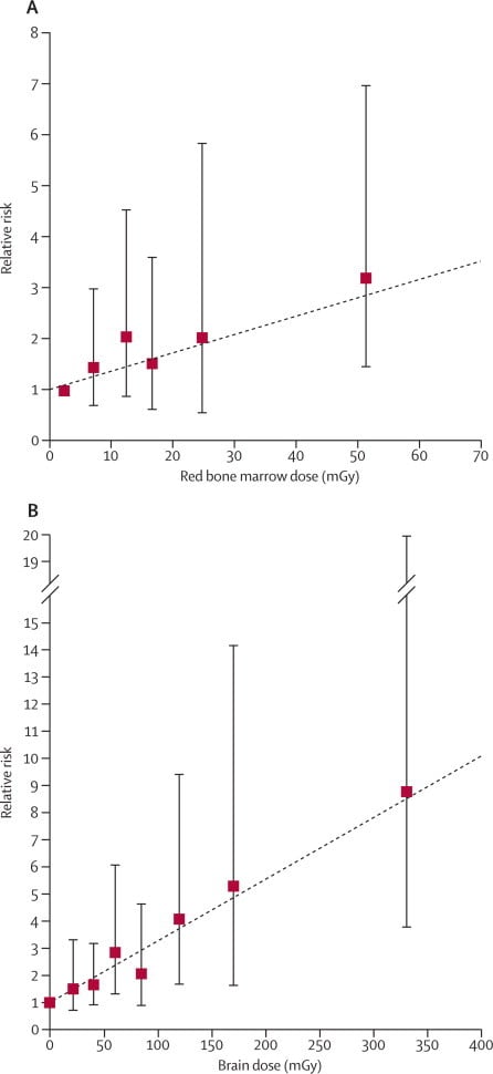
"Protect the young" is not an answer on its own, and a candidate who can quantify it stands out. In a retrospective cohort of around 178,000 patients first scanned before the age of 22 in Great Britain between 1985 and 2002, the dose-response was real: a cumulative red-marrow dose of about 50 mGy carried a relative risk of leukaemia of 3.18 against those receiving under 5 mGy, and about 60 mGy to the brain a relative risk of brain tumour of 2.82. But leukaemia and brain tumours are rare, so the absolute excess was approximately one extra leukaemia and one extra brain tumour per 10,000 head CTs in the ten years after a first scan in a child under 10. That is the number to quote, and the authors' own conclusion follows from it: the clinical benefit of a justified scan outweighs the small absolute risk, so the answer is to justify, then minimise - not to withhold CT from children. Note also what was measured: absorbed dose to the specific organ in mGy, not effective dose in mSv, and the cohort is retrospective, with the earliest scans delivering doses higher than a modern low-dose protocol would.
In a phantom study of CT pulmonary angiography in pregnancy, abdominal shielding increased the mother's effective dose by 47 percent and increased the mean fetal absorbed dose (0.10 to 0.12 mGy) rather than reducing it - because the shield sits within the field seen by automatic exposure control, which responds by driving the tube current up. Shortening the scan to the anatomy actually needed cut fetal dose by 70 percent, from 0.10 to 0.03 mGy. Collimation beats a lead blanket.
That same measurement makes the counselling concrete: the fetal dose from a chest CT is around 0.1 mGy - three orders of magnitude below the 50 to 60 mGy organ doses associated with the tripled childhood cancer risks above. A CT of a limb or shoulder in a pregnant patient is further still from the uterus. The judgement is about justification and scan length, not about refusing an indicated scan.
Q: If you change one acquisition parameter, what happens to dose?
A: mAs is the straightforward one - dose is directly proportional, so halving the tube current-time product halves the dose and raises the noise. kVp raises dose steeply and faster than in proportion, because it increases both the number and the energy of the photons - which is why lowering kVp in a small patient or a child saves a disproportionate amount. Pitch works the other way: at a fixed tube current, increasing pitch spreads the same output over more table travel and reduces dose (and z-axis resolution), which is why many scanners hold quality constant by automatically raising mAs as pitch rises - so pitch must always be read alongside the displayed CTDIvol rather than on its own. Thinner slices improve resolution but need more photons per slice to hold noise constant, so they cost dose. And the parameter that costs nothing to get right is scan length: dose-length product is CTDIvol multiplied by length, so trimming the coverage is the cheapest dose reduction available.
One distinction is worth stating explicitly, because the two situations pull in opposite directions. The shielding finding above is about CT with automatic exposure control, where a lead blanket inside the scanned field misleads the scanner into raising output. It does not transfer to fluoroscopy in theatre, where the operator and staff stand in scattered radiation and personal lead remains the mainstay - occupational protection, dose limits and the pregnant-staff pathway are covered under radiation safety in orthopaedics. The clinical indications for CT, its orthopaedic applications and a fuller treatment of pitch and reconstruction sit in CT imaging principles, and the metal-artefact problem specifically in MRI metal artefact reduction.
Mnemonics & Memory Aids
SCAN
Hook:SCAN: Scale (Hounsfield/windowing), Current (mAs)/kVp, Acquisition (pitch/slice), No more dose than needed (ALARA).
Clinical Decision Scenarios
Practise clinical reasoning and management decisions out loud
“What is a Hounsfield unit, and how do the main CT parameters affect image quality and dose?”
Image formation
- Rotating X-ray tube + detectors measure attenuation from many angles
- Reconstruction (filtered back-projection/iterative) → cross-sectional image
- Helical multidetector CT → rapid volume, multiplanar/3D reconstruction
Hounsfield scale & windowing
- Water = 0 HU, air = -1000 HU, fat ~-100, soft tissue +30-60, bone ~+1000+
- Windowing: level = central HU, width = range across grey scale
- Bone window vs soft-tissue window (fracture best on bone window)
Parameters & dose
- kVp (energy), mAs (photon number/noise), pitch, slice thickness
- More dose → less noise/better resolution but higher dose
- Metrics: CTDIvol (mGy), DLP (mGy.cm), effective dose (mSv)
Safety
- ALARA; automatic exposure control; iterative reconstruction; low-dose protocols
- Paediatric risk with a number: ~50 mGy marrow RR 3.18 leukaemia, ~60 mGy brain RR 2.82 - but ~1 excess of each per 10,000 head CTs under age 10
- Pregnancy: fetal dose from a chest CT ~0.1 mGy; an abdominal shield RAISED maternal dose 47% via AEC - shorten the scan instead (-70%)
- Harmonise protocols (optimise field of view + parameters); trimming scan length is the cheapest dose cut
Evidence & Key Studies
Variability of CT protocols (for THA): a call for harmonisation and low dose
- CT is the principal modality for 3D planning/assessment in total hip arthroplasty, but its image quality carries a radiation penalty, of particular concern in young patients and women of childbearing age due to the greater risk of radiation-induced cancer.
- Across 17 protocols, recommended kV (100-150) and mAs (100-250) varied widely and most protocols were incomplete; CT parameters (kV, mAs, slice thickness) must be optimised with a field of view that includes the relevant bony landmarks.
- A harmonised low-dose CT protocol is needed that provides an optimal balance between image quality and radiation dose.
Dose-monitoring software alerts and CT radiation-dose reduction
- Radiation dose-monitoring software identifies high-dose CT examinations and alerts users, helping to reduce the number of high-dose events and optimise dose.
- High-dose alerts were triggered by patient and technical factors - overweight patients, scan repetition, miscentering, extra studies, orthopaedic hardware in the scan area, and scanning beyond the desired anatomy.
- Most studies showed dose-monitoring tools reduced high-dose events, supporting their role (with proper monitoring and staff training) in CT dose optimisation.
Radiation exposure from CT scans in childhood and subsequent risk of leukaemia and brain tumours: a retrospective cohort study
- Retrospective cohort of patients first examined by CT in NHS centres in Great Britain between 1985 and 2002 while under 22 years old: 74 leukaemias among 178,604 patients and 135 brain tumours among 176,587, with follow-up starting 2 years (leukaemia) and 5 years (brain tumour) after the first scan to exclude scans done for the cancer itself.
- Dose-response was significant for both: excess relative risk per mGy 0.036 for leukaemia and 0.023 for brain tumour. Against a dose under 5 mGy, a cumulative red-marrow dose of about 50 mGy gave a relative risk of leukaemia of 3.18 (95% CI 1.46-6.94) and about 60 mGy to the brain a relative risk of brain tumour of 2.82 (1.33-6.03).
- Because these cancers are rare the absolute risk is small - an estimated one excess leukaemia and one excess brain tumour per 10,000 head CTs in the 10 years after a first scan in a child under 10 - and the authors conclude that clinical benefit should outweigh it, provided dose is kept as low as possible and non-ionising alternatives are considered.
- Read the exposure categories rather than their midpoints: the leukaemia relative risk of 3.18 belongs to a cumulative dose of AT LEAST 30 mGy, whose mean happened to be 51 mGy, and the brain-tumour relative risk of 2.82 to the 50 to 74 mGy band, mean 60 mGy - so the commonly quoted round figures of 50 and 60 mGy are category means, not thresholds.
To use or not use patient shielding on pregnant women undergoing CT pulmonary angiography: a phantom study
- Fetal absorbed dose was measured with thermoluminescent dosemeters in a phantom undergoing CT pulmonary angiography, with maternal effective dose estimated from the dose-length product.
- Abdominal shielding increased the mother's effective dose by 47 percent and increased mean fetal absorbed dose from 0.10 to 0.12 mGy, because the shield entered the field used by automatic exposure control and drove the tube current up.
- Shortening the scan was the effective measure, cutting fetal absorbed dose by 70 percent from 0.10 to 0.03 mGy provided the necessary anatomical coverage was preserved. This is a phantom study, not a patient cohort, and applies to CT with automatic exposure control rather than to fluoroscopic shielding practice.
The radiation penalty of CT image quality (with particular concern for young patients/women of childbearing age), the wide variability of CT protocols and the need for a harmonised low-dose protocol that balances quality against dose come from the cited Ramesh review; the role of dose-monitoring software and the technical/patient factors that drive high-dose events (including orthopaedic hardware and miscentering) from the cited Alanazi review. The paediatric relative and absolute cancer risks come from the Pearce cohort, and the shielding and scan-length findings in pregnancy from the Begano phantom study. The CT image-formation physics, the Hounsfield scale (water 0, air -1000), windowing, the acquisition parameters (kVp/mAs/pitch/slice) and the dose metrics (CTDIvol/DLP/effective dose) with ALARA are standard, well-established teaching, as is the direction in which each parameter moves dose; no single trial establishes an exponent for the kVp-dose relationship, so none is quoted here. Nor is there a validated diagnostic reference level specific to orthopaedic CT that would let a target CTDIvol be given for a given examination.