A Blunt Cerebrovascular Injury in Cervical Trauma
- Vertebral artery injury (VAI) is a form of BLUNT CEREBROVASCULAR INJURY (BCVI) - blunt trauma to the carotid or vertebral arteries - that is an important, potentially PREVENTABLE cause of (posterior-circulation) STROKE after cervical trauma.
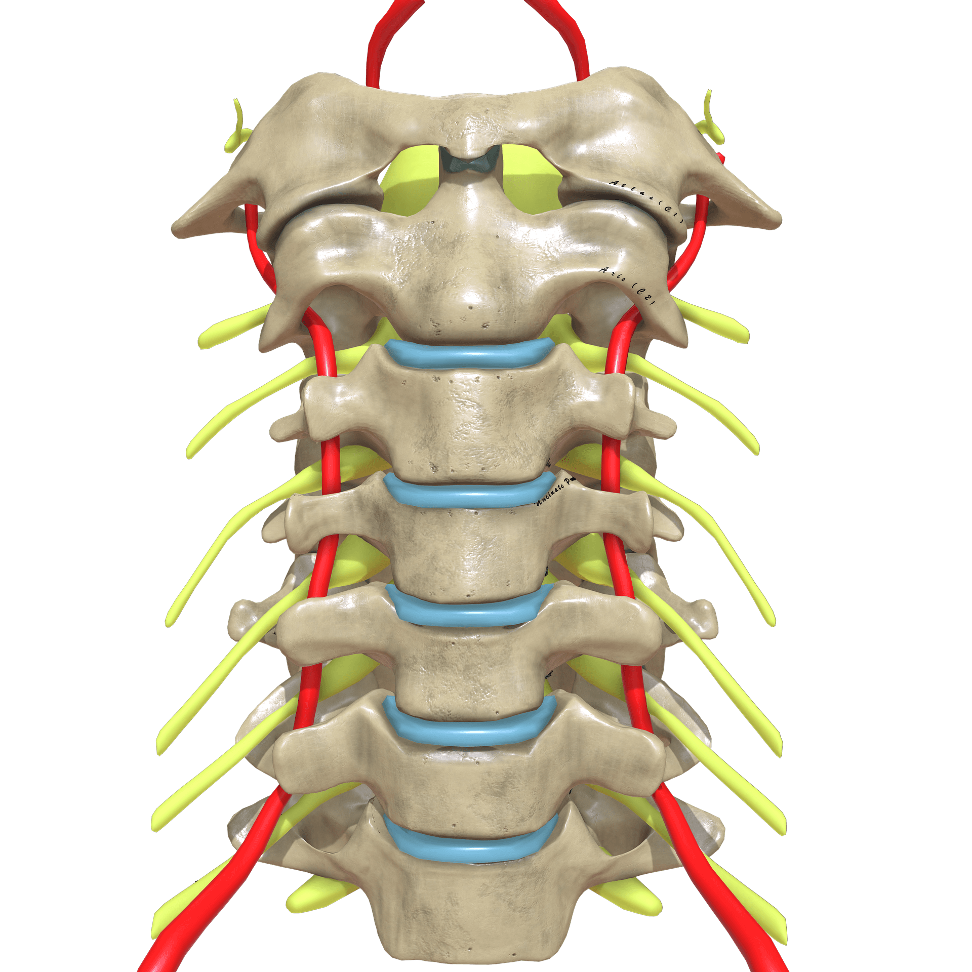
- The artery is vulnerable because its V2 (FORAMINAL) segment runs in a bony tunnel through the TRANSVERSE FORAMINA (C6 up to C2) and the V3 segment loops around the atlas - so cervical injuries that disrupt this anatomy put it at risk.
- High-risk injury patterns: fractures involving the FORAMEN TRANSVERSARIUM, FACET (sub)luxations/dislocations, fracture-subluxations, DISTRACTION injuries, and UPPER cervical (C1-C3) fractures - plus the broader BCVI screening triggers (e.g. severe TBI/GCS, basilar skull fracture, Le Fort II/III, cervical bruit/haematoma, seatbelt sign, or any neurology unexplained by imaging).
- Untreated BCVI causes STROKE in roughly 10-40% of patients, MORE THAN HALF of whom have NO initial stroke symptoms; stroke risk is HIGHEST in the first 7 days (peak in the first 24 hours) - hence early screening and treatment.
- CT ANGIOGRAPHY (CTA) is the SCREENING modality of choice (digital subtraction angiography remains the reference standard in selected cases); injuries are graded by the Biffl/Denver scale (I-V).
- ANTITHROMBOTIC therapy (ASPIRIN or therapeutic anticoagulation) is the MAINSTAY of treatment and is proven safe in trauma patients; ENDOVASCULAR intervention is reserved for selected lesions (e.g. enlarging pseudoaneurysm, grade V); for the ORTHOPAEDIC surgeon the key is to SCREEN high-risk cervical injuries and treat before stroke occurs.
- “Suspect VAI with any cervical fracture through the foramen transversarium, a facet dislocation/subluxation, a distraction injury, or an upper (C1-C3) fracture - screen with CTA.
- “More than half of BCVI patients have NO neurological symptoms at presentation, and stroke risk peaks in the first 24 hours - so screen EARLY rather than wait for symptoms.
- “Antithrombotic therapy (aspirin or anticoagulation) is the mainstay and is safe; grade guides treatment; isolated VAI generally has a low stroke rate with aspirin.
More than half of blunt cerebrovascular injuries present with NO stroke symptoms, yet untreated they cause stroke in 10-40%, with risk highest in the first 7 days (peak in 24 hours). Waiting for symptoms is too late - screen early.
With a high-risk cervical injury, the spine surgeon must screen (CTA) and start treatment, and be mindful that reduction/manipulation of an unstable cervical injury could propagate a dissection - consider the vessel when planning reduction and surgery.
Anatomy & Mechanism
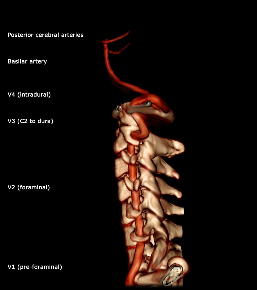
The vertebral artery has four segments: V1 (pre-foraminal) from the subclavian to C6; V2 (foraminal) ascending through the transverse foramina from C6 to C2; V3 (extraspinal) from C2, looping around the atlas to pierce the dura; and V4 (intradural), joining its fellow to form the basilar artery. The V2 segment's bony, enclosed course through the transverse foramina, and the mobile V3 loop at the craniocervical junction, are what make the artery vulnerable in cervical trauma: a fracture that breaches the foramen transversarium, a facet dislocation/subluxation, a distraction injury, or an upper cervical fracture can stretch, compress or tear the vessel - causing intimal injury, dissection, thrombosis, pseudoaneurysm or occlusion.
Who to Screen
Because BCVI is frequently clinically silent initially, screening is based on injury patterns and risk factors (modified Denver/Memphis-type criteria). Screen for VAI/BCVI with:
- Cervical spine fractures - especially through the foramen transversarium, C1-C3 fractures, and facet (sub)luxation/dislocation or fracture-subluxation;
- Severe head injury (low GCS), basilar skull fracture, Le Fort II/III facial fractures;
- Cervical bruit/thrill, expanding neck haematoma, seatbelt sign over the neck, or near-hanging;
- Focal neurology unexplained by brain imaging, or a stroke/TIA pattern. A pragmatic rule: screen any patient whose injuries would otherwise prompt a CT of the neck or chest.
CT angiography (CTA) of the neck is the screening modality of choice (widely available, fast); digital subtraction angiography (DSA) remains the reference standard and is used in selected/equivocal cases or for intervention. Injuries are graded by the Biffl/Denver scale (I-V; see the summary above), which guides treatment and prognosis. Follow-up imaging is often used to track lesion evolution.
Management
The goal is stroke prevention. Antithrombotic therapy - aspirin or therapeutic anticoagulation (heparin) - is the mainstay and has proven safety in trauma patients; treatment is guided by the grade of injury and started as early as the overall injury burden safely allows (given the early stroke window). For isolated vertebral artery injury, the stroke rate is low and aspirin is generally effective (particularly for grade I and IV). Endovascular intervention (stent, coil/occlusion) is reserved for selected lesions - e.g. an enlarging pseudoaneurysm (grade III) or grade V injury. The choice of agent and timing is individualised by balancing stroke risk against bleeding risk (concomitant TBI, solid-organ injury, planned surgery).
| 0 | 1 | 2 |
|---|---|---|
| I | Intimal irregularity / <25% narrowing | Antithrombotic (often aspirin); usually resolves |
| II | Dissection/haematoma >=25% narrowing, thrombus, intimal flap | Antithrombotic; follow-up imaging (can progress) |
| III | Pseudoaneurysm | Antithrombotic; endovascular if enlarging/symptomatic |
| IV | Occlusion | Antithrombotic (aspirin); monitor for posterior-circulation stroke |
| V | Transection / active extravasation | Endovascular/surgical control (often vessel occlusion) |
In a high-risk cervical injury (facet dislocation, foramen-transversarium fracture), remember the vertebral artery when planning closed reduction or operative manipulation - screen with CTA where feasible and weigh the small risk of propagating a dissection, while not unduly delaying urgent reduction of a cord- threatening injury. Coordinate antithrombotic timing with the spinal surgical plan and any associated injuries.
Evidence & Key Studies
Management of blunt cerebrovascular injury
- Untreated BCVI causes stroke in 10-40% of patients, but more than half do not present with stroke symptoms initially; stroke risk is highest in the first 7 days (peak in the first 24 hours).
- CT angiography is the screening modality of choice (DSA in selected cases); screen all patients with injuries that would otherwise prompt CT of the neck or chest.
- Antithrombotic therapy is the mainstay and is safe in trauma patients; endovascular intervention benefits selected patients; treatment is guided by injury grade.
Blunt traumatic vertebral artery injuries: incidence, therapeutic management, and outcomes
- In 156 isolated blunt vertebral artery injuries, most patients were treated with aspirin alone; the risk of stroke after cervical vertebral artery injury was low.
- Aspirin prophylaxis was efficacious in grade I and grade IV injuries; data are limited for grades II and III.
- The strokes that did occur were detected within 24 hours of admission before treatment - reinforcing early screening and treatment.
According to PubMed, the stroke-risk figures, screening (CTA) recommendation and antithrombotic-mainstay management come from the cited Stone BCVI review, and the isolated-vertebral-artery outcomes (low stroke rate, aspirin efficacy) from the cited Zeineddine series. The Biffl/Denver grading and the segmental vertebral-artery anatomy are standard, well-established references. (See also our cervical-trauma topics.)
Clinical Decision Scenarios
Practise clinical reasoning and management decisions out loud
“A patient with a cervical facet dislocation and a fracture through the foramen transversarium is neurologically intact. Why are you concerned about the vertebral artery, who would you screen, and how?”
“CTA confirms a grade II vertebral artery dissection with 30% narrowing. How would you manage it, and how does treatment vary by grade? What if you also need to reduce the cervical injury?”
Mnemonics & Memory Aids
ARTERY
Hook:The ARTERY at risk: at-risk patterns, runs in V2/V3, time-critical, CTA, reduce risk with antithrombotics.
BIFFL
Hook:Biffl I-V: intimal, narrowing/dissection, pseudoaneurysm, occlusion, transection.
Anatomy & risk
- V1 pre-foraminal, V2 foraminal (transverse foramina), V3 atlas loop, V4 intradural -> basilar
- V2/V3 most at risk; injures posterior (vertebrobasilar) circulation
- At-risk injuries: foramen transversarium #, facet (sub)luxation, distraction, C1-C3 #
Why it matters
- Blunt cerebrovascular injury -> stroke in 10-40% if untreated
- More than half SILENT initially; stroke risk peaks first 24h (highest first 7 days)
- Screen early - don't wait for symptoms
Screen & grade
- CTA neck = screening modality of choice (DSA reference standard in select cases)
- Screen Denver-type criteria + anyone needing CT neck/chest
- Biffl/Denver grades I-V guide treatment
Management
- Antithrombotics (aspirin or anticoagulation) = mainstay, safe in trauma; grade-guided
- Isolated VAI: low stroke rate, aspirin effective (esp. I, IV); follow-up imaging for II
- Endovascular for enlarging pseudoaneurysm (III)/grade V; consider vessel before cervical reduction