Weber (Danis-Weber) Ankle Fracture Classification
The Danis-Weber classification
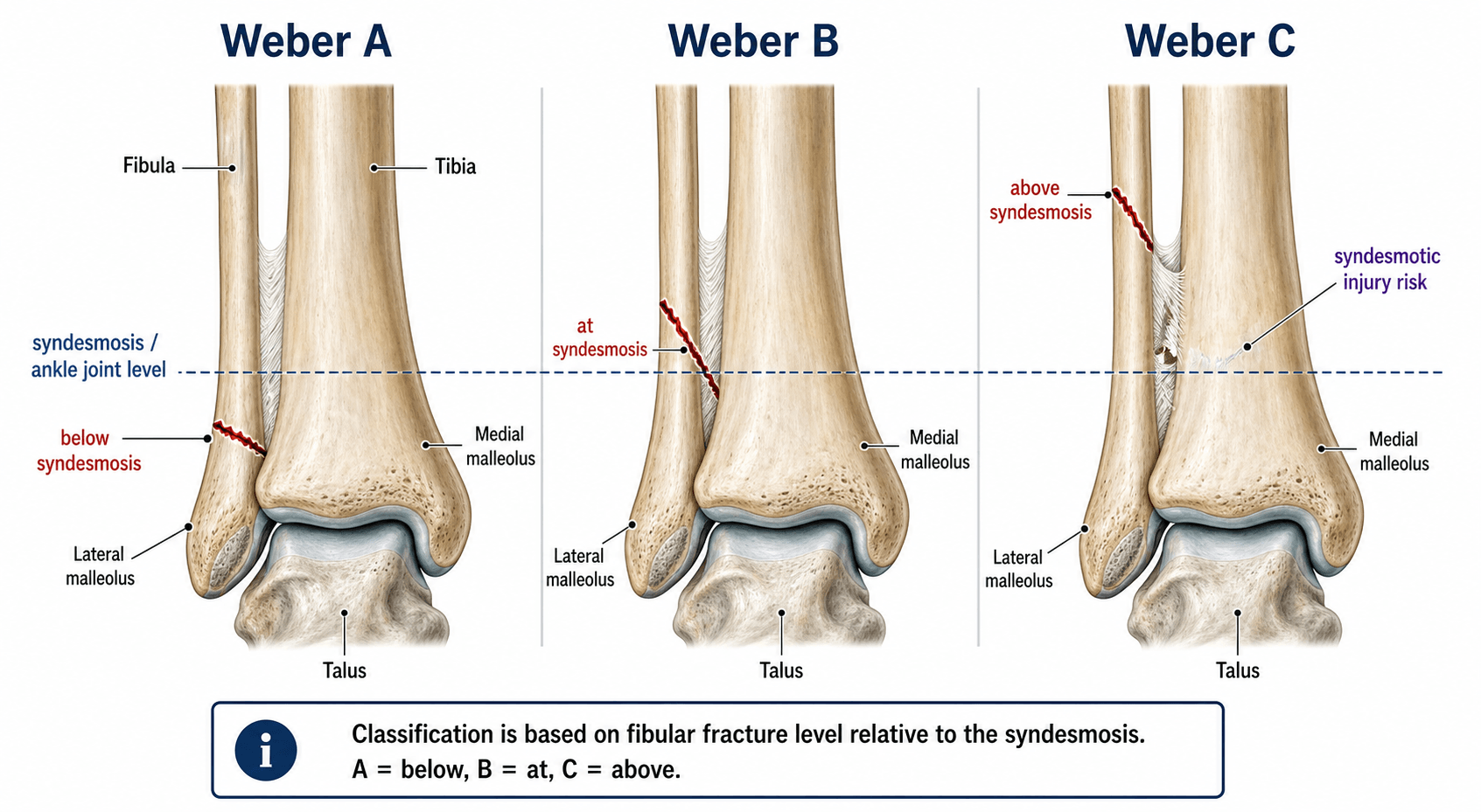
The Danis-Weber system (Weber, 1972) divides lateral malleolar (fibular) fractures by their position relative to the tibial plafond and the tibiofibular syndesmosis — three types. It is deliberately simple and operationally useful because fracture height correlates with the likelihood of syndesmotic injury and therefore with stability.
- Fracture location
- Below the syndesmosis (infrasyndesmotic), often transverse
- Syndesmosis
- Intact
- Typical stability
- Stable
- Usual management
- Non-operative in a walking boot
- Fracture location
- At the level of the syndesmosis (trans-syndesmotic), usually oblique/spiral
- Syndesmosis
- Variable (intact, partial or complete)
- Typical stability
- Variable
- Usual management
- Operative if unstable or if deltoid is ruptured
- Fracture location
- Above the syndesmosis (suprasyndesmotic)
- Syndesmosis
- Always disrupted
- Typical stability
- Unstable
- Usual management
- ORIF of the fibula plus syndesmotic fixation
A below • B at • C aboveABC of the fibula
Hook:A below, B at, C above — the higher the fibular fracture, the higher the syndesmotic risk.
The Weber letter says nothing about the medial malleolus, the posterior malleolus, the deltoid ligament, or the syndesmosis itself. Always look at the mortise view for the medial clear space and tib-fib overlap, and image the whole tibia in a high fibular fracture — a proximal-third fibular fracture is a Maisonneuve until proven otherwise.
Stability, syndesmosis & operative indications
Stability — not the Weber letter — is the gate to theatre. Three principles:
- Medial-side injury converts a lateral-only injury into a bimalleolar-equivalent (Baird-Jackson). A Weber B fibular fracture with a ruptured deltoid and a widened medial clear space behaves like a bimalleolar fracture and must be fixed.
- Mortise congruence is the bedside test of stability. On a well-positioned mortise view the medial clear space should equal the superior clear space (talar dome to plafond). A medial clear space over 4 mm, or greater than the superior clear space, implies deltoid insufficiency and an unstable mortise.
- Weber C means a syndesmotic injury until the operation proves otherwise. Restoring fibular length and rotation is as important as the fixation; the syndesmosis is then tested intra-operatively (hook / external-rotation test) and stabilised if it opens.
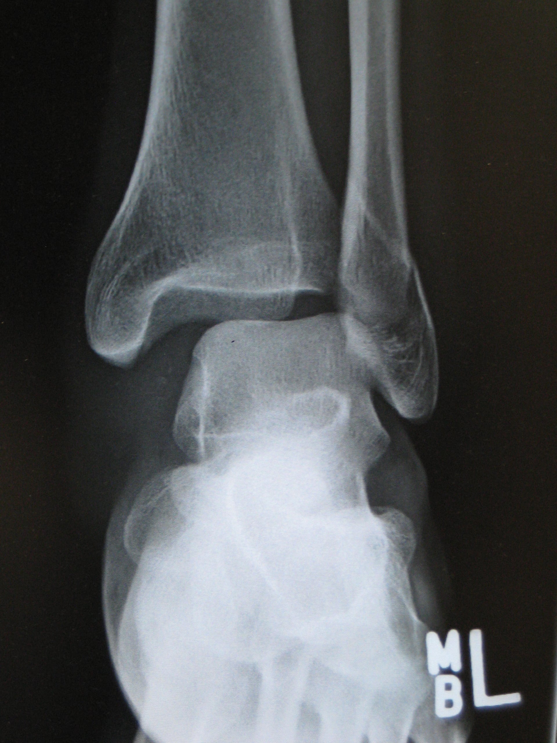
- 1Grade the fibula and image the mortiseAssign the Weber type (A/B/C). Get a mortise view; image the whole tibia in a high fibular fracture (Maisonneuve). CT if the syndesmosis or posterior malleolus is unclear.
- 2Assess the medial side and syndesmosisMedial clear space (should equal the superior clear space; over 4 mm = deltoid insufficiency), tib-fib overlap/clear space, and a stress/weight-bearing view in borderline Weber B.
- 3Treat by stabilityStable (Weber A; Weber B with intact deltoid, congruent mortise) → walking boot. Unstable (Weber B with deltoid rupture/talar shift; Weber C) → ORIF of the fibula restoring length and rotation.
- 4Test and fix the syndesmosisAfter fibular fixation, test the syndesmosis (hook/external-rotation) — almost always disrupted in Weber C — and stabilise with a screw or suture-button if it opens. Fix the posterior malleolus if the fragment is over 25% of the surface or there is a step-off.
- Mortise view
- Congruent, tib-fib overlap preserved
- Medial clear space
- Normal (under 4 mm, equal to superior space)
- Decision
- Non-operative — walking boot, early range of motion
- Mortise view
- Congruent, no deltoid signs
- Medial clear space
- Normal
- Decision
- Non-operative — boot, weight-bear as tolerated
- Mortise view
- Talar shift, medial clear space widened
- Medial clear space
- Over 4 mm, or greater than superior clear space
- Decision
- ORIF of the fibula plus deltoid/syndesmotic assessment
- Mortise view
- Often talar shift, syndesmotic widening
- Medial clear space
- Often widened
- Decision
- ORIF of fibula (restore length/rotation) plus syndesmotic fixation
A Weber C fracture is, by definition, a syndesmotic injury — examine and image the syndesmosis (tib-fib clear space on AP; tib-fib overlap and medial clear space on the mortise; CT if unclear). A high fibular fracture with a tender proximal tibia or proximal fibula is a Maisonneuve until proven otherwise — image the knee and the whole tibia, and expect to stabilise the syndesmosis rather than fix the proximal fibula.
The genuinely hard call is the isolated Weber B (lateral malleolus) fracture with a normal-looking medial clear space on static films — is the deltoid competent (stable, treat in a boot) or incompetent (unstable, fix it)?
- The traditional answer — a stress radiograph. The gravity stress view (patient in lateral decubitus with the injured ankle/leg unsupported, so gravity externally rotates the talus) or a manual external-rotation stress view unmasks deltoid incompetence: a medial clear space over about 4 mm, or greater than the superior clear space, indicates an unstable mortise.
- The modern shift — the weight-bearing mortise view. Stress views over-call instability: a substantial proportion of stress-positive isolated Weber B fractures stay congruent (reduced) under physiological weight-bearing load and do well treated non-operatively, so a weight-bearing radiograph is increasingly preferred to avoid unnecessary surgery.
- The decision: a congruent mortise on a weight-bearing film supports a boot; persistent medial clear space widening / talar shift confirms an unstable injury for ORIF. (The gravity stress view is the more practical screen in the acutely too-painful-to-load patient.)
Limitations of the Weber system
- It only describes the fibula — not the medial malleolus, deltoid, posterior malleolus or syndesmosis directly. Lauge-Hansen and AO/OTA capture more anatomy; in practice most use Weber for the fibula plus a separate description of the medial/posterior sides.
- It does not give a treatment — stability and the medial side do. A Weber B can be operative or non-operative depending on the deltoid.
- Inter-observer agreement is only moderate — the syndesmosis level is hard to define on plain films, and a fracture at the plafond can be read as A, B or C. CT helps when the level is in doubt.
- A Weber B with a deltoid rupture is functionally a bimalleolar injury (Baird-Jackson bimalleolar-equivalent) and should be fixed.
- Posterior malleolar fragments matter — a fragment over 25% of the articular surface, or any step-off, changes the operation regardless of the Weber letter.
Where Weber grades the fibula by height, the Lauge-Hansen system classifies by mechanism — the foot position (supination or pronation) plus the direction of the deforming force — and describes the sequence of structures that fail (staged I–IV), so it captures the medial, posterior and ligamentous injuries that Weber omits. The four patterns and their usual Weber correlate:
- Supination–Adduction (SAD) ≈ Weber A: a transverse avulsion fracture of the fibula below the plafond (the talus pulls the lateral ligaments/fibula), often with a vertical/oblique medial malleolar fracture.
- Supination–External Rotation (SER, "supination–eversion") ≈ Weber B: the commonest pattern; a spiral fibular fracture starting at the plafond/syndesmosis level. Stages run AITFL → spiral fibula → posterior malleolus/PITFL → medial malleolus or deltoid (so the stage predicts the medial and posterior injury).
- Pronation–Abduction (PAB) ≈ Weber C: a suprasyndesmotic fibular fracture, often with lateral comminution (a wedge fragment).
- Pronation–External Rotation (PER, "pronation–eversion") ≈ Weber C: the medial side fails first (medial malleolus or deltoid), then the syndesmosis, then a high fibular fracture — the Maisonneuve is the extreme PER variant with the fibular fracture at the proximal fibula.
- Value & caveat: Lauge-Hansen explains the injury sequence and predicts the associated injuries, but its sub-staging has poor interobserver reliability (Thomsen card) — which is why Weber stays the everyday bedside tool.
Viva practice
Exam viva
Practise clinical reasoning and management decisions out loud
“A 35-year-old twists his ankle playing football. Radiographs show a short oblique fibular fracture at the level of the syndesmosis, with what appears to be widening of the medial clear space. How do you assess and manage this?”
“A 27-year-old is brought in after a fall from height. Imaging shows a fibular fracture 8 cm above the ankle joint, a medial malleolar fracture, and an obvious posterior malleolar fragment. How would you classify and treat this?”
Exam & revision
Everything below condenses the Danis-Weber classification for revision and viva practice.
- A below / B at / C above the syndesmosis (the fibula only).
- Stability — not the letter — drives treatment; read the mortise view (medial clear space, deltoid, syndesmosis).
- Weber B + deltoid rupture (medial clear space over 4 mm) = bimalleolar-equivalent → fix (Baird-Jackson).
- Weber C = syndesmotic injury by definition → fibular ORIF (length & rotation) + syndesmotic stabilisation.
- Restore fibular length and rotation first — the fibula controls talar position.
- High fibular fracture + proximal tenderness = Maisonneuve until proven otherwise; fix the posterior malleolus if over 25% / step-off.
A stable • B variable • C unstableStability descent
Hook:As the fibular fracture rises, syndesmotic injury and mortise instability both rise.
Exam cheat sheet
The three types (fibular fracture height)
- A - below the syndesmosis (infrasyndesmotic), usually transverse
- B - at the level of the syndesmosis (trans-syndesmotic), usually oblique/spiral
- C - above the syndesmosis (suprasyndesmotic)
- Weber describes the fibula only - assess the medial side and syndesmosis separately
Stability and syndesmosis
- A: syndesmosis intact, mortise congruent - stable, non-operative
- B: variable - operate if deltoid ruptured (medial clear space over 4 mm) or mortise incongruent
- C: syndesmosis always disrupted - ORIF of fibula plus syndesmotic fixation
- Baird-Jackson: Weber B + deltoid rupture is functionally a bimalleolar injury
Operative decision
- Restore fibular length and rotation first; the fibula controls talar position
- Fix the posterior malleolus if over 25% of the surface or there is articular step-off
- Test the syndesmosis intra-operatively (hook test, external-rotation stress test) and fix if it opens
- High fibular fracture + proximal tenderness = Maisonneuve until proven otherwise
Pearls & pitfalls
- The Weber letter is not the treatment - stability is
- Mortise view: medial clear space should equal the superior clear space
- Weber C is a syndesmotic injury first and a fibular fracture second
- CT clarifies fragment size, step-off and syndesmotic injury when radiographs are unclear
Evidence Base
Malleolar fractures: nonoperative versus operative treatment. A controlled study
- Randomised controlled study of 92 intra-articular ankle fractures: open reduction and internal fixation versus closed reduction and plaster cast, followed for an average of 7 years.
- The early course was more favourable in the surgically reduced fractures.
- At long-term follow-up, however, there was little difference in results between operative and non-operative treatment.
Fractures of the distal part of the fibula with associated disruption of the deltoid ligament. Treatment without repair of the deltoid ligament
- 24 patients with a distal fibular fracture and deltoid-ligament disruption (a bimalleolar-equivalent injury).
- 19 of 21 (90%) treated WITHOUT deltoid repair had a good or excellent result, with no ankle instability and a near-normal range of motion.
- Repair of the deltoid is unnecessary unless reduction of the lateral malleolus fails to reduce the talus within the mortise.
Quantitative criteria for prediction of the results after displaced fracture of the ankle
- 146 displaced ankle fractures (severity graded by the Lauge-Hansen classification): a prognostic scale was built by multiple regression.
- The significant predictors of outcome were age, the adequacy of post-reduction position of BOTH the medial and lateral malleoli, and restoration of the deltoid ligament and distal tibiofibular syndesmosis.
- Open reduction was superior to closed, and in bimalleolar fractures fixing both malleoli was better than fixing only the medial side.
Observer variation in the radiographic classification of ankle fractures
- Observer-variation study of the Lauge-Hansen and Weber classifications (94 patients, 4 observers, kappa statistics).
- There was an acceptable level of agreement for the overall classification in BOTH systems, but agreement was poor for Lauge-Hansen sub-staging (supination-adduction / supination-eversion).
- The authors concluded that future classification systems should undergo reliability analysis before adoption.
According to PubMed: the operative-versus-non-operative comparison comes from Bauer et al. 1985 (an RCT that found a better early course with surgery but little long-term difference, PMID 3930121); the bimalleolar-equivalent concept and treatment without deltoid repair from Baird & Jackson 1987 (PMID 3440794); the prognostic factors (age, reduction adequacy of both malleoli, deltoid/syndesmosis restoration) from Pettrone et al. 1983 (which graded severity by Lauge-Hansen, not Weber, PMID 6406511); and the reproducibility data from Thomsen et al. 1991 (DOI), which compared Weber and Lauge-Hansen. The medial-clear-space and Maisonneuve principles are standard ankle-trauma teaching.