Compartment Syndrome: Delta-P, Pressure Monitoring and Two-Incision Fasciotomy
Compartment syndrome delta P for clinicians: where the 30 mmHg threshold comes from, how to measure, what BOAST requires, the two-incision release and claims.
By OrthoVellum Editorial TeamPublished 15 min read
Educational content for clinicians, not medical advice. Editorial policy
Key points
- Delta P is diastolic blood pressure minus compartment pressure. McQueen and Court-Brown (1996) recommended decompression when it drops under 30 mmHg; the Edinburgh group later framed the trigger as under 30 mmHg for more than 2 hours.
- An absolute pressure of 30 mmHg over-calls: in the 1996 cohort it would have sent 50 of 116 tibial fractures (43%) to fasciotomy when only 3 had compartment syndrome.
- Pressure is for the patient you cannot examine or cannot decide about. A convincing clinical picture goes straight to theatre, and a palpable pulse never excludes the diagnosis.
- The revised BOAST (July 2025) asks for hourly documented assessment, joint consented decisions on regional anaesthesia, immediate decompression of all involved compartments and re-exploration within 72 hours.
- In closed US claims, fasciotomy within 8 hours of the first symptoms was uniformly associated with a successful defence, and every case with poor communication ended in a payment.
On this page8 sections
A young man with a nailed tibia is asking for more morphine in the small hours, and the ward wants to know whether this is fracture pain or compartment syndrome, and whether a number can settle it. The answer in one line: in a patient you can examine, a convincing clinical picture goes straight to theatre; delta P (diastolic pressure minus compartment pressure) is for the patient you cannot examine or cannot decide about, and the threshold is under 30 mmHg, read as a sustained trend rather than one reading. The treatment is release of all four leg compartments through two long incisions. The full clinical picture is on the acute compartment syndrome of the leg topic page.
Why is compartment syndrome so easy to miss?
Because the patient most at risk looks well. In Edinburgh's series of 164 patients with acute compartment syndrome (opens in a new tab), 69% had an associated fracture, about half of them of the tibial shaft, and most were men under 35. Soft-tissue injury without a fracture was the second commonest cause, and one in ten had a bleeding disorder or was anticoagulated. In a later cohort of 1,388 tibial diaphyseal fractures (opens in a new tab), 160 (11.5%) developed the syndrome and youth was the strongest predictor. The fit young patient with a tibial shaft fracture and good pulses is the high-risk case, not the reassuring one.
The signs are insensitive. Ulmer's review found only four eligible prospective studies. Across them, clinical findings had a sensitivity of 13–19% and a positive predictive value of 11–15%, against specificity and negative predictive value of 97–98%. One finding gave a probability of about 25%; three together gave 93%. The author's own conclusion is that the predictive value "has yet to be defined". The practical reading: findings are cumulative evidence, and a single normal examination never clears an at-risk limb.
The early findings are pain out of proportion to the injury, a rising analgesic requirement, and pain when the muscles of the compartment are passively stretched. A tense compartment supports the diagnosis, but palpation detects and localises a critically raised pressure poorly: about one time in four in a cadaver study. Paraesthesia and weakness mean the nerve is already ischaemic.
The pulse misleads. Whitesides showed in 1975 that there is no effective perfusion within a closed compartment once tissue pressure equals the diastolic pressure, "although distal pulses may be present". Compartment pressure starves the capillaries long before it stops arterial inflow. A pulseless limb points to arterial injury or very advanced compromise; a palpable pulse excludes nothing.
| Compartment | Main muscles | Nerve and sensory check | Passive stretch |
|---|---|---|---|
| Anterior | Tibialis anterior, EHL, EDL, peroneus tertius | Deep peroneal: first web space | Plantarflex the ankle and flex the toes |
| Lateral | Peroneus longus and brevis | Superficial peroneal: dorsum of the foot | Invert the foot |
| Superficial posterior | Gastrocnemius, soleus, plantaris | Sural: lateral foot | Dorsiflex the ankle |
| Deep posterior | Tibialis posterior, FDL, FHL | Tibial: sole of the foot | Extend the toes and dorsiflex the ankle |
The layout of the septa and neurovascular bundles is on the leg compartment anatomy page. Who cannot be examined: the unconscious or sedated, the patient with a regional block or epidural, the intoxicated, and most young children. In children the usual warning is the "three As" of anxiety, agitation and an increasing analgesia requirement. In all of these the decision moves to pressure measurement, monitoring or a low-threshold fasciotomy.
What is delta P, and where does 30 mmHg come from?
Delta P = diastolic blood pressure − compartment pressure. It measures the gradient that drives blood into the tissue, which is why it behaves across the range of blood pressures where an absolute number does not.
Whitesides supplied the principle: perfusion becomes inadequate when tissue pressure rises to within 10–30 mmHg of the diastolic. McQueen and Court-Brown supplied the clinical threshold. They monitored the anterior compartment continuously for 24 hours in 116 patients with tibial diaphyseal fractures. Three (2.6%) developed compartment syndrome. In the first 12 hours, 53 patients had absolute pressures over 30 mmHg and 30 over 40 mmHg, but only one had a differential pressure under 30 mmHg; he had a fasciotomy. In the second 12 hours two more fell under 30 mmHg and were decompressed. None of the 116 had sequelae of compartment syndrome at review six months or more after injury.
| Decision rule (1996 cohort, n = 116) | What it would have done |
|---|---|
| Absolute pressure over 30 mmHg | Fasciotomy in 50 patients (43%) |
| Absolute pressure over 40 mmHg | 27 (23%) considered for an unnecessary fasciotomy |
| Delta P under 30 mmHg | 3 fasciotomies, no missed cases |
The duration came later. The 1996 recommendation was to decompress "if the differential pressure level drops to under 30 mmHg". The Edinburgh review of 2014 is more specific: decompression "primarily on the basis of the differential pressure being less than 30 mmHg for more than 2 h". The same review puts the sensitivity of clinical findings at 13–64% against 94% for monitoring, and specificity at 63–98% against 98%. Quote the threshold with its duration: one transient low reading is not the trigger, and acting on it risks an unnecessary fasciotomy.
How good is monitoring? In 850 monitored tibial diaphyseal fractures, 152 patients had a fasciotomy: 141 true positives, 6 false positives, and 5 with a normal differential pressure but operative findings of compartment syndrome. The estimated sensitivity was 94% and specificity 98%. Those five false negatives are the reminder that the clinical picture keeps the casting vote.
The limits and the arithmetic. The 1996 threshold rests on three events, in one centre, in one compartment, after one injury, so transfer it cautiously to crush injury, a revascularised limb or the forearm. The arithmetic is simple: a diastolic of 65 and a compartment pressure of 40 mmHg give a delta P of 25, under the threshold; a hypotensive patient with a diastolic of 50 reaches the same 25 at a pressure of only 25 mmHg; a hypertensive patient with a diastolic of 110 and a pressure of 32 mmHg has a delta P of 78. No absolute cut-off reads all three correctly: 30 mmHg misses the second and over-calls the third, while 40 mmHg misses the first two.
How do you measure compartment pressure properly?
A single reading is a snapshot of one point in one compartment, so technique decides whether it means anything.
- Device. Use a side-port needle or slit catheter with a calibrated transducer or handheld monitor. A simple straight needle may over-read because tissue can block its tip.
- Zero and insert. Zero the device, insert perpendicular to the skin, and know which muscles lie beneath the needle. A gentle passive stretch of the compartment should produce a brisk rise on the trace.
- Level. Heckman measured all four compartments in 25 closed tibial fractures. The peak pressure was always within 5 cm of the fracture, and falls of 20 mmHg 5 cm from the peak were common. Three of the five patients who needed fasciotomy had readings below the critical threshold within 5 cm of their peak. Measure the anterior and deep posterior compartments at the fracture and above and below it, and use the highest value.
- Record. Write down the compartment, level, device, time and the diastolic pressure taken at the same moment, and the delta P you calculated.
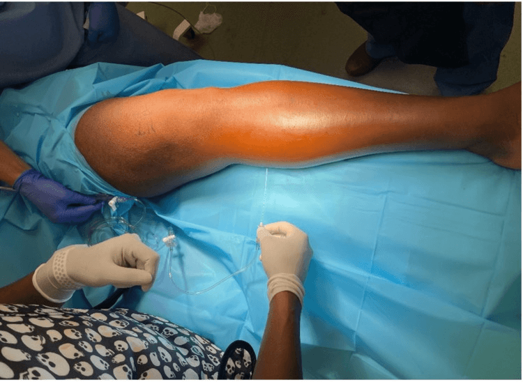
Continuous monitoring is safe, but its benefit is unproven. A retrospective three-centre study compared 171 monitored tibial fractures with 116 unmonitored. Compartment syndrome rates were similar (13 vs 8 patients), with no difference in complications. Time to fasciotomy (19.8 vs 25.8 hours) and split-skin grafting (15% vs 50%) trended in favour of monitoring, but neither difference was significant. A catheter can also block or drift, and a normal anterior trace says nothing about the deep posterior compartment.
What does BOAST say about compartment syndrome?
The British Orthopaedic Association revised its standard, now titled Diagnosis and Management of Compartment Syndrome of the Extremities, in July 2025. The points that change practice:
- Hospitals receiving trauma need training for everyone on the pathway, standardised charts, clear guidance for patients with diagnostic uncertainty or in whom clinical assessment is not possible, and the capability to measure intracompartmental pressure.
- At-risk patients are assessed hourly, recording findings whether present or not, an interpretation and the rationale. The dose and rate of analgesics, particularly opiates, must be recorded.
- Symptoms or signs mean dressings released to skin, the limb raised and re-evaluation within 30 minutes. The operative guide specifies heart level, because raising the limb further can reduce arterial inflow.
- Inconclusive assessment means hourly examination, consideration of pressure measurement in every suspected compartment with simultaneous blood pressure, and senior review.
- A delta P under 30 mmHg "indicates an increased risk", and a consultant orthopaedic surgeon decides between decompression and further monitoring. An absolute pressure over 40 mmHg means urgent decompression should be considered.
- Decompression of all involved compartments is immediate (NCEPOD category 1), with the appearance, viability and debridement of each compartment's contents documented. In the leg a two-incision, four-compartment release is recommended.
The changes from the archived earlier standard (first issued in 2014 as BOAST 10) matter most for regional anaesthesia and timing:
| Issue | Earlier BOAST | Revised BOAST (July 2025) |
|---|---|---|
| Regional anaesthesia | Avoid in high-risk patients; it can mask symptoms | Joint decision with the patient, anaesthetist and surgeon, documented consent, and an agreed policy on who monitors afterwards |
| Timing of surgery | Within an hour of the decision to operate | Immediate, NCEPOD category 1 |
| Re-exploration | At about 48 hours | Within 72 hours, with plastic surgery discussion within 24 hours |
| Late presentation | Over 12 hours: two consultants; non-operative care an option | Two consultants; non-operative care an option, with renal assessment and protection |
| Foot | No consensus | No consensus; the plan must be justified in writing |
How is a two-incision fasciotomy done?
Mubarak and Owen compared the double-incision release with partial fibulectomy in 1977. Both reduced pressure, but they judged the two-incision technique "easier, faster, safer". It remains the standard BOAST recommends. Step-by-step detail with the safety checks is in the four-compartment fasciotomy operative guide.
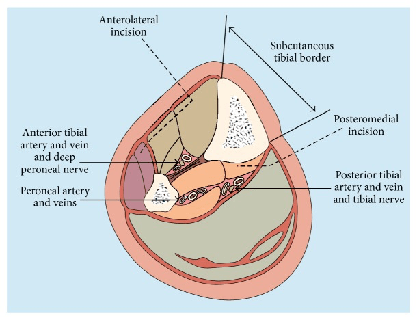
Set-up. Prepare the whole leg from above the knee to the toes. A thigh tourniquet is a contingency only: inflating it adds ischaemia and removes the bleeding you need to judge the muscle.
The anterolateral incision runs about 2 cm anterior to the fibular shaft, long enough to release the full compartments, and stays distal to the fibular head to protect the common peroneal nerve at the neck. Find the anterior intermuscular septum; small transverse windows on each side confirm which compartment is which. Open the anterior fascia in front of the septum and the lateral fascia behind it, along their full length. Distally the superficial peroneal nerve pierces the fascia at a variable level, often near the septum; spread rather than cut blindly there.
The posteromedial incision runs about 2 cm posterior to the posteromedial border of the tibia. Retract the great saphenous vein and saphenous nerve with the anterior flap, then open the superficial posterior fascia over gastrocnemius and soleus.
The deep posterior compartment is the one that gets missed. Seeing gastrocnemius and soleus bulge proves only the superficial release. Return to the tibial border: distally the deep fascia lies immediately behind the tibia; proximally it lies under the soleal bridge, which is detached from the tibia as far as needed. Identify flexor digitorum longus or tibialis posterior before opening the fascia. The posterior tibial neurovascular bundle lies within this compartment, deeper and more posterior, so keep the scissors against the fascial edge under direct vision.
Audit before you leave. Name and point to four fascial openings, anterior, lateral, superficial posterior and deep posterior, and trace each from end to end. Residual bands sit at the skin ends and under the soleal bridge.
Judge the muscle once perfusion and temperature are restored, on colour, consistency, contractility and capacity to bleed. Colour alone is unreliable. Excise clearly dead muscle and leave indeterminate muscle for the planned second look. Major muscle injury needs creatine kinase, potassium, acid-base status, urine output and renal function monitored (see rhabdomyolysis).
Leave the skin open. Closing a fasciotomy at the first operation can recreate the syndrome. Cover the wounds with moist dressings or negative-pressure therapy, keeping foam off exposed nerves and vessels. Vessel-loop "shoelace" dermatotraction can narrow a clean wound once swelling starts to settle. Close directly only when the edges meet without tension; otherwise graft, and plan a flap early for exposed critical structures. Splint the ankle plantigrade to prevent equinus.
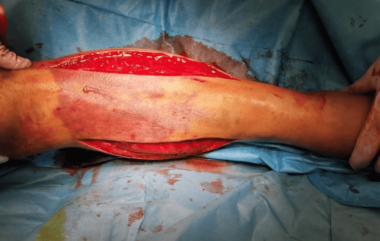
The single-incision parafibular release can decompress all four compartments in experienced hands, reaching the deep posterior compartment from the fibula around the peroneal vessels. It is a different operation, not a shortcut.
Where do compartment syndrome claims come from?
Two closed-claims series show the pattern. Bhattacharyya and Vrahas reviewed 19 closed US claims (16 patients, 1980–2003, total liability US$3.8 million). Time from symptom onset to fasciotomy was linearly associated with a larger payment. A fasciotomy within 8 hours of the first presentation of symptoms was uniformly associated with a successful defence. Poor doctor–patient communication featured in six cases, and all six were paid. In an Italian series of 66 closed claims (opens in a new tab), delay in diagnosis and decompression ran through the series and 48 ended in a verdict of iatrogenic damage. Both datasets are small and jurisdiction-specific, and the 8-hour finding is an association with a successful defence, not with limb outcome.
The pulse note. "Neurovascularly intact, pulses present" records arterial inflow and falsely reassures whoever reads it next. Write down what you examined for.
The analgesia chart is evidence. Record dose and rate, as BOAST requires. When a nurse reports pain that the prescription no longer controls in an at-risk limb, go and examine the patient; do not increase the analgesia over the phone.
Regional anaesthesia is a shared, documented decision. Mar and colleagues' systematic review, of case reports and series only, found classic signs in 32 of 35 patients with epidurals, 18 with documented breakthrough pain. It found no convincing evidence that PCA opioids or regional analgesia delay diagnosis "provided patients are adequately monitored". The 2021 Association of Anaesthetists guideline advises avoiding dense blocks that last well beyond surgery. Its review suggested that lower-concentration peripheral blocks without adjuncts were not associated with delayed diagnosis when surveillance was effective. It asks for scheduled observations by trained staff and documented consent, and neither surgeon nor anaesthetist can veto the other. Pain breaking through a working block is a warning sign until proved otherwise.
The unexaminable patient. In the intubated polytrauma patient, "compartments felt soft" is a weak record. Follow the hospital's pathway: serial or continuous pressure measurement with the delta P recorded, or a decision for fasciotomy.
The note that helps you. "Comfortable overnight, continue analgesia" records nothing about the limb. Compare: "14:00. Pain 3/10, opioid use not increasing. No pain on passive toe flexion or extension. Compartments soft and compressible. First web space and sole sensate; EHL and FHL power full; pulses present. No clinical evidence of compartment syndrome; hourly checks continue." Timed, specific, interpreted and repeated is the BOAST standard. One entry at 08:00 does not cover a limb lost at 16:00.
Consent and the operation note. Where time allows, consent covers fasciotomy, possible debridement, open wounds, a return to theatre, a possible skin graft and scarring, and amputation when presentation is late. The operation note names each compartment released, the muscle's appearance and viability in each, the structures protected and the re-inspection plan.
Talk to the patient and family about what is being watched for and why worsening pain must be reported at once. For the wider duties, see our posts on the medicolegal duties of a surgeon and never events in orthopaedic surgery.
What if the presentation is late?
Fasciotomy is not automatic. Opening a compartment full of established necrotic muscle can raise the risk of infection and systemic toxicity. BOAST asks for two consultants in the decision, with non-operative management an option alongside renal assessment and protection. The operative guide lists exploration with radical debridement, staged salvage, amputation or selective non-operative care, decided on the tissue and the patient rather than the clock alone. Do not declare muscle dead from elapsed time alone, and do not open a dead limb by reflex. What follows a missed release is described on the Volkmann contracture page; the principles for other sites are on the main compartment syndrome page.
In the exam
Examiners want a clinical diagnosis stated first, a threshold quoted with its source and duration, and a release you can describe compartment by compartment. Rehearse the examination itself on the compartment syndrome examination guide.
Frequently asked questions
What delta P indicates compartment syndrome?
Delta P is diastolic blood pressure minus compartment pressure. McQueen and Court-Brown recommended decompression when it drops under 30 mmHg, and the Edinburgh group later specified under 30 mmHg for more than 2 hours on continuous monitoring. The revised BOAST describes a delta P under 30 mmHg as an increased risk, with the decision to decompress or keep monitoring made by a consultant orthopaedic surgeon.
Is a compartment pressure over 30 mmHg an indication for fasciotomy?
Not on its own. In McQueen and Court-Brown's 116 monitored tibial fractures, 53 exceeded 30 mmHg in the first 12 hours, yet only 3 developed compartment syndrome. An absolute threshold of 30 mmHg would have indicated fasciotomy in 43%. Read the number against the diastolic pressure. BOAST advises considering urgent decompression when the absolute pressure exceeds 40 mmHg.
Does a nerve block or epidural mask compartment syndrome?
Dense, long-acting blocks can. A 2009 systematic review found classic signs, often breakthrough pain, in 32 of 35 patients with epidurals, and no convincing evidence that analgesia delays diagnosis when patients are properly monitored. The 2021 Association of Anaesthetists guideline advises avoiding dense blocks that outlast surgery and requires trained, scheduled surveillance and documented consent.
Which compartment is most often missed at leg fasciotomy?
The deep posterior compartment. Opening the superficial posterior fascia and seeing gastrocnemius and soleus bulge does not decompress it. Through the posteromedial incision you return to the tibial border, release the soleal bridge from the tibia proximally as far as needed, identify flexor digitorum longus or tibialis posterior, and open the deep fascia along its length.
How quickly should a fasciotomy be done?
Immediately once the diagnosis is made: the revised BOAST classes it as NCEPOD category 1, the most urgent. There is no reliable clock for tissue death, so do not wait for a nominal six-hour point. In closed US malpractice claims, fasciotomy within 8 hours of the first symptoms was uniformly associated with a successful defence.
References
- McQueen MM, Court-Brown CM. Compartment monitoring in tibial fractures. The pressure threshold for decompression. J Bone Joint Surg Br 1996;78(1):99-104. DOI (opens in a new tab)
- Whitesides TE, Haney TC, Morimoto K, Harada H. Tissue pressure measurements as a determinant for the need of fasciotomy. Clin Orthop Relat Res 1975;(113):43-51. DOI (opens in a new tab)
- McQueen MM, Duckworth AD. The diagnosis of acute compartment syndrome: a review. Eur J Trauma Emerg Surg 2014;40(5):521-528. DOI (opens in a new tab)
- McQueen MM, Duckworth AD, Aitken SA, Court-Brown CM. The estimated sensitivity and specificity of compartment pressure monitoring for acute compartment syndrome. J Bone Joint Surg Am 2013;95(8):673-677. DOI (opens in a new tab)
- Ulmer T. The clinical diagnosis of compartment syndrome of the lower leg: are clinical findings predictive of the disorder? J Orthop Trauma 2002;16(8):572-577. DOI (opens in a new tab)
- Heckman MM, Whitesides TE Jr, Grewe SR, Rooks MD. Compartment pressure in association with closed tibial fractures. J Bone Joint Surg Am 1994;76(9):1285-1292. DOI (opens in a new tab)
- Powell-Bowns MF, Littlechild JE, Yapp LZ, et al. Tibial shaft fractures - to monitor or not? A multi-centre 2 year comparative study. Injury 2021;52(10):3111-3116. DOI (opens in a new tab)
- Mubarak SJ, Owen CA. Double-incision fasciotomy of the leg for decompression in compartment syndromes. J Bone Joint Surg Am 1977;59(2):184-187. DOI (opens in a new tab)
- Bhattacharyya T, Vrahas MS. The medical-legal aspects of compartment syndrome. J Bone Joint Surg Am 2004;86(4):864-868. DOI (opens in a new tab)
- Mar GJ, Barrington MJ, McGuirk BR. Acute compartment syndrome of the lower limb and the effect of postoperative analgesia on diagnosis. Br J Anaesth 2009;102(1):3-11. DOI (opens in a new tab)
- Nathanson MH, Harrop-Griffiths W, Aldington DJ, et al. Regional analgesia for lower leg trauma and the risk of acute compartment syndrome: guideline from the Association of Anaesthetists. Anaesthesia 2021;76(11):1518-1525. DOI (opens in a new tab)
- British Orthopaedic Association. BOA Standard: Diagnosis and Management of Compartment Syndrome of the Extremities. Revised July 2025. Source (opens in a new tab)
Study the full topic
Included in Pro
Go deeper with Pro
- Operative guide: emergency four-compartment fasciotomy of the leg
- Clinical examination: the at-risk limb and compartment syndrome
Share this article
Cite this article
OrthoVellum Editorial Team. Compartment Syndrome: Delta-P, Pressure Monitoring and Two-Incision Fasciotomy [Internet]. OrthoVellum; 2026 Oct 2 [cited 2026 Oct 2]. Available from: https://www.orthovellum.com/blog/compartment-syndrome-delta-p-fasciotomy
About this content
Prepared by the OrthoVellum Editorial Team from cited sources, under our editorial policy.
For education and exam preparation; not medical advice or a substitute for clinical judgement and local guidance.
Spotted an error? Tell us; every report is read.
Get new articles by email
New clinical explainers, landmark-trial summaries and exam guidance from the OrthoVellum blog. No account needed; unsubscribe from any email.