Hawkins Classification of Talar Neck Fractures and the Hawkins Sign
The Hawkins classification of talar neck fractures explained: the four types, osteonecrosis rates by type, the Hawkins sign and how each type changes treatment.
By OrthoVellum Editorial TeamPublished 11 min read
Educational content for clinicians, not medical advice. Editorial policy

Key points
- Hawkins grades a talar neck fracture by how many joints the talus has left: none (I), the subtalar joint (II), the subtalar and ankle joints (III), and, in Canale and Kelly's addition, the talonavicular joint as well (IV).
- Osteonecrosis tracks the subtalar joint. In Vallier's 2014 series it never occurred without subtalar dislocation, and developed in 25% of type IIB and 41% of type III fractures; a meta-analysis pooled 9.8%, 27.4%, 53.4% and 48.0% for types I–IV.
- Osteonecrosis is not collapse: 37% (2004) and 44% (2014) of Vallier's osteonecrosis cases revascularised without dome collapse.
- Reduce a dislocated talus urgently for the skin and the neurovascular structures. Delay to definitive fixation has not been shown to raise the osteonecrosis rate.
- A positive Hawkins sign at 6–8 weeks makes osteonecrosis unlikely; an absent sign is indeterminate. In Tezval's series it was 100% sensitive but only 57.7% specific.
On this page10 sections
A displaced talar neck fracture on the trauma board raises two questions: how urgently must this be reduced, and will the body of the talus survive? The Hawkins classification answers the second by counting the joints the talus has left, because each one marks more of its blood supply torn. It also explains the first, though not in the way it is often taught: the dislocation is an emergency for the skin, while osteonecrosis is largely decided at the moment of injury. This post sets out the four types, the osteonecrosis risk to quote for each, and how to read the Hawkins sign at six to eight weeks. The full clinical picture is on the talus fractures topic page.
What are the four Hawkins types?
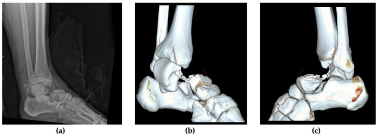
Hawkins described three types in 1970; Canale and Kelly added a fourth. The system grades the injury by the number of joints dislocated, not by fracture displacement alone.
| Type | What the talus has done | Joints out of place | Osteonecrosis: pooled rate (Dodd 2015) |
|---|---|---|---|
| I | Vertical neck fracture, undisplaced | None | 9.8% |
| II | Displaced neck fracture | Subtalar joint subluxed or dislocated; ankle congruent | 27.4% |
| III | Displaced neck fracture; body extruded | Subtalar and ankle | 53.4% |
| IV (Canale and Kelly) | Type III plus talonavicular subluxation or dislocation | Subtalar, ankle and talonavicular | 48.0% |
The pooled figures come from a meta-analysis of the English-language literature by Dodd and Lefaivre, with an overall osteonecrosis rate of 31.2%. Type IV appearing lower than type III probably reflects small numbers: type IV is rare enough that no series has many cases, and the topic page records it as the highest-risk pattern on too few cases to quantify. Type II is the commonest type. Type IV is a very high-energy injury, so check the foot compartments.
Why does counting joints predict osteonecrosis?
Because of where the blood comes in. About 60% of the talus is covered in cartilage and it has no muscle attachments, so vessels reach it only through the narrow non-articular areas of the bone, chiefly around the neck.
- Artery of the tarsal canal, from the posterior tibial artery: the dominant supply to the body (70–80%).
- Artery of the sinus tarsi, from the dorsalis pedis: the lateral body, anastomosing with the artery of the tarsal canal.
- Deltoid branch, from the posterior tibial artery to the medial body: minor and inconsistent, but often the last vessel left after a displaced fracture.
- Superior neck vessels from the dorsalis pedis: the neck and head, preserved in most neck fractures.
The dominant supply enters inferiorly at the neck and runs retrograde into the body. A displaced fracture cuts the body off from its inflow, and each further dislocation strips more of the ligamentous attachments that carry the remaining vessels. That is also why surgery must not finish the job: stripping the neck or injuring the deltoid branch adds to the damage the injury has done.
Is it a neck fracture or a body fracture?
Classify the right bone segment first. Hawkins is for the neck; a body fracture is classified by Sneppen and behaves differently, because it breaks into both the ankle and the subtalar joints (see talar body fractures).
Fractures crossing the anteromedial edge of the trochlea are the hard ones to assign. Inokuchi and colleagues reviewed 215 talar fractures and found that 61 crossed that edge; read from the inferior surface fracture line, they divided into 28 neck and 33 body fractures. Their recommendation was to classify by the inferior fracture line, not the superior. The two other injuries that look like a Hawkins type are dislocations without a neck fracture: a subtalar dislocation leaves the talus in the mortise, and a total talar dislocation extrudes the whole bone from all three joints.
Imaging. Ankle AP, lateral and mortise views and foot views show the joints. The Canale view shows the neck en face: the foot maximally plantarflexed and pronated 15°, with the beam at 75° from the horizontal. CT is indicated for every displaced fracture, because small amounts of displacement change both the Hawkins type and the operation; where an emergency reduction is needed, CT follows it.
Which osteonecrosis figure should you quote?
It depends on which era of series you read, and the older numbers are the ones that stick.
| Series | Patients | Undisplaced (I) | Subtalar out (II) | Ankle and subtalar out (III) |
|---|---|---|---|---|
| Canale and Kelly 1978 | 71 fractures, mean 12.7 years | 2 of 13 | About half | 16 of 19 (complete dislocation of the body) |
| Vallier 2004 | 39 patients with complete radiographic data | — | 9 of 23 (39%) | 9 of 14 (64%) |
| Vallier 2014 | 65 fractures, mean 30 months | 0 | IIA 0; IIB 25% | 41% |
| Dodd 2015 meta-analysis | Pooled literature | 9.8% | 27.4% | 53.4% |
The traditional 80–100% for type III comes from the older series and overstates the modern risk. Two other findings change how the number should be used.
Osteonecrosis is not collapse. In Vallier's 2004 series, 19 of 39 patients showed radiographic osteonecrosis, but 7 of them (37%) revascularised without dome collapse, leaving collapse in 12 (31%). In the 2014 series, 44% of osteonecrosis cases revascularised without collapse. Canale and Kelly made the same point decades earlier: many patients with osteonecrosis treated conservatively had satisfactory results.
Osteonecrosis follows displacement and energy. In 2004 it was associated with comminution of the neck (p < 0.03) and open fracture (p < 0.05). The open-fracture association did not hold in 2014 (30% open against 21% closed, p = 0.55), a reminder of how unstable these subgroup findings are in series of this size.
What is the modified Hawkins type II?
Hawkins's type II includes both a subluxed and a dislocated subtalar joint. Vallier and colleagues split it, in 80 patients with 81 neck and/or body fractures:
- Type IIA: subtalar joint subluxed. Osteonecrosis: none.
- Type IIB: subtalar joint dislocated. Osteonecrosis: 25% (p = 0.03).
Osteonecrosis never occurred without subtalar dislocation, in types I and IIA alike. The series contained 2 type I, 21 type IIA, 23 type IIB, 32 type III and 3 type IV fractures, and 30% were open. The authors' conclusion is hedged to their series: the split "helps to predict" osteonecrosis "as in this series". It is a single-centre retrospective review, and the subdivision is not universally adopted, but it may explain the wide spread of type II figures in older papers that mixed the two.
Does the Hawkins type set the clock for surgery?
It sets the clock for reduction, not for definitive fixation. A dislocated talus threatens the skin over it and the neurovascular bundle, so it is reduced urgently: closed under sedation or anaesthesia if it will go, open in theatre if it will not or if the skin is threatened.
The older teaching that every hour of delay adds osteonecrosis is not supported by the series that tested it:
- Vallier 2004: mean time to fixation was 3.4 days in patients who developed osteonecrosis and 5.0 days in those who did not. The authors' words are careful: "with the numbers available, no correlation could be identified".
- Vallier 2014: 35 patients had delayed ORIF at a mean of 10.6 days, including 20 type IIB and III fractures reduced closed, and 1 of those 20 (5%) developed osteonecrosis.
- Lindvall 2004: in 26 displaced fractures, all closed displaced neck fractures healed regardless of delay, and delay did not appear to affect outcome, union or osteonecrosis.
- Halvorson 2013: a review of 21 reports and 943 talar neck fractures found no demonstrated relationship between the interval to surgery and osteonecrosis.
These are retrospective series, so they show the absence of a detectable timing effect rather than proof that timing never matters. The practical rule from Vallier's 2004 paper is the one to give: reduce dislocations and treat open injuries urgently; the authors suggest that delaying definitive fixation until the swelling has settled may reduce soft-tissue complications.
| Type | Usual management |
|---|---|
| I | Non-weight-bearing cast with close radiographic follow-up, or screw fixation. Any displacement on follow-up films means ORIF. |
| II | Urgent reduction of the subtalar joint, then ORIF when the soft tissues allow. |
| III | Emergency reduction; open reduction if irreducible or the skin is threatened; ORIF. |
| IV | As type III, with attention to the talonavicular joint and the compartments. |
Definitive fixation is through an anteromedial or dual (anteromedial and anterolateral) approach, with lag screws from the neck into the body clear of the joints. The goal is anatomic length with no varus. Medial neck comminution is where varus comes from: compressing a lag screw across it shortens the medial column, so hold length with a medial plate or graft instead. The exposure, reduction sequence and fixation are set out step by step in the talus fracture ORIF operative guide.
What does the Hawkins sign show?
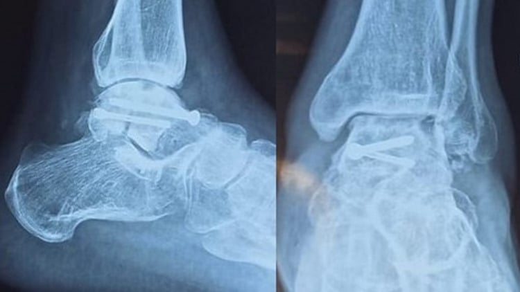
A band of subchondral lucency under the talar dome, seen on the AP or mortise view of the ankle at about 6–8 weeks. It is seldom visible on the lateral view, and in Tezval's series it appeared between the sixth and ninth week. Resorption of subchondral bone during disuse needs a blood supply, so the lucency shows that the body is perfused. An avascular body cannot resorb and stays relatively dense.
How reliable is the Hawkins sign?
Reliable in one direction only. Tezval and colleagues followed 31 patients with operated displaced talar fractures:
- None of the 5 patients who developed osteonecrosis had a Hawkins sign: a sensitivity of 100%.
- Of the 26 who did not, 11 had a full sign, 4 a partial sign and 11 no sign: a specificity of 57.7%.
So a positive sign, full or partial, makes later osteonecrosis unlikely. An absent sign is indeterminate: in this series, more than four in ten patients who kept a viable talus never showed it. Chen and colleagues (44 patients, 48 feet) found the sign in 50%, 30% and 33.3% of types I, II and III–IV, against osteonecrosis rates of 0%, 10% and 50%, and concluded it was a reliable predictor for excluding osteonecrosis.
When the sign is absent. Do not diagnose osteonecrosis from a negative film, and do not plan salvage on it. MRI shows avascular bone before radiographic sclerosis; the topic page advises it at 3–6 months when the sign is absent and the patient is symptomatic, and Chen's group recommends MRI at 12 weeks for sign-negative patients, especially after high-energy injury. Even a positive MRI is not an indication to operate on its own: early MRI changes do not predict the final outcome, and established osteonecrosis is managed by stage, as set out on the talus fractures page.
What matters more than osteonecrosis?
Arthritis. Osteonecrosis dominates the classification, but post-traumatic arthritis is the commoner long-term problem:
- Lindvall 2004: subtalar arthritis in every patient at a mean of 74 months, with more than one joint involved in 16. Osteonecrosis followed 13 of 26 fractures.
- Vallier 2014: post-traumatic arthritis in 54%, rising to 83% with an associated body fracture and 59% in type III.
- Dodd 2015: a mean subtalar arthritis rate of 49%, rising to 81% in studies with more than two years of follow-up.
- Sanders 2004: in 70 displaced neck fractures, secondary reconstructive surgery rose from 24% at one year to 48% at ten years (a life-table estimate), most often for subtalar arthritis or malalignment.
Halvorson's review adds malunion in about 17% and nonunion in about 5%. Varus malunion leaves a stiff, in-toed foot with hindfoot varus, forefoot adduction and loss of subtalar eversion, and loads the medial ankle and peritalar joints. The counselling point from Lindvall's paper is plain: post-traumatic arthritis and chronic pain are expected even after anatomic reduction and stable fixation.
In the exam
Name the type from the joints, give a modern osteonecrosis figure with its source, and separate the emergency (the dislocation) from the timing of fixation. The same structure works for the Weber classification of ankle fractures and for the Garden classification of femoral neck fractures, where displacement also stands in for the blood supply. For holding all of them in memory, see how to memorise orthopaedic classifications.
Frequently asked questions
What is the Hawkins classification?
It classifies talar neck fractures by displacement and the joints the talus has dislocated from. Type I is undisplaced. Type II is displaced with the subtalar joint subluxed or dislocated. Type III adds dislocation of the body from the ankle joint. Type IV, added by Canale and Kelly, adds subluxation or dislocation of the talonavicular joint. The higher the type, the more of the talar blood supply is torn.
What is the Hawkins sign?
A band of subchondral lucency under the talar dome, seen on the AP or mortise view of the ankle at about 6–8 weeks after a talar neck fracture. Resorbing subchondral bone needs a blood supply, so the lucency shows the body is perfused and osteonecrosis is unlikely. It is seldom visible on the lateral view, and in one series it appeared between the sixth and ninth week.
Does an absent Hawkins sign mean avascular necrosis?
No. In Tezval's series every patient who developed osteonecrosis lacked the sign, but so did 11 of the 26 patients whose talus stayed viable, a specificity of 57.7%. An absent sign raises suspicion without making the diagnosis. MRI, typically at about 3 months in a patient without the sign, is the test that shows whether the body is avascular and how much of it is affected.
What is the risk of avascular necrosis for each Hawkins type?
A 2015 meta-analysis pooled osteonecrosis rates of 9.8% for type I, 27.4% for type II, 53.4% for type III and 48.0% for type IV. Vallier's 2014 series found none without subtalar dislocation, 25% in type IIB and 41% in type III. The traditional 80–100% figure for type III comes from older series, and a large share of osteonecrosis revascularises without collapse.
What is the Canale view?
A radiograph that shows the talar neck en face, without the ankle overlapping it. The foot is maximally plantarflexed and pronated 15°, and the beam is angled 75° from the horizontal. It is used to judge displacement, shortening and varus of the neck before fixation and to check the reduction afterwards, alongside CT.
References
- Hawkins LG. Fractures of the neck of the talus. J Bone Joint Surg Am 1970;52(5):991-1002. DOI (opens in a new tab)
- Canale ST, Kelly FB Jr. Fractures of the neck of the talus. Long-term evaluation of seventy-one cases. J Bone Joint Surg Am 1978;60(2):143-156. DOI (opens in a new tab)
- Vallier HA, Nork SE, Barei DP, Benirschke SK, Sangeorzan BJ. Talar neck fractures: results and outcomes. J Bone Joint Surg Am 2004;86(8):1616-1624. DOI (opens in a new tab)
- Vallier HA, Reichard SG, Boyd AJ, Moore TA. A new look at the Hawkins classification for talar neck fractures: which features of injury and treatment are predictive of osteonecrosis? J Bone Joint Surg Am 2014;96(3):192-197. DOI (opens in a new tab)
- Dodd A, Lefaivre KA. Outcomes of talar neck fractures: a systematic review and meta-analysis. J Orthop Trauma 2015;29(5):210-215. DOI (opens in a new tab)
- Halvorson JJ, Winter SB, Teasdall RD, Scott AT. Talar neck fractures: a systematic review of the literature. J Foot Ankle Surg 2013;52(1):56-61. DOI (opens in a new tab)
- Tezval M, Dumont C, Stürmer KM. Prognostic reliability of the Hawkins sign in fractures of the talus. J Orthop Trauma 2007;21(8):538-543. DOI (opens in a new tab)
- Chen H, Liu W, Deng L, Song W. The prognostic value of the Hawkins sign and diagnostic value of MRI after talar neck fractures. Foot Ankle Int 2014;35(12):1255-1261. DOI (opens in a new tab)
- Lindvall E, Haidukewych G, DiPasquale T, Herscovici D Jr, Sanders R. Open reduction and stable fixation of isolated, displaced talar neck and body fractures. J Bone Joint Surg Am 2004;86(10):2229-2234. DOI (opens in a new tab)
- Sanders DW, Busam M, Hattwick E, Edwards JR, McAndrew MP, Johnson KD. Functional outcomes following displaced talar neck fractures. J Orthop Trauma 2004;18(5):265-270. DOI (opens in a new tab)
- Inokuchi S, Ogawa K, Usami N. Classification of fractures of the talus: clear differentiation between neck and body fractures. Foot Ankle Int 1996;17(12):748-750. DOI (opens in a new tab)
Study the full topic
Included in Pro
Go deeper with Pro
- Operative guide: talar neck and body fracture ORIF
- Operative guide: medial malleolar osteotomy to the talar dome
Share this article
Cite this article
OrthoVellum Editorial Team. Hawkins Classification of Talar Neck Fractures and the Hawkins Sign [Internet]. OrthoVellum; 2026 Nov 16 [cited 2026 Nov 16]. Available from: https://www.orthovellum.com/blog/hawkins-classification-talar-neck-fractures
About this content
Prepared by the OrthoVellum Editorial Team from cited sources, under our editorial policy.
For education and exam preparation; not medical advice or a substitute for clinical judgement and local guidance.
Spotted an error? Tell us; every report is read.
Get new articles by email
New clinical explainers, landmark-trial summaries and exam guidance from the OrthoVellum blog. No account needed; unsubscribe from any email.