Weber Classification vs Lauge-Hansen: Ankle Fractures and the Stability Question
The Weber classification of ankle fractures and Lauge-Hansen for clinicians: the types, the stages, and why deltoid and mortise stability decides treatment.
By OrthoVellum Editorial TeamPublished 14 min read
Educational content for clinicians, not medical advice. Editorial policy
Key points
- Weber grades the fibular fracture against the syndesmosis: A below, B at, C above. It says nothing about the medial or posterior structures, and the medial side is what decides stability.
- Lauge-Hansen names the foot position and the deforming force, in four patterns and 13 stages. It explains why a high fibular fracture means the medial side and the syndesmosis have already failed, but it predicted the MRI injury pattern in under half of classifiable fractures in one study.
- The unstable ankle is the one whose talus can shift. A medial clear space over 4 mm on the mortise view, or wider than the superior clear space, means the deep deltoid has failed; 1 mm of lateral talar shift cut tibiotalar contact area by an average of 42% in cadavers.
- The isolated Weber B is the real decision. Stress views over-call instability; in weight-bearing series about 90% of isolated lateral malleolar SER fractures stayed congruent and did well without surgery.
- In adults over 60 with an unstable fracture, the AIM trial found close contact casting equivalent to fixation for 6-month function, with fewer wound problems but more malunion and a 19% crossover to surgery.
On this page9 sections
Every ankle fracture gets a Weber letter in the emergency department and a Lauge-Hansen stage in the viva. Neither answers the question that matters: is the ankle stable? The Weber classification of ankle fractures tells you where the fibula broke and how likely the syndesmosis is to be torn; Lauge-Hansen tells you which way the foot was forced and what else probably failed. Stability is decided on the medial side, by the deep deltoid and the medial clear space, and for the isolated Weber B that usually means a second film. This post covers both systems and the decision they feed; the full clinical picture is on the ankle fractures topic page.
What are Weber A, B and C?
Danis and Weber classified malleolar fractures by one radiographic feature: the level of the fibular fracture relative to the distal tibiofibular syndesmosis. The higher the fibular fracture, the more of the syndesmosis is likely to be disrupted and the less stable the ankle. Weber B is the commonest, about half of ankle fractures.
| Type | Fibular fracture | Syndesmosis | Usual Lauge-Hansen pattern | AO/OTA | Default stability |
|---|---|---|---|---|---|
| A | Below the syndesmosis, usually transverse | Intact | Supination-adduction | 44-A | Usually stable if isolated |
| B | At the syndesmosis, spiral or oblique, starting at the level of the plafond | Intact or partly torn | Supination-external rotation | 44-B | Variable: decided by the medial side |
| C | Above the syndesmosis, up to the proximal fibula | Disrupted | Pronation-external rotation or pronation-abduction | 44-C | Unstable |
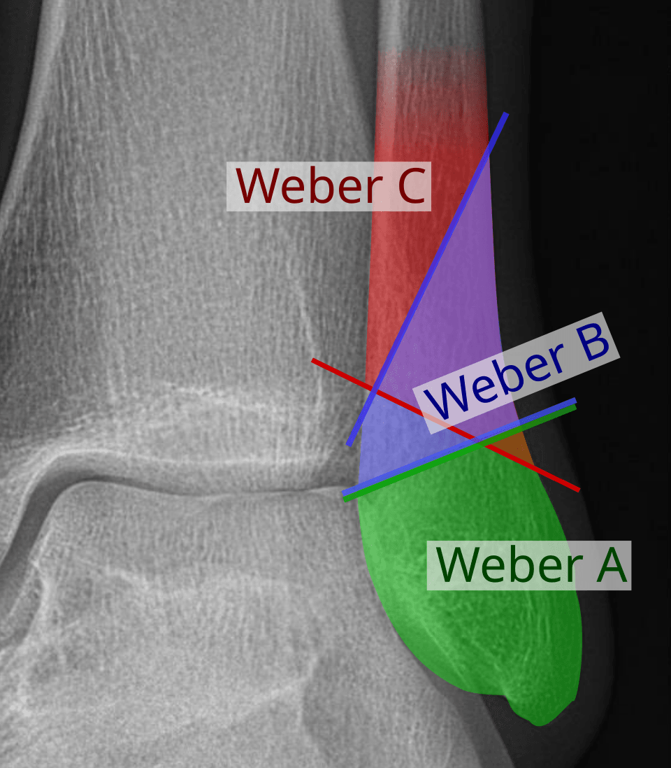
The strength of Weber is that it is simple and reasonably reproducible. In a study quoted in the Clinical Orthopaedics "Classifications in Brief" review, five observers classifying 50 sets of radiographs reached a mean interobserver kappa of 0.61 and intraobserver kappa of 0.74. Its weakness is stated plainly in the same review: the Danis-Weber classification ignores the medial and posterior structures. A Weber B with an intact deltoid and a Weber B with a ruptured deltoid get the same letter and need different treatment.
AO/OTA keeps the three levels and fills that gap with subgroups: 44-B1 is isolated lateral, B2 adds a medial lesion, B3 a medial and a posterior one, and 44-C3 is the proximal fibular fracture of the Maisonneuve injury.
What does Lauge-Hansen add?
Lauge-Hansen classified ankle fractures by mechanism. Working in the 1940s and 1950s on freshly amputated limbs, he fixed the tibia, placed the foot in supination or pronation and applied a deforming force by hand, with three to eight experiments per pattern. The first word is the foot position at injury; the second is the direction of the force. He described four patterns and 13 stages, each stage adding a structure that fails in sequence.
| Pattern | Frequency | Stage 1 | Stage 2 | Stage 3 | Stage 4 |
|---|---|---|---|---|---|
| Supination-external rotation (SER) | 40–75% | AITFL injury | Spiral or oblique distal fibular fracture (Weber B) | PITFL injury or posterior malleolar avulsion | Medial malleolar fracture or deltoid injury |
| Supination-adduction (SAD) | 10–20% | Transverse fibular fracture below the plafond, or lateral ligament avulsion (Weber A) | Vertical medial malleolar fracture or deltoid injury | ||
| Pronation-external rotation (PER) | 5–20% | Medial malleolar fracture or deltoid injury | AITFL injury | Spiral or oblique fibular fracture above the plafond (Weber C) | PITFL injury or posterior malleolar avulsion |
| Pronation-abduction (PAB) | 5–20% | Medial malleolar fracture or deltoid injury | AITFL injury | Transverse or comminuted fibular fracture above the plafond |
Stages follow Tartaglione and colleagues' summary of the 1950 paper, with the classic deltoid alternative added for PAB 1; frequencies are those on the topic page. Two readings of this table carry clinical weight.
In supination-external rotation, the medial side fails last. SER 2 is a lateral malleolar fracture with an intact deltoid and a stable mortise. SER 4 adds a medial malleolar fracture or a deltoid rupture: a bimalleolar fracture, or a bimalleolar-equivalent if the deltoid has gone instead of the bone. The two can look identical on a non-stressed film, which is why the isolated Weber B needs a second look.
In pronation, the medial side fails first. By the time a pronation-external rotation injury has broken the fibula above the syndesmosis, the medial structure and the AITFL have already failed. A high fibular fracture therefore implies a medial injury and a syndesmotic injury. Taken to its end, this is the Maisonneuve pattern: a proximal fibular fracture with a torn syndesmosis and an ankle that can look deceptively benign. Palpate the whole fibula in every ankle injury, and image it if it is tender (see Maisonneuve fractures).
How reliable is Lauge-Hansen?
Less than it is usually taught. Three lines of evidence point the same way.
- It often mispredicts the ligaments. Gardner and colleagues compared radiographic Lauge-Hansen classification with MRI in 59 operatively treated fractures. Ten (17%) could not be classified at all. Of the 49 that could, 26 (53%) had ligament injuries and fracture morphology that did not match the predicted sequence. The authors recommend using the system only as a guide and not relying on it alone for treatment decisions.
- Observers disagree. Five surgeons classified 56 injuries twice (560 classifications). Lauge-Hansen reached interobserver kappa 0.402 and intraobserver 0.398, against 0.708 and 0.608 for AO.
- The experiments have not been reproduced. Michelson's group could not reproduce the SER pattern in 32 cadaveric ankles using Lauge-Hansen's method, and Haraguchi and Armiger produced the short oblique distal fibular fracture with the foot pronated.
The failure that matters is on the medial side. In another MRI comparison quoted on the topic page, the dominant cause of mismatch was a deltoid rupture invisible on plain films, especially in SER stages 2 and 3. A Lauge-Hansen stage cannot tell you the deltoid is intact. Use the system to reason about mechanism and to know where to look; decide stability on imaging.
Weber vs Lauge-Hansen: what each tells you
| Weber | Lauge-Hansen | |
|---|---|---|
| Based on | Fibular fracture level on the radiograph | Foot position and deforming force (inferred from the radiograph) |
| Groups | 3 types | 4 patterns, 13 stages |
| Medial and posterior injury | Not described | Built into the stages, but often wrong against MRI |
| Reproducibility | Kappa about 0.61 interobserver | Kappa about 0.40 interobserver |
| Most useful for | Communication; estimating syndesmotic risk | Knowing where to look for the second lesion; closed reduction |
| Decides stability? | No | No |
Closed reduction reverses the injuring force, which is where the mechanism earns its place. For recall of both systems, see how to memorise orthopaedic classifications.
Why does stability matter more than the letter?
The ankle mortise behaves as a ring of bone and ligament. A ring rarely breaks in one place, so a lateral malleolar fracture should prompt a search for the second break: a medial malleolar fracture, a deltoid rupture, a syndesmotic tear or a proximal fibular fracture.
The structure that holds the talus in place is the deep deltoid, the anterior and posterior tibiotalar ligaments. It is the primary restraint to lateral talar shift. When it fails, the talus can move laterally with the fibula, and small movements matter: in Ramsey and Hamilton's cadaveric study of 23 joints, 1 mm of lateral talar shift reduced tibiotalar contact area by an average of 42%, the largest change of any increment. It is a static study of dissected cadaver joints, without ligaments, muscle tone or cyclic loading: the rationale for anatomic reduction, not proof that 1 mm causes arthritis.
Reading the mortise. The mortise view is taken with the leg in 15–20° of internal rotation. Two measurements answer the stability question; the full set is on the syndesmotic instability page.
- Medial clear space: between the medial malleolus and the medial talus at the level of the talar dome. Over 4 mm, or wider than the superior clear space, means deep deltoid incompetence. Comparing it with the superior clear space, and with the other ankle, is more dependable than any absolute number.
- Tibiofibular clear space: under 6 mm, measured 1 cm above the plafond. It is the least affected by limb rotation.
| Situation | Usual management |
|---|---|
| Isolated Weber A, mortise congruent | Boot, weight-bearing as tolerated |
| Weber A with a vertical medial malleolar fracture (SAD 2) | Fixation; the vertical shear medial fragment is buttressed |
| Isolated Weber B, mortise congruent on a weight-bearing or stress film | Cast or boot, with weekly radiographs for the first 2 weeks |
| Weber B with a medial malleolar fracture, or medial clear space over 4 mm | Open reduction and internal fixation |
| Weber C | Fibular fixation, then intraoperative syndesmotic stress testing; the syndesmosis is usually fixed |
| Maisonneuve | Syndesmotic fixation; the proximal fibula is not fixed |
How do you decide whether an isolated Weber B is stable?
This is the hard call: a spiral fibular fracture at the syndesmosis, perhaps some medial tenderness, and a medial clear space that looks normal on the injury film. The answer comes from a second radiograph, and there are two kinds.
Stress views. A manual external rotation stress view, or a gravity stress view with the patient lateral and the injured ankle hanging unsupported so that gravity externally rotates the talus. Widening of the medial clear space means deltoid incompetence.
Stress views over-call instability. Koval and colleagues sent 21 patients with an isolated Weber B and a positive manual stress test (widening of 5 mm or more) for MRI of the deep deltoid. Nineteen (90%) had only a partial tear and were treated in a walking boot with weight-bearing as tolerated; all united without residual medial widening, and 14 of the 15 reviewed at a year scored 100 on the AOFAS scale. Stress-view widening did not correlate with complete rupture on MRI. It is a small retrospective series.
Where MRI is not available, the threshold can be raised. Van Leeuwen and colleagues studied 39 SER fractures with no medial or posterior fragment against MRI. A medial clear space of 4 mm or more on the plain mortise had a sensitivity of 66.7% for complete deltoid rupture. A gravity stress medial clear space of 6 mm or more had a sensitivity and negative predictive value of 100% and specificity of 91.7%, but a positive predictive value of only 50%. It rules a complete tear out well and rules it in poorly.
Weight-bearing views. The alternative is to load the ankle and see whether the mortise stays congruent.
- Weber and colleagues (2010): of 57 isolated lateral malleolar SER fractures, 51 (90%) were stable on weight-bearing radiographs and treated without surgery. In the 47 patients followed up (mean 62 months), the mean AOFAS score was 96.1. The surgical decision waited 3–10 days for the weight-bearing film, a delay the authors report patients accepted.
- Seidel and colleagues (2017): of 104 SER fractures, 14 were clearly displaced and fixed. Among the rest, 44 had borderline widening on gravity stress (medial clear space 4–7 mm) but a congruent weight-bearing film, and were treated without surgery. At a mean of 23 months their scores matched those stable on both tests, and every fracture healed with a congruent mortise. The authors' conclusion is that gravity stress views appear to overrate the need for surgery. This was a level III prospective comparison.
- Hoshino and colleagues (2012): 38 stress-positive lateral malleolar fractures with an intact mortise went into a walking cast and had weight-bearing films at 7 days. Three (8%) showed widening and had surgery; two of those three were pronation-external rotation injuries. The authors recommend weight-bearing assessment for ligamentous SER 4 fractures and advise against non-operative treatment of PER 3–4 injuries with this protocol.
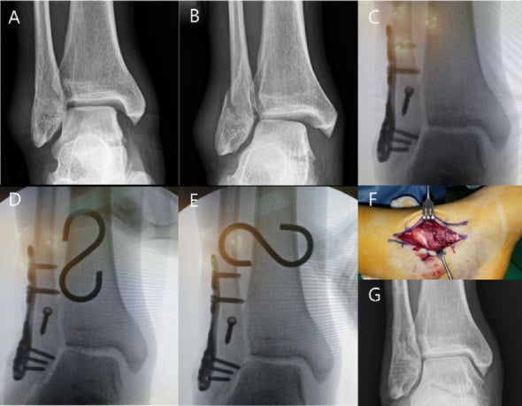
A working sequence. Read the static mortise first. A medial clear space over 4 mm, or wider than the superior clear space, is unstable and needs no further test. If the mortise is congruent, protect the ankle and repeat the film weight-bearing at about a week; if it stays congruent, continue non-operative treatment with weekly films for the first two weeks, because the risk is late displacement. A gravity stress view remains the practical screen for the patient too sore to load. The deltoid ligament injuries page covers the MRI findings for the cases that stay uncertain.
What happens to the unstable ankle in theatre?
The fibula is fixed first, to length and rotation, because a short or externally rotated fibula is the classic cause of a malreduced mortise. The step-by-step technique is in the ORIF of Weber B/C fibula with medial malleolus operative guide.
Then the syndesmosis is stress-tested, and the tests are weaker than most trainees assume. In Pakarinen's prospective study of 140 unstable SER fractures, only 24 (17%) were still unstable on a standardised 7.5 Nm external rotation stress test after malleolar fixation. Against that reference the hook test had a sensitivity of 0.25 and specificity of 0.98, and the fluoroscopic external rotation test 0.58 and 0.96. A positive test is trustworthy; a negative one does not clear the syndesmosis. The population was SER injuries, so the 17% does not describe Weber C fractures. Fixation options and the reduction checks are in the syndesmosis fixation guide.
On the medial side, routine deltoid repair has not been shown to improve outcomes once the fibula and syndesmosis are anatomically stabilised. If the medial clear space stays wide after both, the deep deltoid is incompetent and is repaired.
What about older adults? The AIM trial
The AIM trial asked whether an older patient with an unstable fracture needs the operation at all. It randomised 620 adults over 60 (mean age 71, 74% women) with an overtly unstable ankle fracture, across 24 UK hospitals, to fixation or close contact casting: a moulded below-knee cast with minimal padding, applied in theatre under general or spinal anaesthesia by a trained surgeon. Patients with serious limb or concomitant disease, or substantial cognitive impairment, were excluded.
At 6 months the Olerud-Molander score was 66.0 after surgery and 64.5 after casting (mean difference −0.6, 95% CI −3.9 to 2.6), inside the prespecified equivalence margin of ±6 points. The two arms failed in opposite directions:
| Outcome | Surgery | Close contact casting |
|---|---|---|
| Infection and wound breakdown | 10% | 1% |
| Radiological malunion | 3% | 15% |
| Converted to surgery | — | 19% (52 of 275) |
The trial's wording is that close contact casting may be an appropriate treatment. Read it as a technique-specific result: the cast was applied under anaesthesia by trained surgeons with close radiographic surveillance, the primary endpoint was at 6 months, too early for post-traumatic arthritis, and one patient in five in the casting arm ended up having the operation. It supports offering casting to an older patient who fits the trial population, and the tenfold difference in wound complications is the trade to discuss. It does not license ordinary casting of an unstable ankle.
For how classification feeds the operative decision in the other high-volume fracture of older adults, see our post on the Garden classification of femoral neck fractures.
In the exam
Examiners want both systems named and placed, their limits acknowledged, and a stability decision justified from the medial side.
Frequently asked questions
What is the Weber classification of ankle fractures?
The Danis-Weber classification grades the lateral malleolar fracture by its level relative to the distal tibiofibular syndesmosis. Weber A is below the syndesmosis and is usually stable. Weber B is at the level of the syndesmosis and has variable stability. Weber C is above the syndesmosis, which is disrupted, and is treated as unstable. AO/OTA uses the same three levels as 44-A, 44-B and 44-C.
What is the difference between the Weber and Lauge-Hansen classifications?
Weber is radiographic: it describes where the fibula breaks relative to the syndesmosis. Lauge-Hansen is mechanistic: it names the foot position at injury (supination or pronation) and the deforming force (adduction, abduction or external rotation), then stages the sequence of structures that fail. Roughly, Weber A matches supination-adduction, Weber B supination-external rotation, and Weber C pronation-external rotation or pronation-abduction.
Is a Weber B ankle fracture stable?
It depends on the medial side. A Weber B with a medial malleolar fracture, or a medial clear space over 4 mm or wider than the superior clear space, is unstable and usually fixed. An isolated Weber B with a congruent mortise is often stable. Weight-bearing radiographs at about a week, or a gravity stress view if the patient cannot load, separate the two.
What medial clear space means an unstable ankle fracture?
On a mortise view taken in 15–20° of internal rotation, a medial clear space over 4 mm, or one wider than the superior clear space, indicates deep deltoid incompetence and lateral talar shift. It is measured between the medial malleolus and the medial talus at the level of the talar dome. On gravity stress views, one MRI-referenced study used 6 mm or more to identify complete deltoid rupture.
Does every Weber C ankle fracture need surgery?
Weber C fractures are treated as unstable because the syndesmosis is disrupted, and they are usually fixed: the fibula is reduced and plated, then the syndesmosis is stress-tested and fixed if it is unstable. In a Maisonneuve injury the syndesmosis is fixed but the proximal fibula is not. Non-operative care is generally reserved for non-ambulatory or severely comorbid patients.
References
- Lauge-Hansen N. Fractures of the ankle. II. Combined experimental-surgical and experimental-roentgenologic investigations. Arch Surg 1950;60(5):957-985. PubMed (opens in a new tab)
- Tartaglione JP, Rosenbaum AJ, Abousayed M, DiPreta JA. Classifications in Brief: Lauge-Hansen classification of ankle fractures. Clin Orthop Relat Res 2015;473(10):3323-3328. DOI (opens in a new tab)
- Gardner MJ, Demetrakopoulos D, Briggs SM, et al. The ability of the Lauge-Hansen classification to predict ligament injury and mechanism in ankle fractures: an MRI study. J Orthop Trauma 2006;20(4):267-272. DOI (opens in a new tab)
- Yin MC, Yuan XF, Ma JM, et al. Evaluating the reliability and reproducibility of the AO and Lauge-Hansen classification systems for ankle injuries. Orthopedics 2015;38(7):e626-e630. DOI (opens in a new tab)
- Ramsey PL, Hamilton W. Changes in tibiotalar area of contact caused by lateral talar shift. J Bone Joint Surg Am 1976;58(3):356-357. PubMed (opens in a new tab)
- Koval KJ, Egol KA, Cheung Y, et al. Does a positive ankle stress test indicate the need for operative treatment after lateral malleolus fracture? A preliminary report. J Orthop Trauma 2007;21(7):449-455. DOI (opens in a new tab)
- van Leeuwen C, Haak T, Kop M, et al. The additional value of gravity stress radiographs in predicting deep deltoid ligament integrity in supination external rotation ankle fractures. Eur J Trauma Emerg Surg 2019;45(4):727-735. DOI (opens in a new tab)
- Weber M, Burmeister H, Flueckiger G, Krause FG. The use of weightbearing radiographs to assess the stability of supination-external rotation fractures of the ankle. Arch Orthop Trauma Surg 2010;130(5):693-698. DOI (opens in a new tab)
- Seidel A, Krause F, Weber M. Weightbearing vs gravity stress radiographs for stability evaluation of supination-external rotation fractures of the ankle. Foot Ankle Int 2017;38(7):736-744. DOI (opens in a new tab)
- Hoshino CM, Nomoto EK, Norheim EP, Harris TG. Correlation of weightbearing radiographs and stability of stress positive ankle fractures. Foot Ankle Int 2012;33(2):92-98. DOI (opens in a new tab)
- Pakarinen H, Flinkkilä T, Ohtonen P, et al. Intraoperative assessment of the stability of the distal tibiofibular joint in supination-external rotation injuries of the ankle: sensitivity, specificity, and reliability of two clinical tests. J Bone Joint Surg Am 2011;93(22):2057-2061. DOI (opens in a new tab)
- Willett K, Keene DJ, Mistry D, et al. Close contact casting vs surgery for initial treatment of unstable ankle fractures in older adults: a randomized clinical trial. JAMA 2016;316(14):1455-1463. DOI (opens in a new tab)
Study the full topic
Included in Pro
Go deeper with Pro
- Operative guide: ORIF of Weber B/C fibula with medial malleolus
- Operative guide: ankle syndesmosis fixation
Share this article
Cite this article
OrthoVellum Editorial Team. Weber Classification vs Lauge-Hansen: Ankle Fractures and the Stability Question [Internet]. OrthoVellum; 2026 Oct 21 [cited 2026 Nov 16]. Available from: https://www.orthovellum.com/blog/weber-classification-ankle-fracture
About this content
Prepared by the OrthoVellum Editorial Team from cited sources, under our editorial policy.
For education and exam preparation; not medical advice or a substitute for clinical judgement and local guidance.
Spotted an error? Tell us; every report is read.
Get new articles by email
New clinical explainers, landmark-trial summaries and exam guidance from the OrthoVellum blog. No account needed; unsubscribe from any email.