Intra-articular Hindfoot Injuries with a Precarious Blood Supply
- A talar BODY fracture is an intra-articular fracture of the body of the talus that, unlike a talar NECK fracture, disrupts BOTH the tibiotalar (ankle) AND the subtalar (posterior facet) articular surfaces - so anatomic reduction of the joint surfaces is the priority and post-traumatic arthritis of both joints is common.
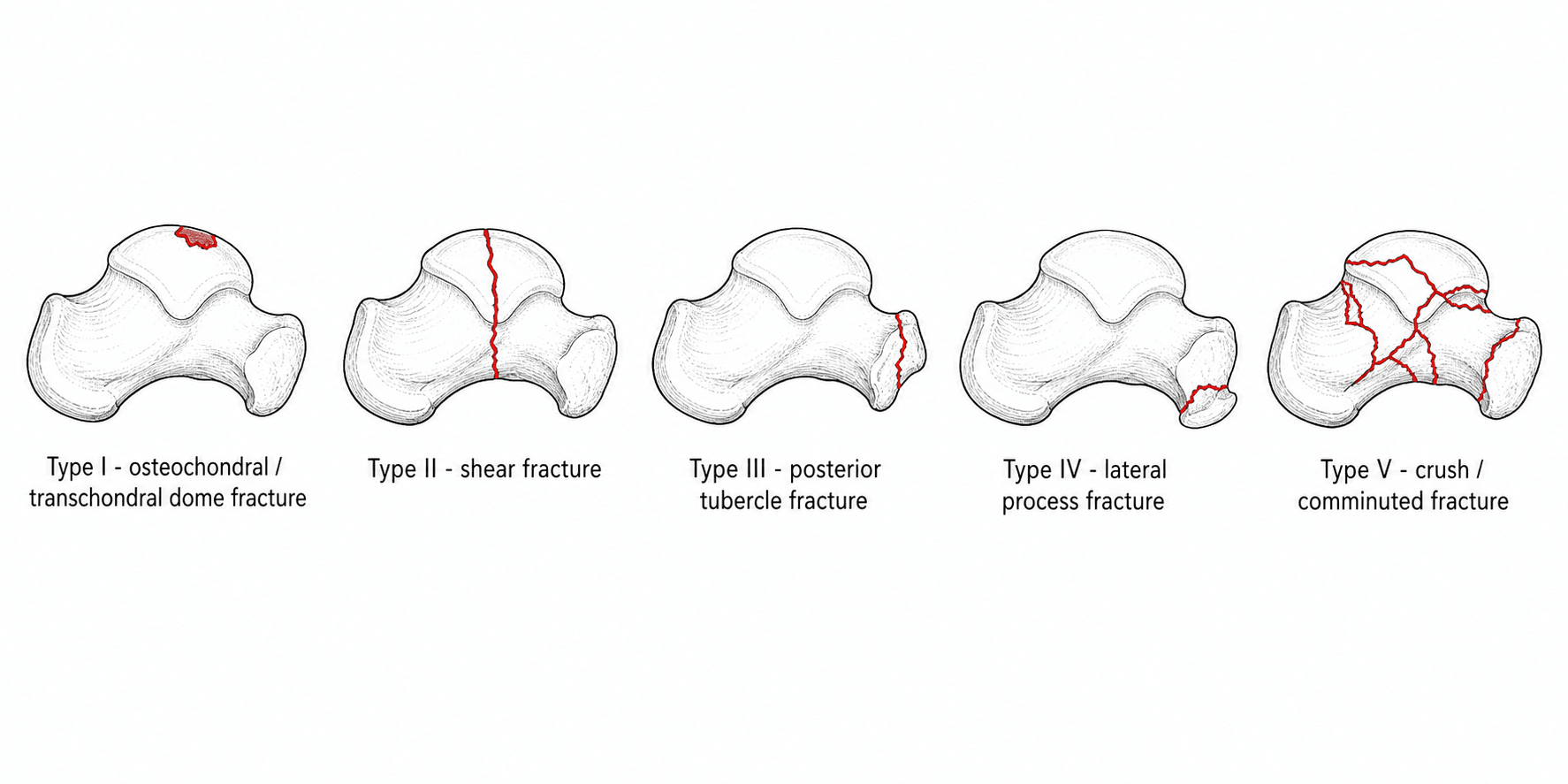
- Fractures are described by the SNEPPEN classification: I osteochondral/transchondral dome, II coronal/sagittal/horizontal SHEAR (the main body fracture), III posterior tubercle, IV lateral process, V crush/comminuted. Lateral process, posterior process and osteochondral dome lesions each have a dedicated OrthoVellum topic.
- The talus has NO muscular attachments and is largely covered by articular cartilage, so its blood supply enters through a limited non-articular area and runs largely RETROGRADE (artery of the tarsal canal from the posterior tibial, deltoid branches, and the artery of the sinus tarsi from the dorsalis pedis/peroneal). Displaced body fractures therefore carry a HIGH risk of AVASCULAR NECROSIS.
- CT is MANDATORY: plain radiographs underestimate talar body fractures, and CT defines fracture planes, articular comminution, displacement and associated injuries to plan the approach and fixation.
- Treatment of a displaced talar body fracture is URGENT anatomic reduction and RIGID internal fixation (lag screws +/- mini-fragment plates); adequate exposure of the body usually requires a MEDIAL or LATERAL MALLEOLAR OSTEOTOMY, taking care to preserve the deltoid (and its blood supply). Truly non-displaced fractures may be treated non-operatively in a non-weight-bearing cast with close radiographic surveillance.
- Complications are frequent: AVASCULAR NECROSIS (watch for the Hawkins sign - subchondral lucency at 6-8 weeks indicating revascularisation; its ABSENCE suggests AVN), POST-TRAUMATIC ARTHRITIS of the tibiotalar and subtalar joints, malunion, and wound problems - these injuries are best managed at, or referred to, a foot-and-ankle/trauma centre.
- “Talar BODY fracture = intra-articular, involves BOTH tibiotalar and subtalar surfaces (vs neck fracture); Sneppen classification.
- “Retrograde blood supply (artery of tarsal canal etc.) -> high AVN risk; Hawkins sign (subchondral lucency ~6-8 wks) = revascularising; absent = AVN likely.
- “CT mandatory; displaced -> urgent anatomic ORIF, usually needing a malleolar osteotomy for exposure.
Intra-articular through the body; disrupts both the tibiotalar and the subtalar (posterior facet) surfaces. Sneppen classification. Priority = anatomic articular reduction; both joints at risk of arthritis.
Extra-articular to the ankle (involves subtalar); Hawkins classification; AVN risk rises with displacement/dislocation. (See our Talus Fractures topic.)
Anatomy, Blood Supply & Why AVN Happens
About 60% of the talus is covered by articular cartilage and it has no muscular or tendinous attachments, so blood can only enter through the limited non-articular surfaces. The supply comes mainly from the artery of the tarsal canal (a branch of the posterior tibial artery) which is the dominant supply to the body, supplemented by deltoid branches (medially) and the artery of the sinus tarsi (from the dorsalis pedis and peroneal arteries). Much of this flow runs retrograde (distal to proximal) into the body, so a displaced body fracture readily strips the supply and causes avascular necrosis. This is the anatomical reason talar body and neck fractures behave so differently from most other fractures.
Assessment & Imaging
Talar body fractures follow high-energy mechanisms (falls from height, motor-vehicle crashes, axial loading) and are frequently part of a polytrauma or associated with other hindfoot/malleolar injuries, so a careful soft-tissue and neurovascular assessment is essential. Plain AP, lateral and mortise radiographs underestimate these fractures; CT is mandatory to define the fracture planes, the degree of articular comminution and displacement, and any associated lateral/posterior process or malleolar fractures - which together determine the surgical approach and fixation strategy.
Management
- Non-displaced fractures: a truly non-displaced talar body fracture (confirmed on CT) can be managed non-operatively in a non-weight-bearing cast/boot with close radiographic surveillance, because even minimally displaced intra-articular fractures may need fixation.
- Displaced fractures: require urgent anatomic reduction of the articular surfaces and rigid internal fixation with lag screws, often supplemented by mini-fragment plates for comminution. Buried/headless screws are used where they would otherwise be prominent in the articular surface.
- Exposure: the body of the talus is deep and largely articular, so adequate visualisation usually requires a medial or lateral MALLEOLAR OSTEOTOMY (e.g. a medial malleolar osteotomy reflected on the deltoid ligament, preserving the deltoid blood supply to the body). The osteotomy is then fixed.
- Severe comminution (Sneppen V): anatomic reconstruction may be impossible; options include limited fixation, primary or delayed arthrodesis (tibiotalar or tibiotalocalcaneal), with the patient counselled about stiffness and AVN.
These are high-energy injuries: soft-tissue swelling and fracture blisters often dictate timing (definitive fixation may be staged after temporary stabilisation), an open fracture or an irreducible dislocation demands emergency treatment, and the surgeon must protect the deltoid ligament and its branches during a medial malleolar osteotomy, since stripping it removes one of the few remaining sources of blood to the body and worsens the risk of AVN. Complex hindfoot injuries are best referred to a foot-and-ankle/trauma centre.
Complications & Outcomes
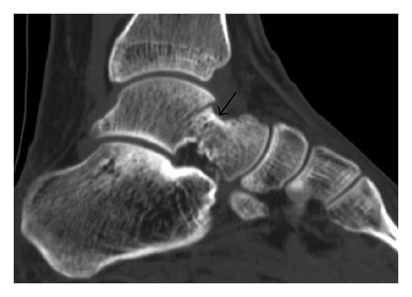
- Avascular necrosis is the feared complication of displaced body fractures (because of the retrograde supply). The Hawkins sign - a subchondral lucency in the talar dome seen at about 6-8 weeks - reflects subchondral bone resorption that requires intact vascularity, so its presence is reassuring (revascularisation) and its absence raises concern for AVN (MRI clarifies). Not all AVN collapses; AVN without dome collapse may remain functional.
- Post-traumatic arthritis of the tibiotalar and/or subtalar joints is common after these double-articular injuries and is the main long-term outcome determinant; salvage is by arthrodesis of the affected joint(s).
- Other complications: malunion (altered hindfoot mechanics), non-union, wound problems/infection, and stiffness. Overall functional outcome correlates with the severity of the original fracture and the quality of articular reduction.
Evidence & Key Studies
Surgical management of talus fractures: mid-term functional and radiographic outcomes
- In a prospective series of 42 surgically treated talar neck and body fractures, 28.5% were ISOLATED talar BODY fractures (a further 21.5% combined neck-and-body), classified by Hawkins, Sneppen and Marti.
- Avascular necrosis affected the talar body in 14.3% (three requiring secondary arthrodesis) and arthritis developed in 23.8%; AVN incidence without dome collapse did not necessarily cause functional impairment.
- Functional/radiographic outcome correlated with fracture severity; osteosynthesis was advocated even for non-displaced patterns, with staged treatment for dislocated/open injuries and referral of complex injuries to foot-surgery centres.
Combined talus fracture with medial malleolar fracture: a case report with review of literature
- Describes a Sneppen type II talar body fracture with an ipsilateral medial malleolar fracture - illustrating the association of body fractures with malleolar injury and the value of the Sneppen classification.
- Treatment was open anatomic reduction of the talar body with a Herbert (headless) screw and cannulated-screw fixation of the medial malleolus.
- Emphasises early diagnosis and fixation to optimise outcome, while noting the unpredictable risks of AVN, wound complications and post-traumatic ankle arthritis.
According to PubMed, the proportion of body fractures and the AVN/arthritis rates come from the cited Kopp series, and the Sneppen-type-II body-with-malleolar example and fixation principles from the cited Moger case report. The Sneppen classification, the retrograde talar blood supply and the Hawkins sign are standard, well-established orthopaedic teaching. (See also our Talus Fractures, Lateral Process Talus Fractures, Posterior Process Talus Fractures and Osteochondral Lesion of the Talus topics.)
Clinical Decision Scenarios
Practise clinical reasoning and management decisions out loud
“How does a talar body fracture differ from a talar neck fracture, how are body fractures classified, and why are they prone to avascular necrosis?”
“A young man falls from a height and CT shows a displaced shear (Sneppen II) fracture of the talar body with intact skin. How would you manage him and what complications would you counsel him about?”
Mnemonics & Memory Aids
DSPLC
Hook:Sneppen I-V = Dome, Shear, Posterior tubercle, Lateral process, Crush.
TALUS
Hook:TALUS body fracture = two joints, AVN, lag screws, urgent, Sneppen + CT.
Definition & classification
- Intra-articular body fracture - disrupts tibiotalar AND subtalar surfaces
- Sneppen: I dome, II shear, III posterior tubercle, IV lateral process, V crush
- Lateral process / posterior process / OCD have dedicated topics
Blood supply & AVN
- No muscle attachments; ~60% cartilage; retrograde supply
- Artery of tarsal canal (post. tibial) dominant + deltoid + sinus tarsi (DP/peroneal)
- Hawkins sign (subchondral lucency ~6-8 wks) present = revascularising; absent = AVN
Imaging & management
- CT mandatory (plain films underestimate)
- Non-displaced: NWB cast + surveillance | Displaced: urgent anatomic ORIF
- Exposure usually via malleolar osteotomy (protect deltoid); lag/headless screws +/- plate
Complications
- AVN; post-traumatic tibiotalar + subtalar arthritis
- Malunion, non-union, wound problems, stiffness
- Salvage = arthrodesis; refer complex injuries to a foot-and-ankle centre