Salter-Harris Classification: Types I–V, Growth-Arrest Risk and the Exam Traps
The Salter-Harris classification for clinicians: types I–V, why site and displacement predict growth arrest as much as type, and the exam traps to avoid.
By OrthoVellum Editorial TeamPublished 13 min read
Educational content for clinicians, not medical advice. Editorial policy

Key points
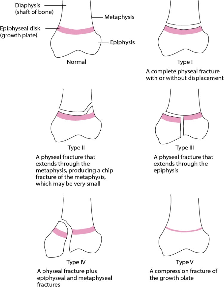
- Salter-Harris sorts physeal fractures by the path of the fracture line: I through the physis, II out through the metaphysis, III out through the epiphysis into the joint, IV across all three, V a crush.
- The type predicts arrest because III, IV and V cross or crush the germinal layers, but site and initial displacement predict arrest at least as well. A displaced Type II at the distal femur or distal tibia is not a benign injury.
- In 124 distal tibial physeal fractures, 67% of the premature closures followed Type II fractures, and each millimetre of initial displacement carried a relative risk of 1.15.
- Types III and IV need anatomical reduction; about 2 mm of articular step or gap is the working threshold for fixation, a figure that comes from 23 triplane fractures.
- Union does not exclude arrest. Follow a displaced Type II and any distal femoral injury to maturity, and read the Harris line on every follow-up film.
On this page8 sections
A child twists an ankle or falls on a knee, and the radiograph shows a line through the growth plate. The question that matters is whether this child will keep growing evenly. The short answer: the Salter-Harris classification describes the path of the fracture line through the physis, and that path tells you whether the germinal layers were crossed, so Types III, IV and V carry the most risk. But site and initial displacement predict arrest at least as well as the type, and a displaced Type II at the distal femur or distal tibia is not a benign injury. The full clinical reference is the physeal injuries topic page.
Salter-Harris types I–V at a glance
Salter and Harris published the system in 1963. It sorts physeal fractures by where the fracture line runs relative to the physis, metaphysis and epiphysis, and the numbering runs roughly from the best prognosis to the worst.
| Type | Fracture line | Joint surface | Usual treatment | Prognosis (conventional teaching) |
|---|---|---|---|---|
| I | Through the physis only; often occult on radiographs | Spared | Cast; gentle reduction if displaced | Good |
| II | Through the physis, exiting the metaphysis (Thurston-Holland fragment) | Spared | Closed reduction and cast | Good at forgiving sites |
| III | Through the physis, exiting the epiphysis into the joint | Involved | Anatomical reduction; fixation for more than about 2 mm step or gap | Fair; good if anatomically reduced |
| IV | Across metaphysis, physis and epiphysis | Involved | Anatomical reduction and fixation | Poor; high arrest risk |
| V | Crush of the physis, no discrete fracture line | Spared | Immobilise if suspected; surveillance | Very poor; often recognised only when growth stops |
The usual mnemonic is SALTR: Slip, Above, Lower, Through, Ruin (or Ram). "Above" and "Lower" assume the joint is at the bottom of the picture, as in the classic distal femoral drawing. Read them as away from the joint (metaphysis) and towards it (epiphysis) and the mnemonic works at every physis.
How common is each type? Type II is the commonest, and it is often quoted at 75%. The population figure is lower. Peterson's Olmsted County study identified 951 physeal fractures in 850 children over ten years: 54% were Type II, the finger phalanges were the commonest site (37%), and 149 fractures (16%) did not fit the Salter-Harris system at all.
That gap is why Peterson proposed his own classification, which adds previously unclassified patterns. The one examiners ask about is Peterson Type VI, in which part of the physis is missing; it is always an open fracture, almost always leads to premature physeal closure and almost always needs late reconstruction. Do not confuse it with Rang's Type VI, an injury to the peripheral perichondral ring, which can produce a peripheral bar even when the physeal cartilage looks intact.
Why does the type predict growth arrest?
Because the fracture line tells you which cells were damaged. From epiphysis to metaphysis the physis runs through the reserve, proliferative and hypertrophic zones, then the zone of provisional calcification. The hypertrophic zone is the weakest: its chondrocytes are enlarging and dying and its matrix is in transition, so Type I and II fractures typically propagate through it and leave the germinal reserve and proliferative layers on the epiphyseal side.
Types III and IV cross those germinal layers vertically. In a Type IV the fracture line itself becomes a template for a bony bar that bridges metaphysis to epiphysis, which is why residual displacement threatens growth as well as the joint. Type V crushes the germinal cells directly. The periphery matters too: the ring of LaCroix and the perichondral ring stabilise the physis, and damage to them, as in a severe Type I, can contribute to instability and arrest.
This is the right way to understand the classification, but it is a mechanism, not an epidemiology. It describes what a fracture line does to cells. It does not tell you how often a bar forms at a given physis, and for that the site and the energy of the injury matter as much as the number.
Why does the site matter as much as the type?
Because physes are not all the same shape. The distal radius is flat, so a Type I or II shears cleanly through the hypertrophic zone and arrest is rare, well under 5% for a Type II there. The distal femur is the opposite: prominent mamillary processes interdigitate with the metaphysis, so the physis cannot fail along one clean plane, and a shear injury tears across the germinal layers even in a nominal Type II.
The distal femur is also the physis with the most to lose. It is the largest in the body and contributes about 70% of femoral length and 35% of total leg length, roughly 1 cm a year near peak growth. Arrest after distal femoral physeal injury is conventionally quoted at 30–50% whatever the type; that range is teaching rather than measurement. The measured anchor is Arkader's series of 73 distal femoral physeal fractures from two level I paediatric centres: an overall complication rate of 40%, growth arrest the commonest, with Salter-Harris type (P = 0.031) and displacement (48.8% vs 26.6% complications) both predictive. Complications were higher when hardware crossed the physis (65% vs 30%), a difference that fell short of significance (P = 0.06). The detail is on the distal femoral physeal injuries topic page.
The proximal tibia is the other high-risk lower-limb physis: it contributes about 55% of tibial growth, and displacement can injure the popliteal artery, so examine the pulses before anything else.
| Site | What the evidence shows | Source |
|---|---|---|
| Distal radius | Arrest rare, well under 5% for a Type II | Conventional teaching |
| Distal femur | 40% complications, arrest the commonest; arrest quoted at 30–50% at any type | Arkader 2007 (n = 73); teaching range |
| Distal tibia | Premature physeal closure in 12.1% to 27.2% | Leary 2009 (n = 124); Barmada 2003 (n = 92) |
| Medial malleolus (SH III/IV) | Physeal bar in 22 of 41 (53.7%) with more than 3 months of follow-up | Abbot 2024 |
At the distal tibia, why do most arrests follow Type II fractures?
Leary and colleagues followed 124 children with distal tibial physeal fractures until symmetric growth was confirmed on Harris lines or the physis had closed. Premature physeal closure occurred in 15 (12.1%). Of those, 67% followed Salter-Harris II fractures, 13% Type III, 13% Type IV and 7% triplane fractures; none followed a Type I or Tillaux fracture.
Two things explain it. The first is base rates: Type II is by far the commonest pattern, so a modest per-fracture risk produces most of the arrests. The second is energy. The amount of initial displacement predicted closure, with a relative risk of 1.15 for every millimetre, and the mechanism of injury was also predictive; residual displacement after reduction and the number of reduction attempts showed only non-significant trends.
Barmada's distal tibial series points the other way on that last point. In 92 fractures, 27.2% closed prematurely: 36% of Type I and II injuries and 38% of the Type III and IV (medial malleolar) injuries, against 21% of triplane and none of the Tillaux fractures. Initial displacement did not predict closure; more anatomical reduction did. A residual physeal gap of more than 3 mm after reducing a Type I or II raised the closure rate to 60%, against 17% with no gap, and in all five Type II fractures opened for a residual gap the periosteum was trapped in the physis.
The two series disagree on whether the injury or the reduction matters more, and neither is randomised. The paediatric ankle ORIF operative guide draws the cautious conclusion: a residual gap may mean interposed periosteum and a higher arrest risk, but retrospective evidence has not proved that opening every gap prevents arrest, so do not promise a family that surgery will.
The medial malleolus is its own problem. In Abbot's series, 22 of 41 children (53.7%) with medial malleolar fractures developed a bar. Every one had been reduced to within 2 mm, yet mean residual displacement was 1.2 mm in those who formed a bar and 0.8 mm in those who did not (P = 0.03). Bars were diagnosed at a mean of 4.9 months, and 6 of 22 after six months. The 53.7% is an upper estimate: 37 of 78 consecutive patients were excluded for less than three months of radiographic follow-up, and bars were called on plain films.
When is a physeal fracture fixed?
Types I and II. Closed reduction and a cast is the mainstay. Reduce gently by reversing the mechanism, using the Thurston-Holland fragment as a guide, and stop after one or two controlled attempts, because repeated forceful manipulation adds physeal damage and rarely overcomes interposed periosteum. An unstable reduction is held with smooth K-wires, removed at about 3–4 weeks. Open reduction is for the reduction that is blocked or unacceptable, not for every displaced Type II.
Types III and IV. These are intra-articular, and anatomical reduction is required for the joint as much as for the physis. About 2 mm of residual step or gap is the working threshold for fixation, with screws or smooth wires placed within the epiphysis and parallel to the physis, never into the joint. Avoid threaded screws across an open physis where the geometry allows, because they create a tether that can cause asymmetric growth.
Where the 2 mm comes from. The figure traces to Ertl (1988): 23 triplane fractures, in which residual displacement of 2 mm or more after reduction was associated with a less than optimum result, unless the epiphyseal fracture lay outside the primary weight-bearing area of the ankle. Results also worsened with time, from 20 of 23 patients asymptomatic at 18–36 months to 8 of 15 at 38 months to 13 years. Choudhry's CT-based series of 78 transitional fractures found no functional penalty with residual displacement up to 2.4 mm at a minimum of two years. So state the location with the measurement, and treat 2 mm as a working threshold in the weight-bearing plafond rather than a universal law.
The transitional fractures. The distal tibial physis closes centrally first, then medially, and laterally last, over roughly 18 months, typically completing at about 14 in girls and 16 in boys. The Tillaux fracture is a Type III avulsion of the anterolateral epiphysis by the anterior inferior tibiofibular ligament; the triplane is a multiplanar Type IV-equivalent. Both are planned on CT, and because the physis is closing, a screw that crosses it is often acceptable where it would be avoided in a younger child. The Tillaux and triplane fractures page covers the patterns, and the paediatric ankle fractures page the whole ankle.
CT helps you plan the operation, not name the fracture. Thawrani had five observers classify 50 distal tibial physeal fractures. Interobserver agreement on the Salter-Harris type was kappa 0.67 on radiographs and 0.57 once CT was added; surgeons rated CT most useful for planning screw placement, and it changed the treatment decision in about a fifth of cases. Displacement measurements, by contrast, were highly reproducible. Let the measured step and gap, not your confidence in the type, drive the decision.
How long should a physeal fracture be followed?
A healed fracture can still arrest. A partial arrest declares itself as asymmetric growth and progressive angulation, often months later, and the earliest radiographic sign is the Harris (Park) line. A transverse sclerotic line that runs parallel to the physis and moves away from it with growth means even, resumed growth. A line that converges on one point of the physis, or is tethered to it, points at a forming bar.
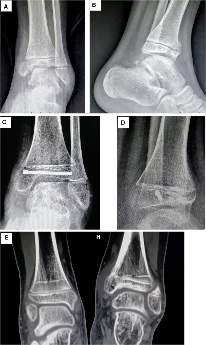
Minimum surveillance. Any Type III or IV, or any injury at a high-risk site, is followed for at least a year; the distal femur for at least two; the medial malleolus for at least 12 months, since bars there were diagnosed up to 11.8 months after injury. A significantly displaced Type II and any distal femoral physeal injury are best followed to skeletal maturity.
When a bar forms. Map it with MRI or CT and express it as a percentage of the physeal area. A bar under 50% of the physis in a child with at least 2 years of growth left can be excised, with fat or PMMA interposed to stop it reforming; a larger bar, or one with little growth remaining, is managed by completing the epiphysiodesis, with contralateral epiphysiodesis and osteotomy as needed. The technique is in the physeal bar resection operative guide, and the angular correction options on the guided growth page.
What are the exam traps?
Examiners use the classification to find out whether you understand the physis, not whether you can recite five lines. These are the errors that cost marks:
- "The X-ray is normal, so it is a sprain." In a child the physis is weaker than the ligaments. Point tenderness over the physis means a Type I injury until proven otherwise; immobilise it.
- "Type II is the benign type." It is benign at the distal radius. At the distal tibia it causes most arrests in absolute terms, and at the distal femur it carries a high arrest risk like every other type; follow a displaced one.
- "Type II is 75% of physeal fractures." Quote the 75% if you must, but the population figure is 54%, and 16% of fractures in that study fitted no Salter-Harris type.
- "Type V is diagnosed on the first film." It is usually diagnosed retrospectively, when growth arrest appears.
- "Five types cover everything." Know Peterson Type VI (open loss of part of the physis, near-certain arrest) and Rang's Type VI (perichondral ring injury and its peripheral bar).
- "Two millimetres everywhere." The threshold came from triplane fractures in the weight-bearing area, and at the medial malleolus a bar formed with residual displacement averaging 1.2 mm.
- "Fix it with a big screw across the physis." Fix within the epiphysis, parallel to the physis; smooth wires if the physis must be crossed, removed early. The closing physis of a transitional fracture is the accepted exception.
In the exam
Describe the fracture line, name the type, then move straight to site, displacement and follow-up. For keeping the types straight under pressure, see how to memorise orthopaedic classifications; for the adult ankle that these injuries are mistaken for, see the Weber classification.
Frequently asked questions
What are the five Salter-Harris types?
Type I runs through the physis only. Type II runs through the physis and exits through the metaphysis, leaving a Thurston-Holland fragment. Type III runs through the physis and exits through the epiphysis into the joint. Type IV crosses metaphysis, physis and epiphysis. Type V is a crush injury of the physis with no discrete fracture line, usually recognised only when growth stops.
Which Salter-Harris type is the most common?
Type II. Textbooks often quote about 75%, but the population-based Olmsted County study of 951 physeal fractures found 54% were Type II, and 16% did not fit the Salter-Harris system at all. The commonest site in that study was the finger phalanges, at 37% of all physeal fractures.
Which Salter-Harris fractures cause growth arrest?
Types IV and V carry the highest risk because they cross or crush the germinal layers, and Type III is intra-articular and needs anatomical reduction. But arrest also follows Type II injuries at high-risk sites: at the distal tibia two thirds of premature closures in one 124-patient series followed Type II fractures, and the distal femur behaves badly whatever the type.
Can a Salter-Harris type I fracture have a normal X-ray?
Yes. A Type I fracture may show no fracture line and only subtle physeal widening, or nothing at all. The diagnosis is clinical: point tenderness over the physis after an injury in a skeletally immature child means a physeal injury until proven otherwise. MRI is reserved for the child whose radiograph is negative but in whom suspicion stays high.
What does the SALTR mnemonic stand for?
Slip (Type I, through the physis), Above (Type II, the metaphysis is involved), Lower (Type III, the epiphysis is involved), Through (Type IV, through all three layers) and Ruin or Ram (Type V, a crush). Above and Lower assume the joint is at the bottom of the picture, so read them as away from and towards the joint.
References
- Salter RB, Harris WR. Injuries involving the epiphyseal plate. J Bone Joint Surg Am 1963;45(3):587-622. DOI (opens in a new tab)
- Peterson HA, Madhok R, Benson JT, Ilstrup DM, Melton LJ 3rd. Physeal fractures: Part 1. Epidemiology in Olmsted County, Minnesota, 1979-1988. J Pediatr Orthop 1994;14(4):423-430. DOI (opens in a new tab)
- Peterson HA. Physeal fractures: Part 2. Two previously unclassified types. J Pediatr Orthop 1994;14(4):431-438. DOI (opens in a new tab)
- Peterson HA. Physeal fractures: Part 3. Classification. J Pediatr Orthop 1994;14(4):439-448. DOI (opens in a new tab)
- Leary JT, Handling M, Talerico M, Yong L, Bowe JA. Physeal fractures of the distal tibia: predictive factors of premature physeal closure and growth arrest. J Pediatr Orthop 2009;29(4):356-361. DOI (opens in a new tab)
- Barmada A, Gaynor T, Mubarak SJ. Premature physeal closure following distal tibia physeal fractures: a new radiographic predictor. J Pediatr Orthop 2003;23(6):733-739. DOI (opens in a new tab)
- Arkader A, Warner WC Jr, Horn BD, Shaw RN, Wells L. Predicting the outcome of physeal fractures of the distal femur. J Pediatr Orthop 2007;27(6):703-708. DOI (opens in a new tab)
- Abbot MD, Siebert MJ, Wimberly RL, Wilson PL, Riccio AI. Physeal bar formation after pediatric medial malleolus fractures. Orthopedics 2024;47(1):e33-e37. DOI (opens in a new tab)
- Ertl JP, Barrack RL, Alexander AH, VanBuecken K. Triplane fracture of the distal tibial epiphysis. Long-term follow-up. J Bone Joint Surg Am 1988;70(7):967-976. PubMed (opens in a new tab)
- Choudhry IK, Wall EJ, Eismann EA, Crawford AH, Wilson L. Functional outcome analysis of triplane and tillaux fractures after closed reduction and percutaneous fixation. J Pediatr Orthop 2014;34(2):139-143. DOI (opens in a new tab)
- Thawrani D, Kuester V, Gabos PG, et al. Reliability and necessity of computerized tomography in distal tibial physeal injuries. J Pediatr Orthop 2011;31(7):745-750. DOI (opens in a new tab)
Study the full topic
Included in Pro
Go deeper with Pro
See what Pro includesShare this article
Cite this article
OrthoVellum Editorial Team. Salter-Harris Classification: Types I–V, Growth-Arrest Risk and the Exam Traps [Internet]. OrthoVellum; 2026 Oct 28 [cited 2026 Oct 28]. Available from: https://www.orthovellum.com/blog/salter-harris-classification
About this content
Prepared by the OrthoVellum Editorial Team from cited sources, under our editorial policy.
For education and exam preparation; not medical advice or a substitute for clinical judgement and local guidance.
Spotted an error? Tell us; every report is read.
Get new articles by email
New clinical explainers, landmark-trial summaries and exam guidance from the OrthoVellum blog. No account needed; unsubscribe from any email.