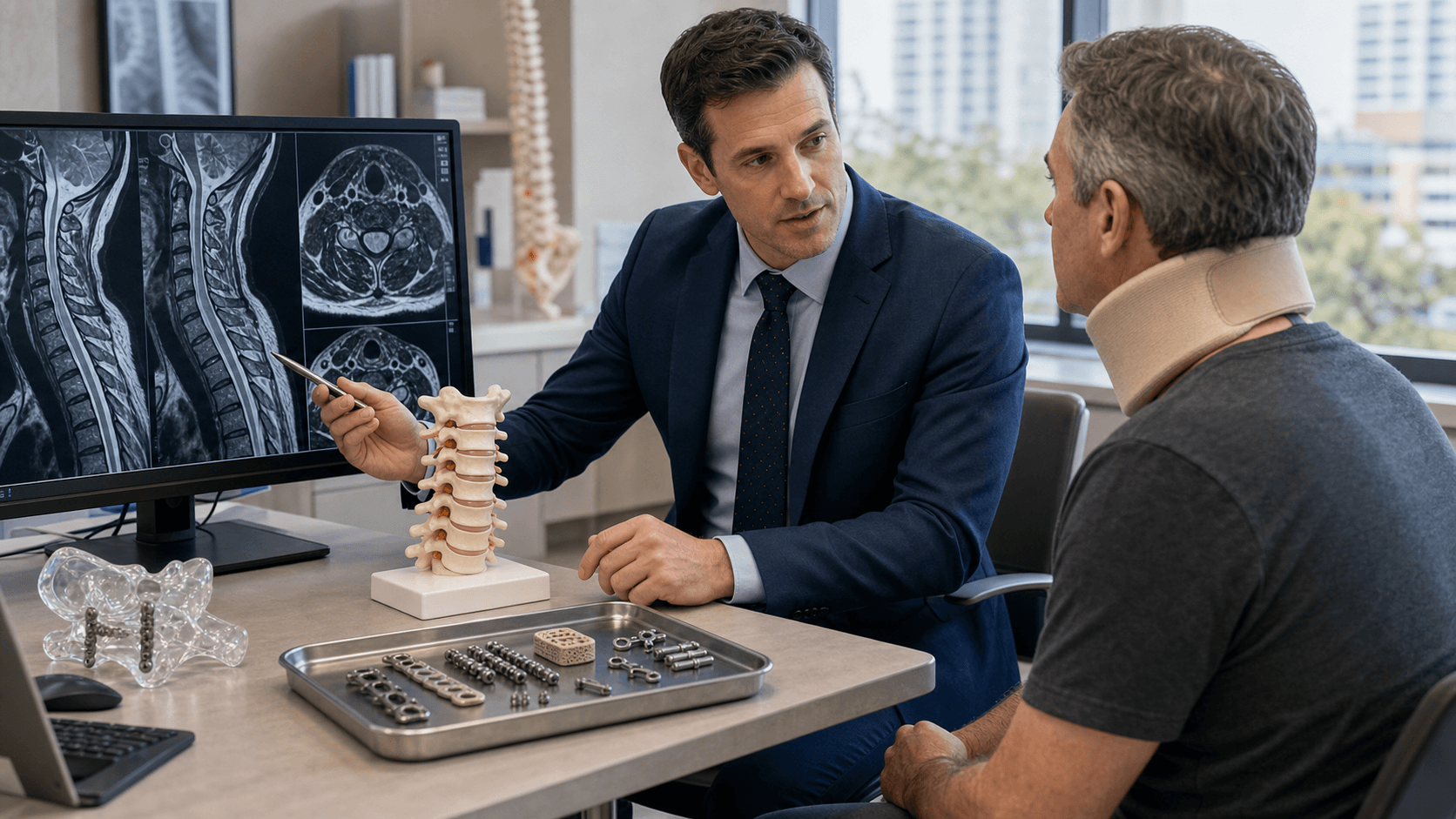
Smith-Robinson Approach | Disc Excision | Interbody Fusion | Plate Fixation
- Smith-Robinson approach is standard anterior cervical exposure
- Longus colli dissection protects vertebral artery and sympathetic chain
- Recurrent laryngeal nerve is at risk on right side (loops around subclavian)
- Dysphagia is commonest complication (transient in most)
- Anterior plate increases fusion rate especially in multilevel
- “Left-sided approach preferred to avoid RLN (loops around aorta - longer protected course)
- “Esophagus lies behind trachea - retract together medially
- “Vertebral artery lies in transverse foramen from C6 upward
- “Superior laryngeal nerve at risk with retraction above C3
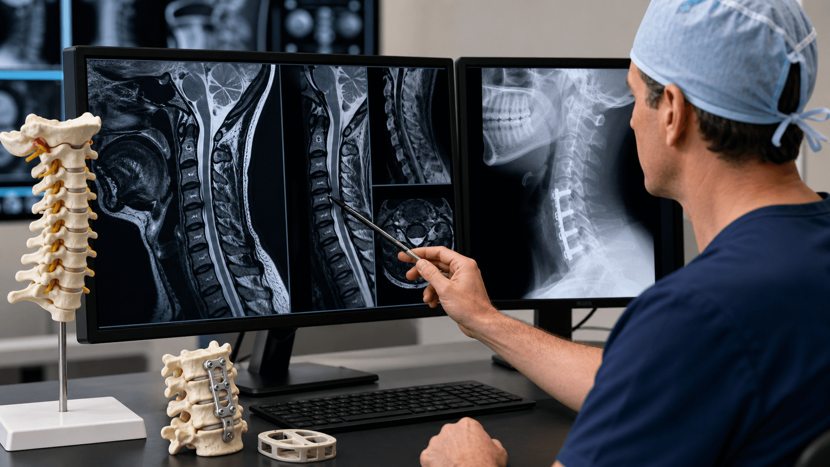
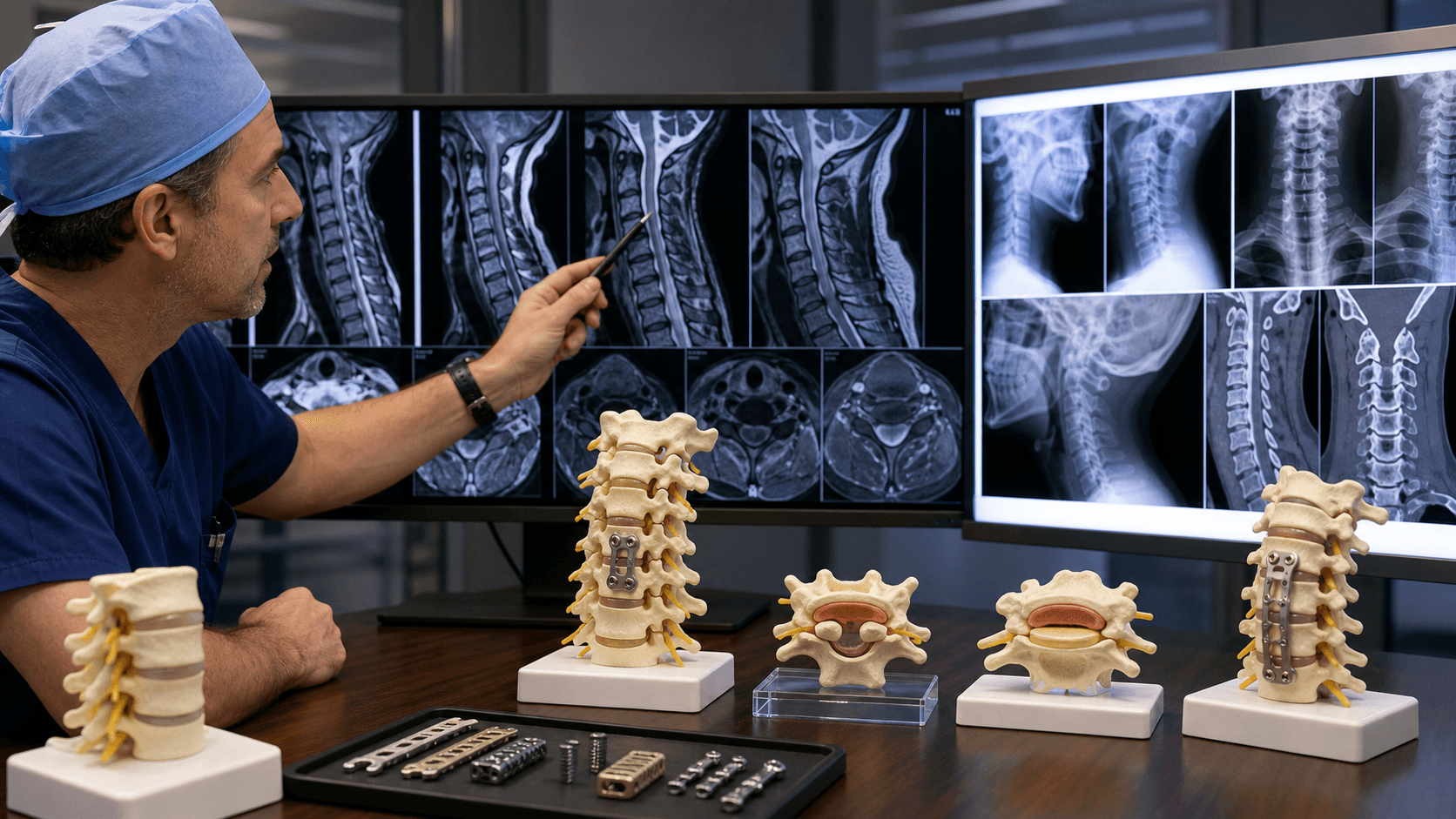
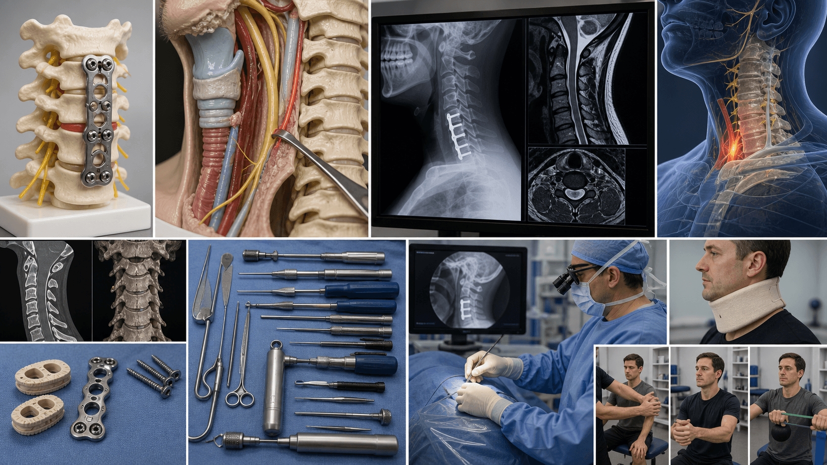
Clinical Imaging
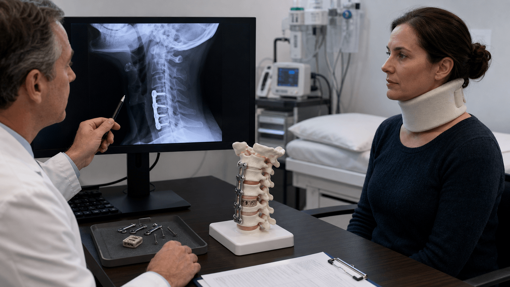
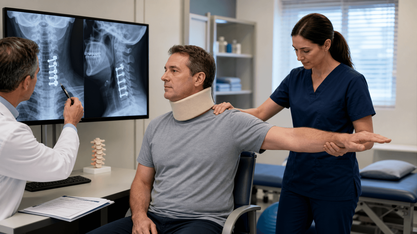
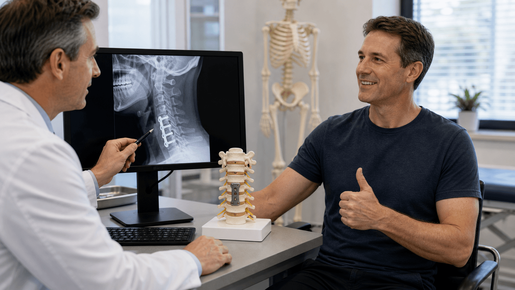
Post-Operative ACDF Radiographs

ACDF Complications: Adjacent Segment Disease and Subsidence

Left-sided approach preferred as recurrent laryngeal nerve has longer protected course around aortic arch. On the right side, RLN loops around subclavian artery and is more variable and vulnerable. Some surgeons prefer right for better angle to right-sided pathology.
The longus colli muscles must be dissected to expose disc space and protect vertebral artery laterally. Carotid sheath (carotid, IJV, vagus) retracted laterally. Esophagus and trachea retracted medially.
Dysphagia is the most common complication (50% transient, 2-5% persistent). Minimize with: deflating endotracheal cuff during retraction, limiting retraction pressure, shorter surgical time, and avoiding high retractor blade placement.
Anterior plate increases fusion rates especially in multilevel surgery. Cage height should restore disc height and lordosis but avoid over-distraction. Bone graft options include autograft, allograft, or cage with bone substitute.
| Parameter | Details | Clinical Relevance |
|---|---|---|
| Common indications | Radiculopathy, myelopathy, disc herniation | Failed conservative treatment or progressive neuro deficit |
| Most common level | C5-6 (60%), C6-7 (30%) | Correlates with degenerative disease pattern |
| Approach side | Left preferred (RLN protection) | Right may be used for right-sided pathology |
| Fusion rate | 95% single level, 85% two level | Plate increases rate in multilevel |
| Adjacent segment disease | 2.5-4% per year | Similar to lumbar spine |
| Common complications | Dysphagia (most common), hoarseness, hematoma | Most transient, rare but serious permanent |
ACDFACDF - Indications
Hook:ACDF is indicated for Arm pain, Cord compression, Disc herniation, and Failed conservative care
SMITHSMITH - Approach Layers
Hook:SMITH-Robinson approach layers from superficial to deep
DANGERDANGER - Complications
Hook:DANGER zones in ACDF - watch for these complications
LEFTLEFT Side Approach
Hook:LEFT side preferred for recurrent laryngeal nerve protection
Overview and Epidemiology
Anterior cervical discectomy and fusion (ACDF) is one of the most commonly performed spinal procedures. First described by Robinson and Smith in 1955, it revolutionized cervical spine surgery by providing anterior access for neural decompression.
Historical development:
- 1955: Smith and Robinson describe anterior approach with iliac crest graft
- 1958: Cloward describes cylindrical dowel graft technique
- 1970s-80s: Introduction of anterior cervical plating
- 1990s-2000s: Development of cage technology
- 2000s-present: Total disc replacement as alternative
Indications:
- Cervical radiculopathy: Failed conservative treatment (6-12 weeks)
- Cervical myelopathy: Any progressive or significant myelopathy
- Disc herniation: Symptomatic soft or hard disc
- Cervical instability: Traumatic or degenerative
- Tumor/infection: Anterior column pathology
For cervical radiculopathy without myelopathy, 6-12 weeks of conservative treatment is recommended before surgery. Myelopathy with progressive symptoms or significant cord compromise should proceed to surgery without delay.
Pathophysiology and Mechanisms
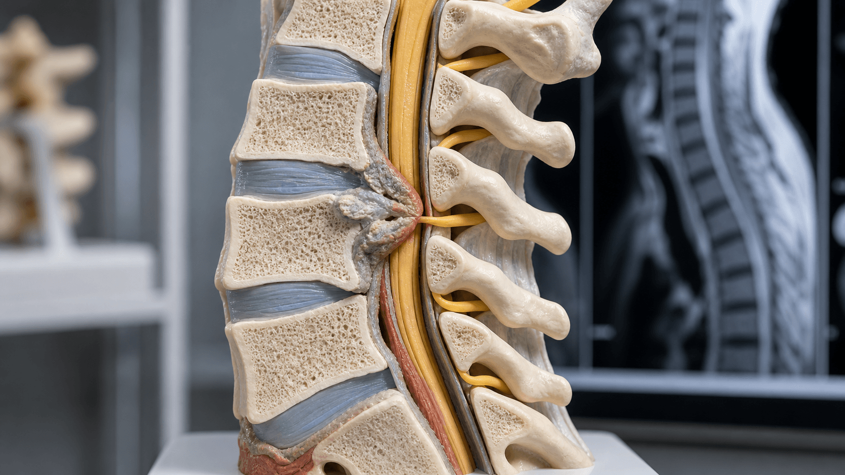
Surgical anatomy of the anterior cervical spine:
Superficial structures (anterior to posterior):
- Skin and platysma muscle
- Superficial cervical fascia
- Sternocleidomastoid muscle (lateral landmark)
Deep structures:
- Carotid sheath (lateral): Contains carotid artery, internal jugular vein, vagus nerve
- Trachea and esophagus (medial): Retracted together
- Pretracheal fascia: Dissected to reach spine
- Longus colli muscles: Cover anterior vertebral bodies
The vertebral artery enters the transverse foramen at C6 (variable, can be C5-C7). During lateral dissection and foraminotomy, stay medial to the uncovertebral joint to avoid vertebral artery injury.
Key neural structures:
-
Recurrent laryngeal nerve (RLN):
- Left: Loops around aortic arch (longer protected course)
- Right: Loops around subclavian artery (shorter, more vulnerable)
- Supplies all intrinsic laryngeal muscles except cricothyroid
- Injury causes hoarseness, aspiration
-
Superior laryngeal nerve (SLN):
- External branch at risk with retraction above C3
- Supplies cricothyroid muscle
- Injury affects voice quality/projection
-
Sympathetic chain:
- Lies lateral on longus colli
- Injury causes Horner syndrome
Disc space anatomy:
- Anterior longitudinal ligament (superficial)
- Annulus fibrosus
- Nucleus pulposus
- Posterior longitudinal ligament
- Uncovertebral joints (Joints of Luschka) - lateral margin
Classification Systems
When to perform ACDF
| Indication | Key Features | Timing |
|---|---|---|
| Radiculopathy | Dermatomal arm pain, motor/sensory deficit | After 6-12 weeks failed conservative |
| Myelopathy | Long tract signs, gait disturbance, hand dysfunction | Urgent - avoid delay |
| Soft disc herniation | Acute or subacute, may be single level | Failed conservative or significant deficit |
| Spondylotic radiculopathy | Hard disc, osteophytes, foraminal stenosis | Failed conservative treatment |
| Traumatic instability | Flexion-distraction, facet dislocation | Urgent |
Myelopathy with progression or significant cord compression should not be delayed for conservative treatment.
Clinical Assessment
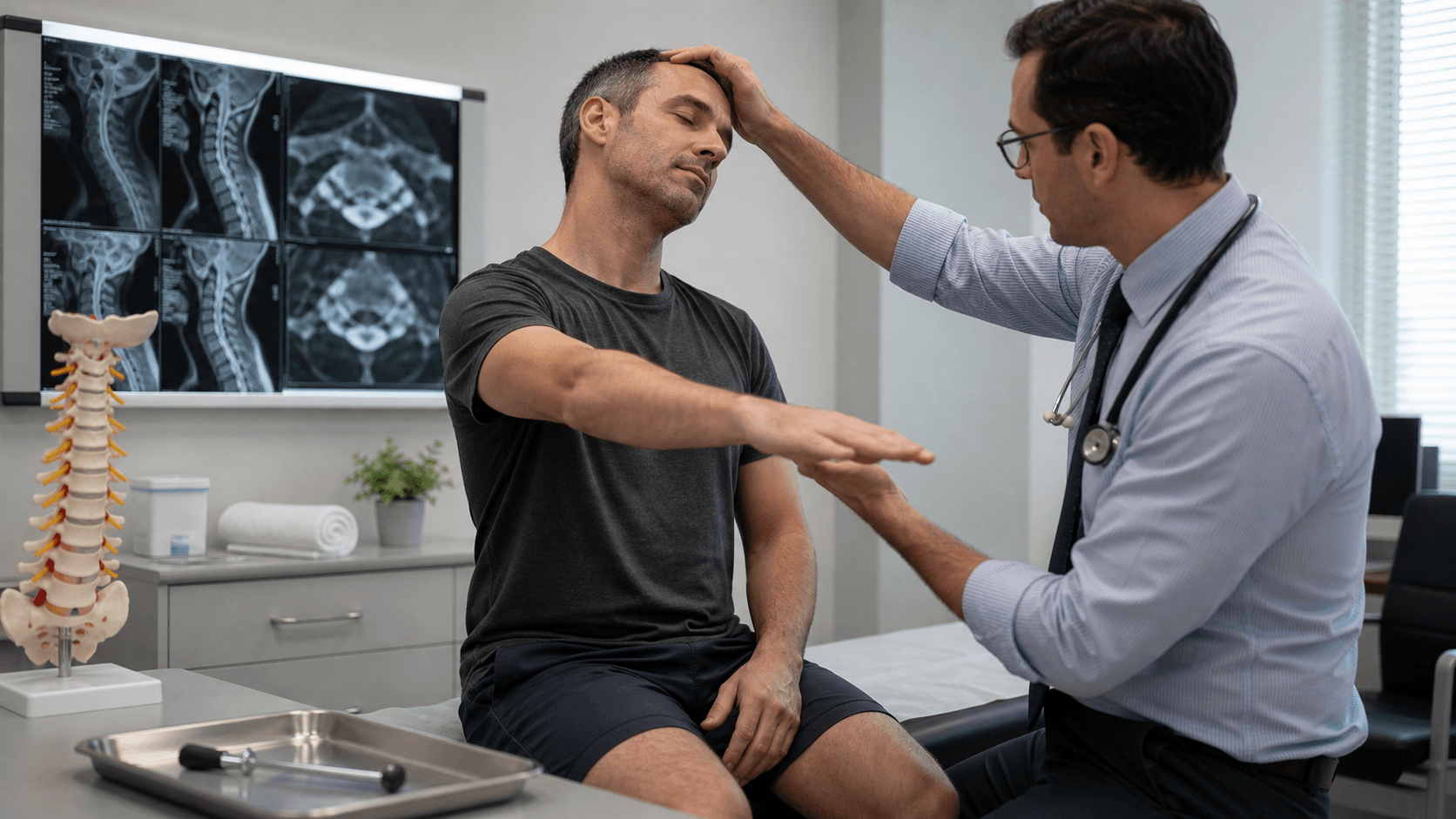
History:
- Neck pain (axial)
- Arm pain (radicular - dermatomal distribution)
- Numbness/tingling (dermatomal)
- Weakness (myotomal)
- Hand clumsiness (myelopathy)
- Gait disturbance (myelopathy)
- Bladder/bowel symptoms (severe myelopathy)
- Duration and progression
- Response to conservative treatment
Examination:
| Root | Motor | Sensory | Reflex |
|---|---|---|---|
| C5 | Deltoid, biceps | Lateral arm | Biceps |
| C6 | Wrist extensors, biceps | Lateral forearm, thumb | Brachioradialis |
| C7 | Triceps, wrist flexors | Middle finger | Triceps |
| C8 | Finger flexors, intrinsics | Medial forearm, ring/little finger | None reliable |
| T1 | Intrinsics | Medial arm | None reliable |
Myelopathy signs:
- Upper motor neuron signs (hyperreflexia, clonus, Babinski)
- Lhermitte sign (electric shock with neck flexion)
- Hoffmann sign (thumb/index flexion with middle finger flick)
- Gait disturbance (broad-based, spastic)
- Hand dysfunction (grip and release test)
- Inverted radial reflex
The C5-6 disc compresses the C6 nerve root (exiting above the disc). Similarly, C6-7 disc affects C7 root. The nerve root exits above the pedicle of the same numbered vertebra (unlike lumbar spine where root exits below).
Differential diagnosis - what mimics cervical radiculopathy/myelopathy:
| Condition | Distinguishing Features | Key Discriminator |
|---|---|---|
| Cervical radiculopathy | Dermatomal arm pain, myotomal weakness, reflex loss | Spurling test positive; MRI root compression matches level |
| Carpal tunnel syndrome | Night pain, thumb/index/middle numbness, thenar wasting | Tinel/Phalen positive; NCS shows median neuropathy at wrist |
| Cubital tunnel (ulnar at elbow) | Ring/little finger numbness, intrinsic wasting | Sensory loss spares forearm (vs C8/T1); NCS localises to elbow |
| Thoracic outlet syndrome | Positional arm symptoms, vascular or lower-trunk pattern | Provocative manoeuvres; vascular studies; often normal MRI |
| Parsonage-Turner (neuralgic amyotrophy) | Severe shoulder pain then patchy weakness, often post-viral | Multifocal/non-dermatomal; MRI spine normal |
| Shoulder pathology (cuff, OA) | Pain with shoulder movement, no distal neurology | Reproduced by shoulder, not neck, examination |
| Intracranial/other UMN lesion | Bilateral UMN signs, cranial nerve or cerebellar signs | Brain MRI; cord signal absent or beyond cervical level |
| Motor neurone disease | Mixed UMN and LMN signs, fasciculations, no sensory loss | No sensory level; EMG diffuse denervation |
The most dangerous trap is operating on imaging that does not match the clinical picture. Painless progressive weakness with fasciculations and no sensory loss suggests motor neurone disease - decompression will not help and the diagnosis may be missed. Always confirm clinico-radiological correlation before committing to ACDF.
Investigations
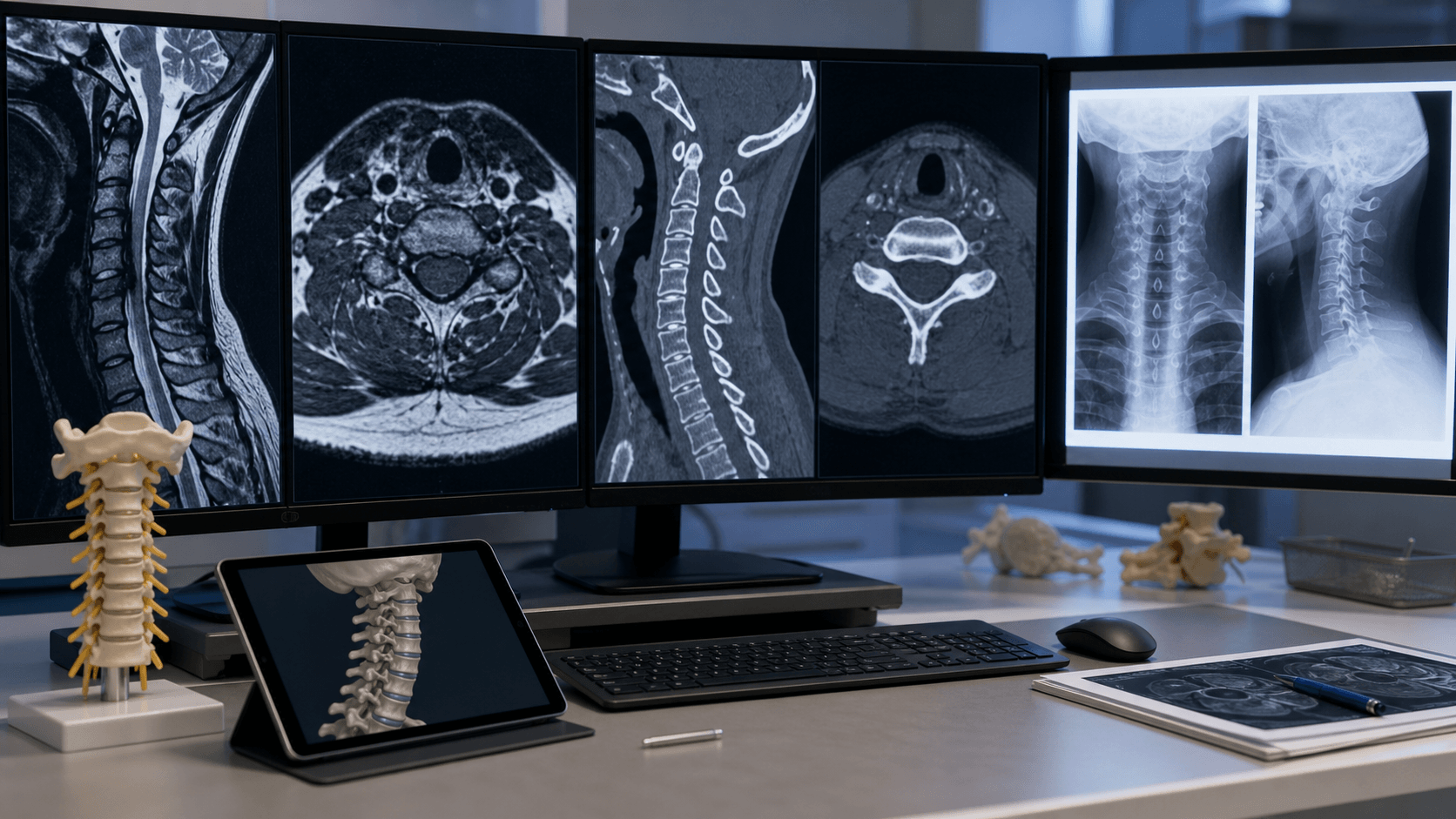
Gold standard imaging
Key sequences:
- T1-weighted: Anatomy, bone marrow changes
- T2-weighted: Cord signal (myelomalacia), disc pathology
- Gradient echo (T2*): Metal artifact reduction, better disc visualization
Key findings:
- Disc herniation (soft disc)
- Osteophyte complex (hard disc)
- Cord compression
- Cord signal change (poor prognostic sign)
- Foraminal stenosis
MRI should correlate with clinical findings to confirm surgical level(s).
Management Algorithm
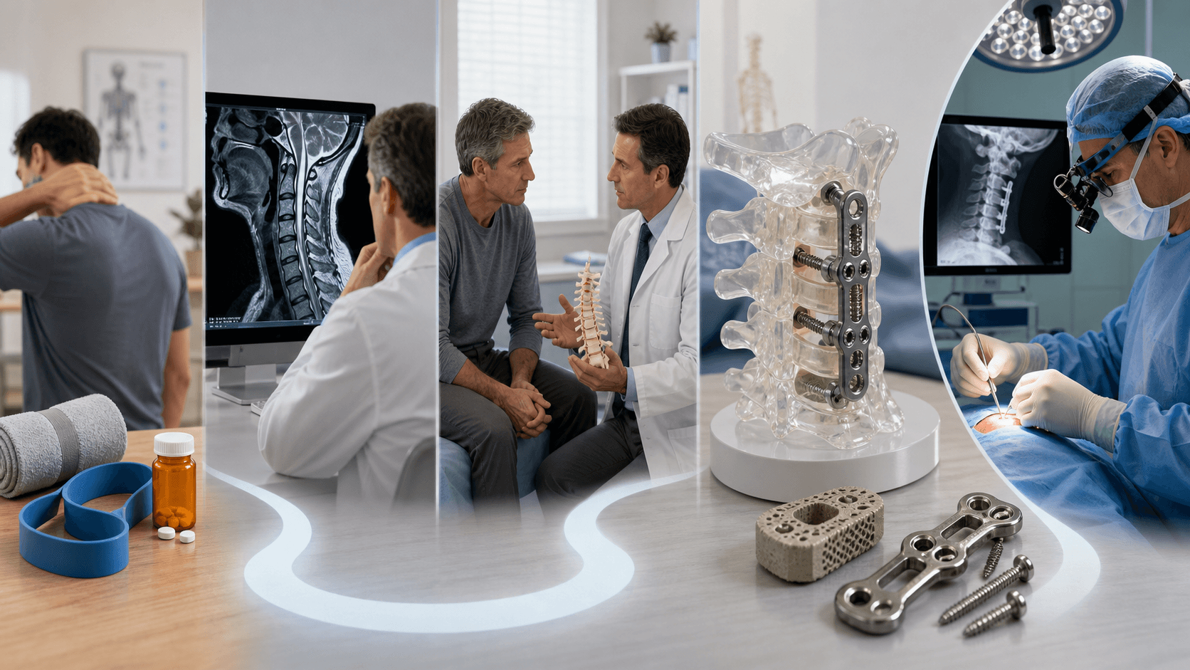
For radiculopathy without myelopathy
Medications:
- NSAIDs
- Oral corticosteroids (short course for acute)
- Neuropathic pain agents (gabapentin, pregabalin)
- Muscle relaxants
Physical therapy:
- Cervical traction (controversial)
- Strengthening exercises
- Postural training
- Activity modification
Injections:
- Cervical epidural steroid injection
- Selective nerve root blocks
- Transforaminal vs interlaminar approach
Expected outcomes:
- 50-70% improve with conservative treatment
- Natural history of radiculopathy is generally favorable
Give adequate conservative trial unless progressive deficit or myelopathy.
Surgical Technique
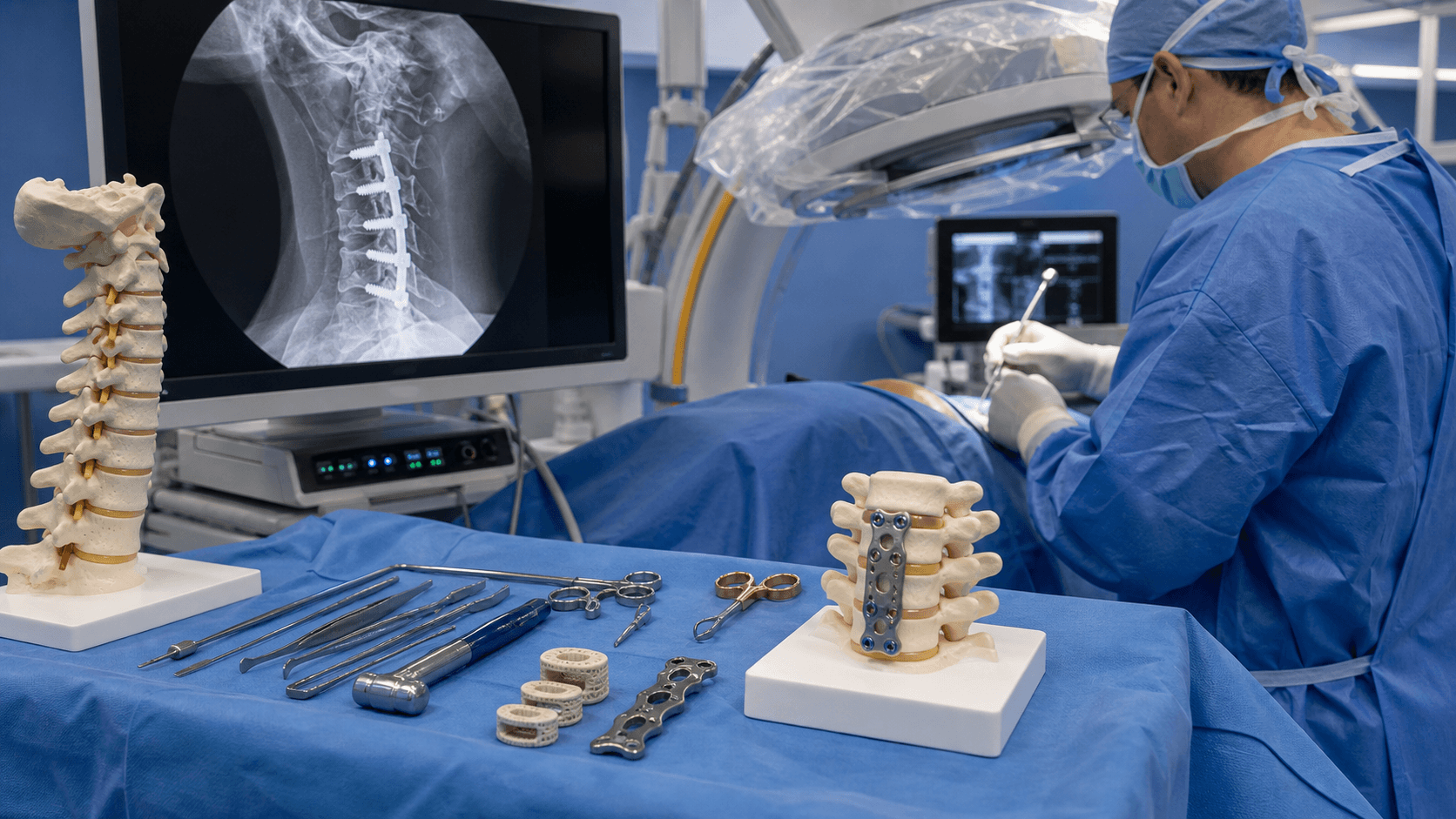
Smith-Robinson anterior cervical approach
Positioning:
- Supine with neck in neutral or slight extension
- Head on gel ring or Mayfield
- Arms tucked at sides
- Fluoroscopy to confirm level
Approach Steps
Transverse skin crease incision (cosmetic) or longitudinal incision for multilevel. Left side preferred. Incise platysma in line with skin.
Identify medial border of sternocleidomastoid. Develop plane between carotid sheath laterally and trachea/esophagus medially. Blunt dissection to pretracheal fascia.
Divide pretracheal fascia. Identify anterior spine covered by longus colli. Confirm level with fluoroscopy using needle in disc.
Subperiosteal elevation of longus colli off anterior vertebral bodies. Elevate to expose uncovertebral joints (lateral limit of dissection). Place self-retaining retractor under longus colli.
The longus colli must be elevated carefully to protect the vertebral artery and sympathetic chain laterally.
Complications
Frequently encountered issues
| Complication | Incidence | Prevention/Management |
|---|---|---|
| Dysphagia | 30-50% transient, 2-5% persistent | Minimize retraction, deflate ETT cuff, shorter surgery |
| Hoarseness (RLN injury) | 2-5% transient, 0.2-1% permanent | Left-sided approach, gentle retraction, protect nerve |
| Hematoma | 1-2% | Meticulous hemostasis, consider drain |
| Pseudarthrosis | 5% single level, 15% multilevel | Use plate, cage with bone graft, no smoking |
| Adjacent segment disease | 2.5-4% per year | Minimize levels, consider disc replacement |
Dysphagia is the most common complication but usually resolves within weeks.
Postoperative Care
First 24-48 hours
Monitoring:
- Airway assessment (watch for hematoma)
- Neurological checks
- Pain management
- Dysphagia screening before oral intake
Red flags:
- Increasing neck swelling
- Stridor or respiratory distress
- New neurological deficit
Hematoma protocol:
- If airway compromise developing
- Open wound at bedside to decompress
- May need emergency intubation (can be very difficult)
- Return to OR for evacuation
First 48 hours critical for hematoma detection.
Outcomes and Prognosis
| Indication | Success Rate | Recovery Timeline | Factors Affecting Outcome |
|---|---|---|---|
| Radiculopathy | 85-95% | Arm pain improves within days/weeks | Duration of symptoms, motor deficit |
| Myelopathy | 70-80% stabilize/improve | May take 6-12 months | Preop severity, cord signal change |
| Disc herniation | 90-95% | Rapid improvement typical | Soft disc better than hard disc |
Prognostic factors:
| Factor | Better Prognosis | Worse Prognosis |
|---|---|---|
| Symptom duration | Short (less than 6 months) | Prolonged (greater than 2 years) |
| Pathology type | Soft disc | Hard disc, severe spondylosis |
| Myelopathy signs | Mild, recent onset | Severe, long-standing, cord signal change |
| Number of levels | Single level | Multiple levels |
| Patient factors | Non-smoker, no diabetes | Smoker, diabetic, workers comp |
ACDF has excellent outcomes for radiculopathy; myelopathy outcomes depend on severity and duration.
Evidence Base

Surgery vs Physiotherapy vs Collar for Radiculopathy (Persson RCT)
- Surgery gave faster relief of pain, sensory loss and weakness at 4 months
- All three arms converged by 1-year follow-up
- Conservative care has a generally favourable natural history
- Reserve surgery for failed conservative care or progressive deficit
Cervical Disc Arthroplasty vs ACDF - 7-Year IDE Trial (Mobi-C)
- Single-level TDR non-inferior; two-level TDR superior at 7 years
- Lower index-level and adjacent-level reoperation with TDR
- Motion preserved with TDR
- TDR contraindicated in significant facet arthrosis, kyphosis or instability
Zero-Profile Spacer vs Cage-Plate - Dysphagia (Meta-analysis)
- Anterior plates contribute to early dysphagia
- Zero-profile/low-profile devices lower early dysphagia rates
- No difference in fusion, function or late dysphagia
- Plate still favoured where lordosis correction or maximal multilevel stability is needed
Surgery for Cervical Spondylotic Myelopathy (AOSpine North America)
- Surgery improves function and quality of life at all severity grades
- Benefit seen even in moderate and severe myelopathy
- Complication rate ~19%, comparable to historical series
- Do not delay - myelopathy is progressive and recovery is duration-dependent
Adjacent-Segment Disease After Anterior Cervical Arthrodesis (Hilibrand)
- Adjacent-segment disease ~2.9% per year, ~25% at 10 years
- Highest risk at C5-6 and C6-7
- Single-level fusion carried higher adjacent-level risk than multilevel
- Two-thirds of affected patients ultimately required further surgery
Incidence and Natural History of Dysphagia After Anterior Cervical Surgery (Bazaz)
- Dysphagia is common early (~50% at 1 month) but largely resolves
- Persistent moderate/severe dysphagia uncommon (~5% at 6 months)
- Female sex and multilevel surgery increase risk
- Most dysphagia is self-limiting and managed expectantly
The classic teaching of a left-sided approach to protect the recurrent laryngeal nerve rests on the more constant, longer left RLN course around the aortic arch (the right RLN loops around the subclavian and may run non-recurrently in roughly 1 in 200). However, large series and meta-analyses have not consistently shown a difference in symptomatic RLN palsy between left and right approaches, so surgeon familiarity and the side of pathology are legitimate determinants. Quote the anatomical rationale in the viva, but acknowledge the evidence is not definitive.
Controversies and Areas of Uncertainty
These are high-yield discussion points where an examiner probes judgement rather than recall - state both sides and where the evidence currently sits.
Long-term IDE data show TDR is non-inferior at one level and clinically superior at two levels with lower reoperation, yet ACDF remains the default in many systems on grounds of cost, surgeon familiarity and concerns about heterotopic ossification and long-term implant behaviour. The debate is about patient selection, not whether TDR works.
The left-versus-right RLN argument is anatomically logical but not consistently supported by clinical series. Defensible positions exist for both sides; the unwary candidate who states left is "always safer" can be pushed.
For mild degenerative cervical myelopathy (mJOA 15-17), AOSpine permits either surgery or structured non-operative care with close monitoring. The uncertainty is which mild patients will progress - cord signal change, large cord compression and clinical deterioration push toward surgery.
Anterior plating improves lordosis and multilevel stability but contributes to early dysphagia; zero-profile and stand-alone cages reduce early dysphagia with comparable fusion. The trade-off is alignment/stability versus swallowing morbidity.
Routine drainage after ACDF is not evidence-based for preventing airway-threatening haematoma (which is usually arterial and rapid); meticulous haemostasis and vigilant post-op monitoring matter more. Practice varies widely.
For multilevel myelopathy, anterior (ACDF/corpectomy), posterior (laminoplasty, laminectomy and fusion) and combined approaches all have advocates. Sagittal alignment (kyphosis favours anterior), number of levels, OPLL and bone quality drive the decision.
Exam Viva Scenarios
Practise clinical reasoning and management decisions out loud
“A 45-year-old man presents with 8 weeks of right arm pain radiating to the thumb with numbness. He has weakness of wrist extension. MRI shows a C5-6 disc herniation compressing the C6 nerve root.”
“A 62-year-old woman presents with difficulty writing and buttoning clothes, unsteady gait, and electric shocks down her spine when she flexes her neck. Examination shows hyperreflexia and Hoffmann sign bilaterally. MRI shows multilevel stenosis C4-C7 with cord compression and T2 signal change in the cord at C5-6.”
“You are called to see a patient 6 hours after ACDF who is developing stridor and has increasing neck swelling. The patient is becoming increasingly distressed.”
“A fit 38-year-old non-smoker has single-level C5-6 soft-disc radiculopathy that has failed three months of conservative care. He asks whether he should have a fusion or a disc replacement. How do you counsel him and decide?”
MCQ Practice Points
Q: Which side is preferred for the anterior cervical approach and why?
A: Left side is preferred. The recurrent laryngeal nerve (RLN) on the left loops around the aortic arch, giving it a longer and more protected course, while the right RLN loops around the subclavian artery, making it shorter and more vulnerable to injury during retraction.
Q: A C5-6 disc herniation will compress which nerve root?
A: C6 nerve root. In the cervical spine, the nerve root exits ABOVE the pedicle of the same-numbered vertebra, so the C5-6 disc affects the C6 root. This is opposite to the lumbar spine pattern.
Q: What is the recommended timing for surgical intervention in cervical myelopathy?
A: Early surgery is recommended - do not delay for conservative treatment. Evidence shows better outcomes when surgery is performed within 6 months of symptom onset. Myelopathy with progressive symptoms is urgent.
Q: What is the rate of dysphagia after ACDF?
A: 50% transient dysphagia (usually resolves within weeks), with 2-5% persistent dysphagia. Dysphagia is the most common complication of ACDF.
Q: What is the annual rate of adjacent segment disease after cervical fusion?
A: 2.5-4% per year. This is similar to lumbar spine fusion. Total disc replacement may reduce this rate but evidence is limited.
Q: What is the immediate management of suspected postoperative hematoma with airway compromise?
A: Open the wound at the bedside to evacuate hematoma and relieve pressure on the airway. This should be done before or while attempting intubation, as intubation may be extremely difficult due to swelling.
Guidelines, Registries & Global Practice
ACDF is one of the most frequently performed spine operations worldwide and is a core procedure across FRCS (Tr&Orth), FRACS, EBOT/FEBOT, ABOS, DNB/MS and SICOT curricula. Evidence and recommendations are drawn from international guidelines and registries to give a global picture rather than any single country's practice.
Global epidemiology
- Cervical radiculopathy has an annual incidence of roughly 80-100 per 100,000, peaking in the fifth and sixth decades; the great majority improve with non-operative care.
- Degenerative cervical myelopathy is the commonest cause of non-traumatic spinal cord dysfunction in adults worldwide, and its prevalence is rising with ageing populations.
- C5-6 and C6-7 are consistently the most commonly operated levels across all regions, mirroring the distribution of degenerative load.
Major guidelines, side by side
| Body | Radiculopathy | Myelopathy | Arthroplasty vs ACDF |
|---|---|---|---|
| AOSpine / AO Foundation (global) | Trial of non-operative care first if no progressive deficit | Operate for moderate-severe DCM; offer surgery or structured non-op for mild DCM | Both acceptable for 1-2 level disease without instability |
| NASS (North America) | Surgery superior for short-term relief of refractory radiculopathy | Decompression recommended for symptomatic myelopathy | TDR non-inferior to ACDF for selected 1-2 level disease |
| NICE / BOA (UK) | Conservative care first; refer if red flags or persistent deficit | Urgent referral for suspected myelopathy | Arthroplasty an option in appropriately selected patients |
| EFORT / European consensus | Emphasis on clinico-radiological correlation before surgery | Early decompression to limit irreversible cord injury | Motion preservation favoured in young, single-level, no facet arthrosis |
Registry and outcome evidence
- FDA IDE randomised trials (Mobi-C, Prestige, ProDisc-C, Bryan) underpin the global acceptance of cervical arthroplasty as non-inferior to ACDF for selected 1-2 level disease, with lower index-level reoperation in long-term follow-up.
- Spine registries and large administrative datasets (for example the Quality Outcomes Database in North America and national spine registries in Europe and Australasia) consistently report high patient-reported improvement and low major-complication rates after ACDF, supporting its status as a workhorse procedure.
High- vs limited-resource practice variation
- In well-resourced settings, PEEK/titanium cages, anterior plating, intra-operative fluoroscopy and (increasingly) arthroplasty are routine.
- In limited-resource settings, structural allograft or autograft (Smith-Robinson tricortical iliac crest) without a plate remains a valid, low-cost technique with good single-level fusion rates; intra-operative imaging and disc replacement may be unavailable.
- Across all settings the principles are identical: confirm clinico-radiological correlation, decompress adequately, restore disc height and alignment, and do not delay surgery for progressive myelopathy.
Exam Cheat Sheet
Key Numbers
- C5-6 most common level (55-65%)
- Fusion rate: 95% single, 85% multilevel
- Dysphagia: 50% transient, 2-5% persistent
- Adjacent segment disease: 2.5-4%/year
Approach Anatomy
- Left side preferred (RLN protection)
- Carotid sheath retracted laterally
- Trachea/esophagus retracted medially
- Longus colli protects vertebral artery
Nerve Root Levels
- C5-6 disc = C6 root compression
- C6-7 disc = C7 root compression
- Root exits ABOVE pedicle of same number
- Different from lumbar spine pattern
Complications
- Dysphagia (most common)
- Hematoma (airway emergency)
- RLN injury (hoarseness)
- Pseudarthrosis (especially multilevel)
Exam Traps
- Not knowing left vs right approach rationale
- Wrong nerve root level correlation
- Delaying surgery for myelopathy
- Not recognizing hematoma emergency