Column Concept | Judet-Letournel Classification | Approach Selection
- Two-column concept - anterior and posterior columns meet at iliac crest
- 3 Judet views - AP, obturator oblique, iliac oblique
- Dome concept - weight-bearing 10cm arc must be reduced
- Approach dictated by column - KL for posterior, IL/Stoppa for anterior
- Anatomic reduction (under 2mm step) = 80% good outcomes
- “Both-column = 'spur sign' on obturator oblique
- “Posterior wall = most common pattern
- “Sciatic nerve at risk in posterior approaches
- “Delay surgery 3-5 days to reduce blood loss
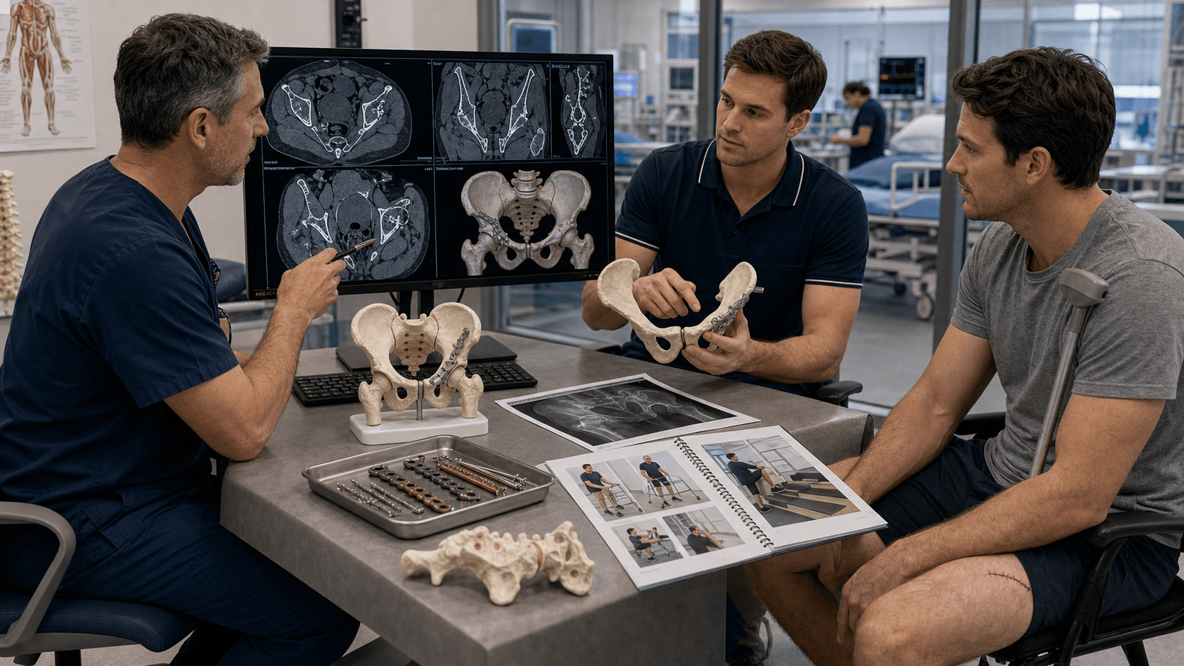
Clinical Imaging
Imaging Atlas


Anterior column: Pubis to iliac crest via pelvic brim. Posterior column: Ischium to iliac crest via greater sciatic notch. They meet at the iliac crest like an inverted Y.
Obturator oblique (45° toward injured hip): Shows anterior column and posterior wall. Iliac oblique (45° away): Shows posterior column and anterior wall.
Weight-bearing dome = superior 10cm arc. Must be anatomically reduced. Peripheral fractures may be treated conservatively.
Kocher-Langenbeck: Posterior column/wall. Ilioinguinal/Stoppa: Anterior column. Extended iliofemoral: Both columns (rarely used).
| Pattern | Approach | Key Structures at Risk | Priority |
|---|---|---|---|
| Posterior wall | Kocher-Langenbeck | Sciatic nerve | Urgent if dislocated |
| Posterior column | Kocher-Langenbeck | Sciatic nerve, SGA | Urgent |
| Anterior column | Ilioinguinal/Stoppa | LFCN, femoral vessels | Semi-urgent |
| Transverse | Based on displacement | Column-specific | Assess dome |
| Both column | Usually ilioinguinal | Corona mortis, LFCN | Complex - delay OK |
PAPATElementary Patterns
Hook:PAPAT - 5 elementary patterns, Posterior Wall is most common!
BATTPAssociated Patterns
Hook:5 Associated patterns combine elements - Both Column is the hallmark!
OAPJudet Views
Hook:OAP - Obturator sees Anterior column, iliac oblique sees Posterior column
SAFESciatic Nerve Protection
Hook:Keep the hip SAFE during posterior approaches!
Overview and Epidemiology
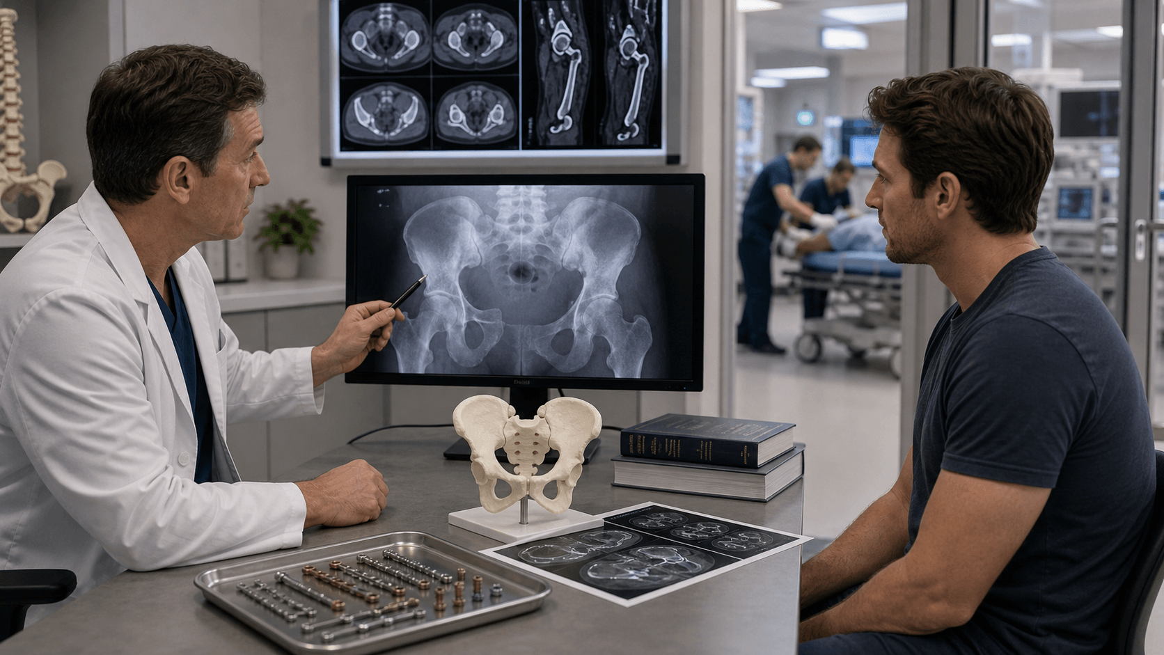
Acetabular fractures are complex injuries requiring detailed anatomical understanding. The Judet-Letournel classification is the gold standard and must be known for the exam. Approach selection is directly tied to fracture pattern.
- Young adults: High-energy MVA, falls from height
- Elderly: Low-energy falls, osteoporotic bone
- Male predominance: 3:1 ratio
- Associated injuries: 80% have hip dislocation
- Dashboard injury: Knee strikes dashboard, force through femur
- Position determines pattern: Flexed hip = posterior wall
- Lateral compression: Direct blow to trochanter
- Force magnitude: Determines comminution
Historical Context
Judet and Letournel revolutionized acetabular surgery in the 1960s-1980s. Before their work, most acetabular fractures were treated non-operatively with poor results. Their classification and surgical approaches remain the foundation today.
Anatomy and Biomechanics
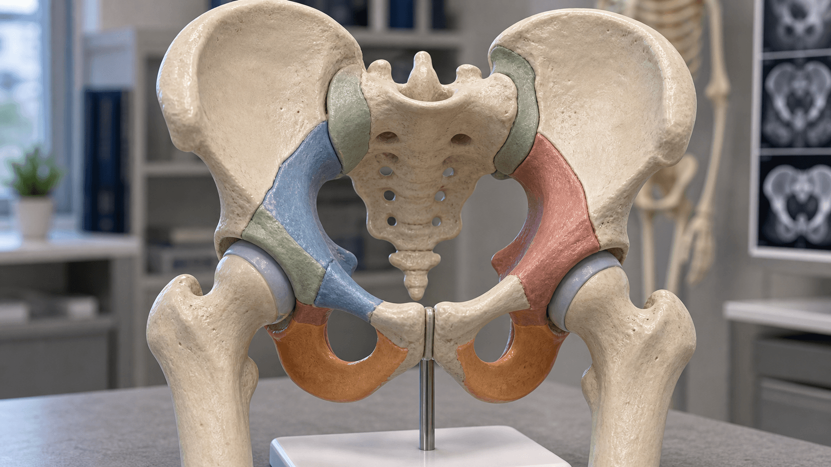
The Two-Column Concept
The acetabulum is supported by two columns that meet at the iliac crest like an inverted Y:
- Anterior column: Anterior half of iliac wing + pelvic brim + superior pubic ramus
- Posterior column: Posterior half of iliac wing + greater/lesser sciatic notches + ischial tuberosity + inferior pubic ramus
Key Anatomical Landmarks
| Landmark | Location | Clinical Significance |
|---|---|---|
| Iliopectineal line | Anterior column | Disruption = anterior column fracture |
| Ilioischial line | Posterior column | Disruption = posterior column fracture |
| Acetabular roof/dome | Superior 10cm arc | Weight-bearing - must reduce |
| Anterior wall | Medial curve on AP | Anterior wall outline |
| Posterior wall | Lateral curve on AP | Posterior wall outline |
| Teardrop | Floor of acetabular fossa | Medial wall/quadrilateral plate |
Neurovascular Anatomy
- Sciatic nerve: Courses 1cm inferior to piriformis
- Superior gluteal artery: Above piriformis
- Inferior gluteal artery: Below piriformis
- Piriformis: Key landmark
- Lateral femoral cutaneous nerve: Medial to ASIS
- Femoral nerve/vessels: In iliac fossa
- Corona mortis: Anastomosis on pubic ramus
- External iliac vessels: Medial retraction risk
Corona mortis ("crown of death") is an aberrant obturator vessel crossing the superior pubic ramus. Present in 30-70% of patients. Must be identified and ligated during Stoppa or ilioinguinal approaches.
Classification Systems
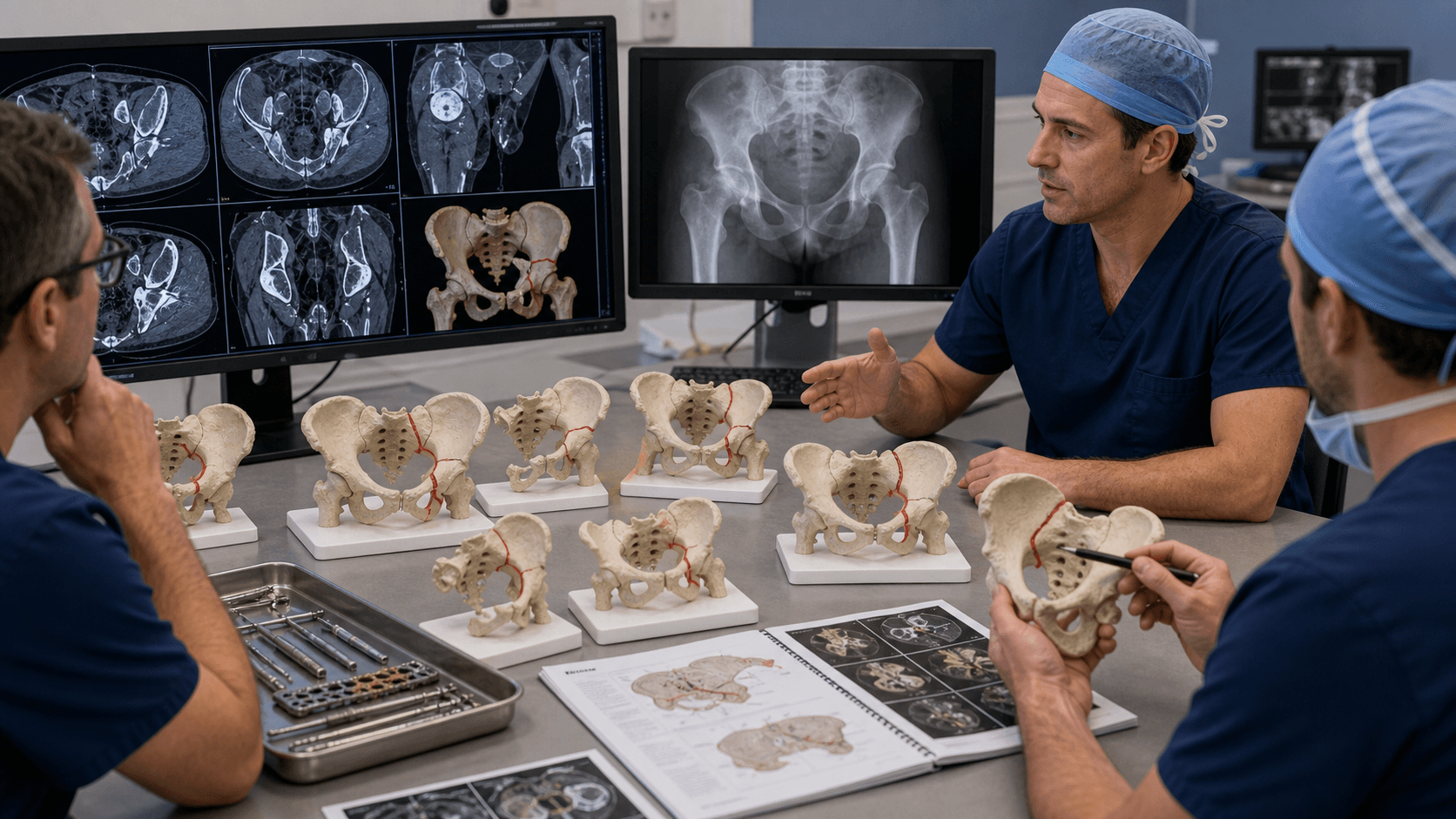
Elementary Patterns (5)
| Pattern | Frequency | Key Feature | Approach |
|---|---|---|---|
| Posterior wall | 25% (most common) | Fragment from posterior rim | Kocher-Langenbeck |
| Posterior column | 4% | Through greater sciatic notch to obturator foramen | Kocher-Langenbeck |
| Anterior wall | 2% (rare) | Anterior rim fragment | Ilioinguinal/Stoppa |
| Anterior column | 4% | Through pelvic brim | Ilioinguinal/Stoppa |
| Transverse | 8% | Divides acetabulum horizontally | Based on displacement |
Posterior wall fractures are the most common elementary pattern (~25%). They typically occur with dashboard injuries when the hip is flexed. The sciatic nerve is at risk in up to 30% of cases.

Clinical Assessment
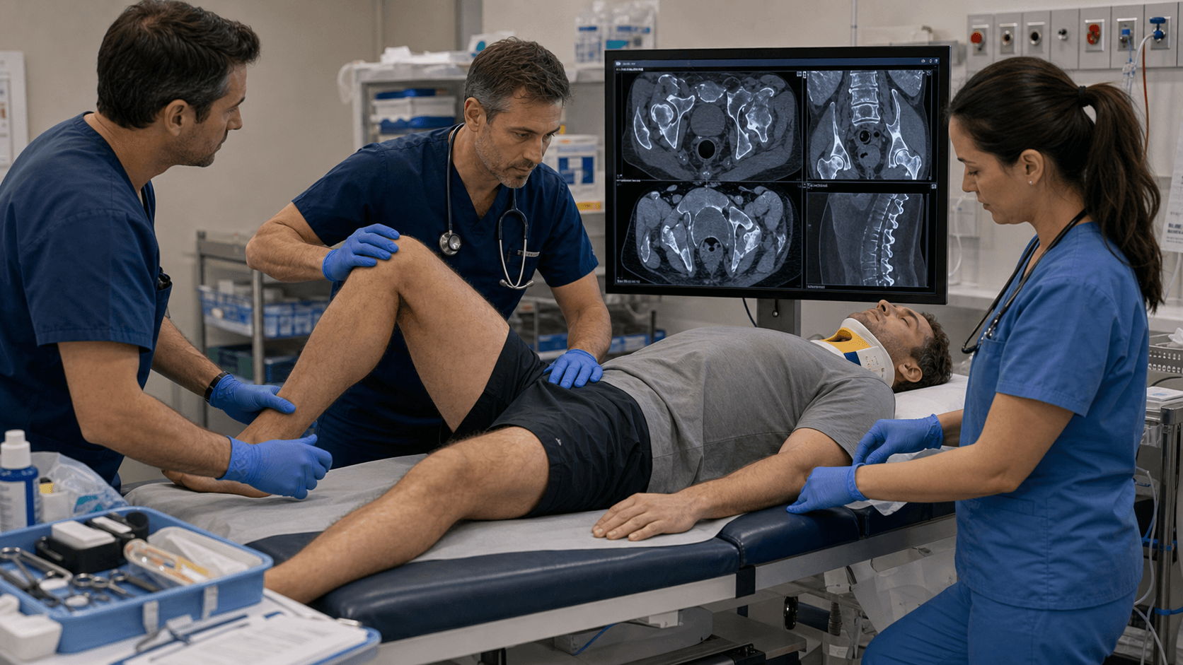
- Mechanism: Dashboard injury, fall from height, lateral blow
- Position at impact: Determines fracture pattern
- Associated injuries: Head, chest, abdomen, pelvis
- Pre-injury function: Critical for decision-making
- Inspection: Hip position (posterior dislocation = flexed, adducted, IR)
- Palpation: Tenderness over greater trochanter
- ROM: Limited and painful
- Neurovascular: Sciatic nerve (dorsiflexion, plantarflexion, sensation)
30% of posterior wall/column fractures have sciatic nerve injury. Always document:
- Peroneal division (more common): Ankle/toe dorsiflexion, foot eversion, sensation dorsal foot
- Tibial division: Ankle/toe plantarflexion, sensation plantar foot Document BEFORE and AFTER reduction/surgery!
Associated Injuries
| Injury | Incidence | Assessment |
|---|---|---|
| Hip dislocation | Up to 80% | Hip position, urgent reduction |
| Sciatic nerve injury | 30% (posterior) | Motor/sensory exam |
| Femoral head fracture | 10% | CT scan |
| Knee ligament injury | Dashboard mechanism | Examine knee |
| Ipsilateral femur fracture | 10% | Full femur X-ray |
Differential Diagnosis of the Painful, Loaded Hip After Trauma
| Diagnosis | Discriminating Feature | Confirming Test |
|---|---|---|
| Acetabular fracture | Disruption of iliopectineal/ilioischial line; dome involvement | AP + Judet views, CT |
| Pure hip dislocation (no fracture) | Concentric on post-reduction films, intact lines | Post-reduction CT excludes fragment/wall fracture |
| Femoral head fracture (Pipkin) | Fragment infero-medial to head, head incongruity | CT - assess Pipkin type |
| Femoral neck fracture | Disruption of Shenton line, shortening/ER of limb | AP pelvis, CT if occult |
| Pelvic ring injury | Pubic rami/SI joint disruption, not the dome | Inlet/outlet views, CT |
| Quadrilateral plate / medial wall fracture | Central protrusion of head, teardrop displaced | Obturator oblique, CT |
A concentric post-reduction radiograph does not exclude an acetabular fracture. An incarcerated osteochondral or wall fragment can sit in the joint despite an apparently reduced hip - post-reduction CT is mandatory after every traumatic hip dislocation.
Investigations
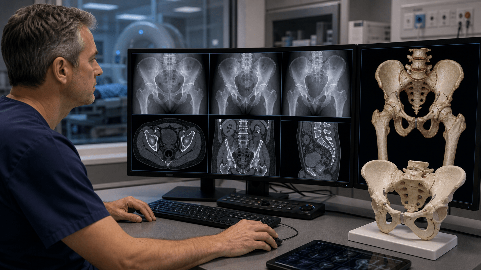
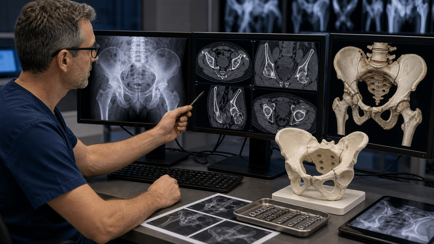
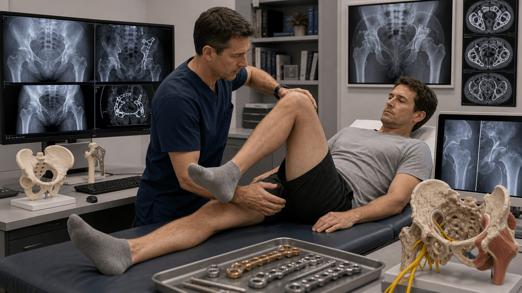
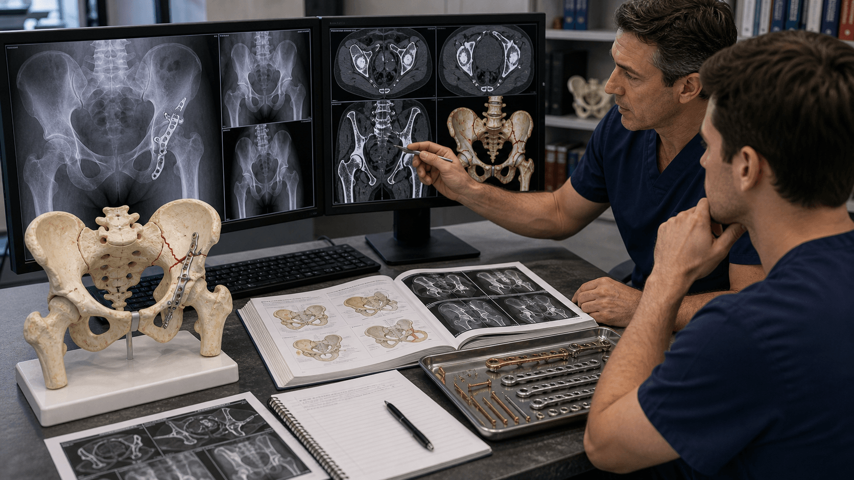
Imaging Protocol
AP Pelvis: Assess both columns, dome, teardrop. Obturator oblique (45° toward): Anterior column + posterior wall. Iliac oblique (45° away): Posterior column + anterior wall.
Mandatory for all acetabular fractures. Defines fracture pattern, comminution, impaction, loose bodies. 3D reconstructions show pattern clearly. Subtract femoral head for better visualization.
MRI: Labral injury, femoral head cartilage. CTA: If vascular injury suspected (rare).
Radiographic Lines
6 key lines to assess on AP pelvis:
- Iliopectineal line = anterior column
- Ilioischial line = posterior column
- Acetabular roof = dome
- Anterior wall = medial curve
- Posterior wall = lateral curve
- Teardrop = medial wall/quadrilateral plate


Management Algorithm
Indications for Non-operative Treatment
| Criterion | Threshold | Rationale |
|---|---|---|
| Displacement | Less than 2mm | Acceptable articular congruity |
| Roof arc | Greater than 45° | Dome not involved in fracture |
| Both column | Secondary congruence | Head moves with medial fragment |
| Low anterior column | Below sourcil | Non-weight bearing area |
Low anterior column fractures that exit below the weight-bearing dome may be treated non-operatively if stable.
Protocol: Traction (4-8 weeks) or touch-down weight bearing with close radiographic follow-up.
Surgical Technique
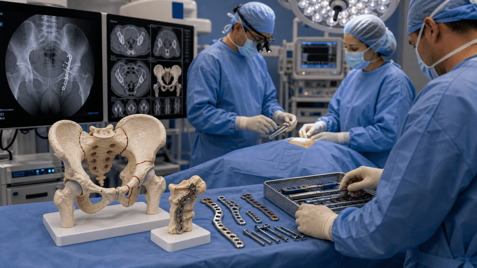
Kocher-Langenbeck Approach
Indications: Posterior wall, posterior column, transverse with posterior displacement
Surgical Steps
Lateral decubitus with hip flexed 20-30°, or prone with bolsters. Knee flexed to relax sciatic nerve.
From PSIS curving over greater trochanter, extending distally along femoral shaft, or straight for Gibson approach.
Split gluteus maximus in line with fibers. Identify and protect sciatic nerve inferior to piriformis.
Detach piriformis, obturator internus, gemelli from greater trochanter (leave quadratus femoris to protect MFCA). Capsulotomy to access joint.
Reduce fragments under direct vision. Buttress plate for posterior wall (spring plate). Reconstruction plate for column.
Knee flexion reduces tension on sciatic nerve. Limit hip flexion beyond 60°. External rotation relaxes piriformis. Use retractors carefully - avoid persistent traction.
Complications
| Complication | Incidence | Risk Factors | Management |
|---|---|---|---|
| Post-traumatic arthritis | 20-30% | Malreduction, cartilage damage | THA when mature |
| AVN femoral head | 5-10% | Dislocation duration, posterior injury | Core decompression, THA |
| Heterotopic ossification | 20-50% | Posterior approach, head injury | Prophylaxis (indomethacin or XRT) |
| Sciatic nerve injury | 10-15% | Posterior approach, retraction | Observation, most recover |
| DVT/PE | Variable | Pelvic surgery, immobility | Thromboprophylaxis |
| Infection | 3-5% | Open fracture, prolonged surgery | Debridement, antibiotics |
Heterotopic Ossification (HO)
Brooker Grade III-IV HO occurs in 20-50% of posterior approaches. Prophylaxis options:
- Indomethacin 25mg TDS for 6 weeks (most common)
- Radiation therapy: Single fraction 700cGy within 72 hours Both reduce severe HO to under 5%.
Post-Traumatic Arthritis
The most significant long-term complication. Risk factors:
- Articular step greater than 2mm
- Femoral head cartilage damage
- Delayed reduction of dislocation
- Age at injury
THA after acetabular fracture is technically challenging with higher complication rates. Delay at least 3-6 months for fracture healing.
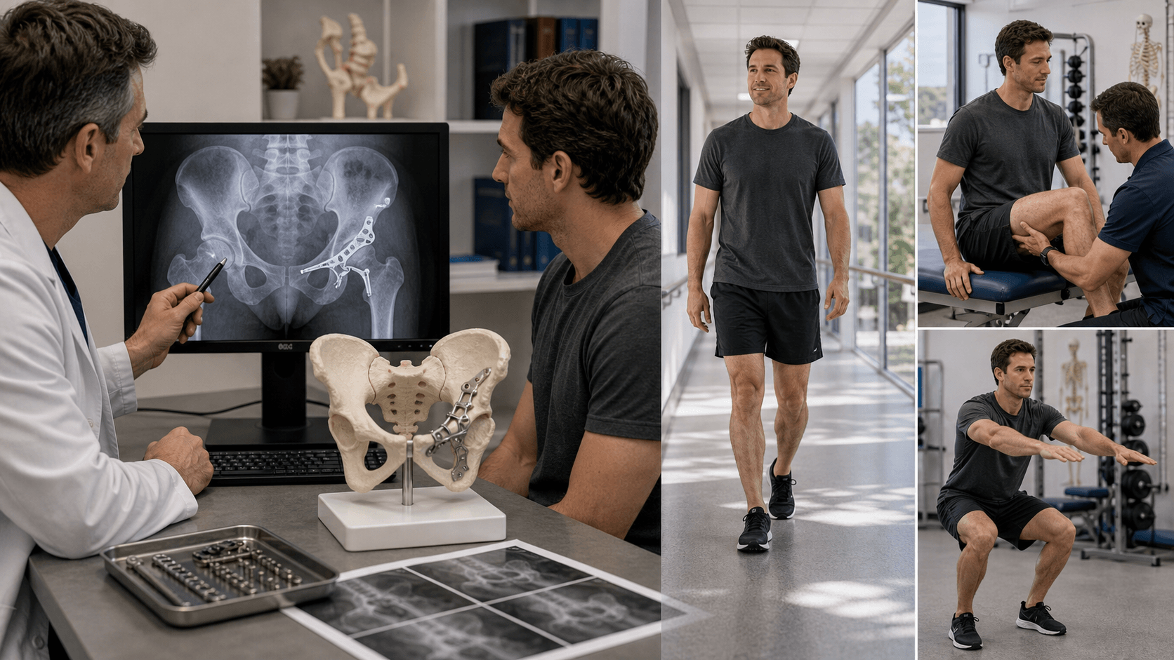
Postoperative Care
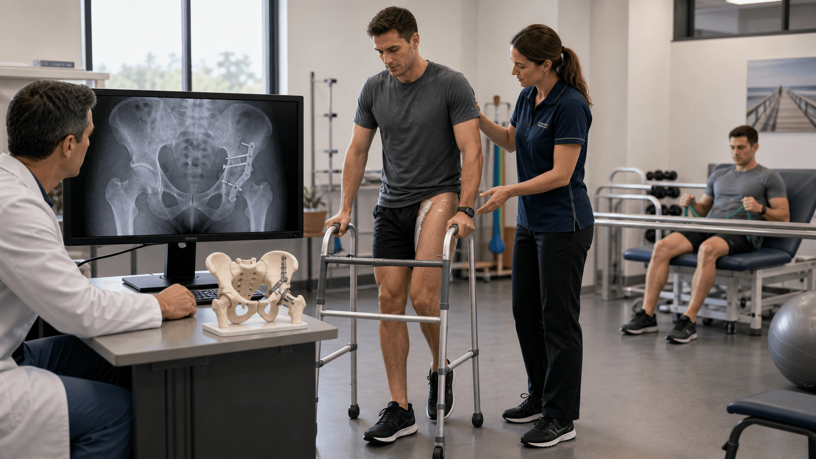
Postoperative Protocol
DVT prophylaxis (mechanical + LMWH). Monitor sciatic nerve function. Drain management. Pain control.
Touch-down weight bearing (TDWB) with frame/crutches. PT for ROM and strengthening.
X-rays to assess healing. Continue TDWB. Remove sutures/staples.
Partial weight bearing if healing. Repeat X-rays. Continue PT.
Full weight bearing when radiographic union. Return to activities. Monitor for HO, arthritis.
HO Prophylaxis Protocol
- Indomethacin 25mg TDS for 6 weeks (start within 48h)
- Or single-dose radiation 700cGy within 72h post-op
- Continue DVT prophylaxis for 4-6 weeks
Outcomes and Prognosis
Radiographic Outcomes
| Reduction | Grade | Good/Excellent Outcome |
|---|---|---|
| Anatomic (0-1mm) | Excellent | 80-85% |
| Imperfect (2-3mm) | Satisfactory | 65-75% |
| Poor (over 3mm) | Poor | 40-50% |
Functional Outcomes
- Anatomic reduction (under 2mm)
- Simple fracture pattern
- Short dislocation time (under 6 hours)
- Young age
- No femoral head damage
- Articular comminution
- Femoral head impaction (Gull sign)
- Posterior dislocation over 12 hours
- Age over 60
- Both-column fractures (complex)
Matta's criteria for outcome assessment:
- Excellent: No pain, normal ROM, no limp
- Good: Mild pain with activity, slight limp
- Fair: Moderate pain, limp, uses cane
- Poor: Severe pain, marked limp, disability
Anatomic reduction = 80% excellent/good outcomes
Controversies and Areas of Uncertainty
No consensus on the best strategy for osteoporotic, dome-involving fractures in older patients. Options span non-operative care, ORIF (often with quadrilateral-plate buttressing), and acute fix-and-replace (ORIF plus same-sitting total hip arthroplasty). Registry and systematic-review data favour acute arthroplasty for poor-prognosis hips, but randomised evidence is still lacking.
Indomethacin and single-fraction radiotherapy are of similar efficacy, but the absolute benefit of routine prophylaxis is debated given NSAID risks (GI, renal, possible effect on bone healing) and the logistics/cost of radiotherapy. Many units now reserve prophylaxis for extensile/posterior approaches in high-risk patients.
The modified Stoppa / anterior intrapelvic approach has largely displaced the classical ilioinguinal in many centres for anterior and quadrilateral-plate access, but the extent of its advantage and the role of combined approaches for complex transverse and T-type patterns remain debated.
The classic 40-50% fragment-size threshold for instability is imperfect. Examination under anaesthesia and CT-based fragment mapping are increasingly used because intermediate-size fragments (20-50%) have unpredictable stability and may still require fixation.
Evidence Base

Accuracy of Reduction Drives Outcome (Landmark Series)
- 262 displaced acetabular fractures (255 hips, mean 6-year follow-up) treated by open reduction and internal fixation within 21 days. Anatomic reduction was achieved in 71% of hips and the rate fell with increasing fracture complexity, older age and delayed surgery. Overall clinical result was excellent in 40%, good in 36%, fair in 8% and poor in 16% - clinical outcome tracked closely with radiographic reduction. Both-column fractures were the most common pattern (35%).
Postoperative CT Predicts Posterior Wall Outcome
- 67 surgically treated posterior wall fractures assessed with postoperative 2D CT. Plain films graded 65 of 67 reductions as anatomic, yet CT revealed greater than 2mm offset in 11 hips and gaps of 2mm or more in 52. A residual gap of 10mm or more, or total gap area of 35mm2 or more, was associated with a poor result. CT-assessed reduction was highly predictive of clinical outcome.
HO Prophylaxis: Indomethacin vs Radiation (RCT)
- Prospective randomised trial of 166 patients operated through posterior or extensile approaches. Brooker grade III-IV heterotopic ossification occurred in 11% of the indomethacin group (25mg three times daily for 6 weeks) versus 4% of the radiation group (800 cGy within 72h) - no significant difference. All 16 untreated patients developed HO (38% grade III-IV).
Long-Term Joint Survival After Fixation
- 61 hips treated with open reduction and internal fixation via surgical hip dislocation (mean follow-up 12.4 years). Ten-year cumulative survivorship was 82%. Independent predictors of an unfavourable outcome were femoral chondral lesions, marginal impaction, longer operative duration and older patient age.
Acute vs Delayed THA (Systematic Review)
- Systematic review and meta-analysis of 5 studies (255 patients) comparing acute 'fix-and-replace' total hip arthroplasty with delayed THA after initial fixation. Functional outcomes, complications and mortality were comparable, but delayed THA had a significantly higher revision rate (17.1% vs 4.3%, p=0.002).
Geriatric Fix-and-Replace vs ORIF
- Retrospective comparison of 17 fix-and-replace versus 11 ORIF-alone patients aged 55 and over. Acute outcomes (length of stay, disposition, 90-day readmission, time to mobilisation and HOOS Jr. scores) did not differ, but more fix-and-replace patients were permitted earlier weight-bearing (70% vs 9%).
Judet-Letournel Classification & Surgical Approaches
- Foundational text defining the two-column concept, the 10-pattern classification (5 elementary, 5 associated) and the standard surgical approaches (Kocher-Langenbeck, ilioinguinal, extended iliofemoral). Established that operative reduction of displaced fractures yields markedly better results than non-operative treatment.
Exam Viva Scenarios
Practise clinical reasoning and management decisions out loud
“28-year-old driver in MVA. X-ray shows posterior hip dislocation with posterior wall fracture. Sciatic nerve intact. How do you manage this?”
“45-year-old fell from roof. CT shows both-column acetabular fracture with spur sign visible. No dislocation. How do you approach this?”
“32-year-old motorcyclist with acetabular fracture. CT shows transverse pattern with associated posterior wall fragment. Which approach?”
“78-year-old woman, low-energy fall onto the side. CT shows a displaced anterior-column fracture with quadrilateral-plate comminution, marginal impaction and pre-existing hip osteoarthritis. How do you manage her?”
MCQ Practice Points
Q: What is the most common elementary acetabular fracture pattern?
A: Posterior wall - accounts for approximately 25% of all acetabular fractures. Typically caused by dashboard injury with hip in flexed position.
Q: Which radiographic line represents the anterior column on AP pelvis?
A: Iliopectineal line - the ilioischial line represents the posterior column. Remember: "Pectineal = Anterior, Ischial = Posterior"
Q: What approach is used for posterior column fractures?
A: Kocher-Langenbeck - this posterior approach gives direct access to posterior column and wall. The ilioinguinal/Stoppa is used for anterior column.
Q: What is pathognomonic for a both-column acetabular fracture?
A: Spur sign - a fragment of intact ilium "floating" above the acetabulum visible on obturator oblique view. Indicates both columns separated from axial skeleton.
Q: What is the most important factor for good outcome in acetabular fractures?
A: Anatomic reduction (under 2mm) - Matta's studies showed 83% excellent/good outcomes with anatomic reduction vs 50% with poor reduction greater than 3mm.
Q: What is the corona mortis?
A: Aberrant obturator vessel crossing the superior pubic ramus. Present in 30-70% of patients. Must be ligated during ilioinguinal/Stoppa approaches to prevent catastrophic hemorrhage.
Guidelines, Registries & Global Practice
Global Epidemiology
- Bimodal incidence: high-energy injuries in young men (road traffic, falls from height) and a rising low-energy group in osteoporotic elderly
- Ageing shift: the fastest-growing cohort worldwide is patients over 60 with anterior-column and quadrilateral-plate involvement
- Male predominance overall (~3:1) but narrows with age
- 80% of high-energy fractures are associated with hip dislocation
- High-resource: CT-based planning, dedicated pelvic-acetabular surgeons, fix-and-replace pathways
- Limited-resource: plain-film classification, traction-based non-operative care, delayed or no fixation
- Transfer to a specialist centre improves reduction quality and is recommended for displaced/complex patterns everywhere
Guidelines & Society Positions (Side by Side)
| Body | Region | Emphasis |
|---|---|---|
| AO Foundation / OTA | Global / US | Two-column concept and Judet-Letournel as the operative framework; anatomic reduction of the weight-bearing dome; approach dictated by column |
| BOA / BOAST (Pelvic & Acetabular) | UK | Network model - timely transfer of complex fractures to a specialist pelvic-acetabular unit; definitive surgery by a named specialist team |
| EFORT / European consensus | Europe | Early CT, dedicated theatre lists, growing role of acute total hip arthroplasty in the elderly |
| AAOS / OTA evidence reviews | US | Reduction accuracy and articular congruity as the dominant outcome determinants; HO prophylaxis for extensile approaches |
There is broad global consensus on the essentials: CT is mandatory for classification and planning; displaced fractures of the weight-bearing dome warrant anatomic operative reduction (under 2mm); approach is dictated by the fractured column; and complex patterns are best managed in specialist pelvic-acetabular units. The main area of practice variation is the management of the geriatric fracture - non-operative care, ORIF, or acute fix-and-replace.
Registry & Outcome Notes
- Joint registries (NJR UK, AJRR US, AOANJRR Australia, Swedish/Norwegian) track total hip arthroplasty performed after acetabular fracture as a distinct, higher-risk indication, with elevated dislocation and revision rates compared with primary osteoarthritis THA.
- This registry signal underpins the interest in acute fix-and-replace for poor-prognosis fractures in older patients, where revision rates appear lower than for delayed conversion.
High- vs Limited-Resource Practice Variation
- High-resource: routine 3D CT planning, intra-operative fluoroscopy/navigation, dedicated pelvic-acetabular surgeons, and ready access to acute arthroplasty.
- Limited-resource: greater reliance on skeletal traction and accepted non-operative management of borderline patterns, with fixation reserved for clearly unstable or dome-involving fractures; outcomes remain acceptable when secondary congruence is preserved.
Classification
- 5 Elementary: PW, PC, AW, AC, Transverse
- 5 Associated: Both-column, T-type, Trans+PW, PC+PW, AC+PHT
- Posterior wall = most common (25%)
- Both column = spur sign pathognomonic
Radiographic Lines
- Iliopectineal = anterior column
- Ilioischial = posterior column
- Obturator oblique: anterior column + posterior wall
- Iliac oblique: posterior column + anterior wall
Approach Selection
- Kocher-Langenbeck: Posterior wall/column
- Ilioinguinal/Stoppa: Anterior wall/column
- Extended iliofemoral: Both column (rare)
- Sciatic nerve at risk in KL approach
Key Numbers
- Under 2mm step = acceptable reduction
- Greater than 40% wall = needs fixation
- 3-5 days delay = less blood loss
- 80% good outcome with anatomic reduction
Complications
- HO: 20-50% (indomethacin prophylaxis)
- Sciatic nerve: 10-15%
- Post-traumatic arthritis: 20-30%
- Corona mortis: ligate in anterior approaches