The Groin Pain Muscle and the Landmark of the Medial Thigh
- Origin is a narrow, strong tendon from the anterior surface of the body of the pubis in the angle between the crest and the symphysis, immediately below the pubic tubercle; insertion is the middle third of the medial lip of the linea aspera.
- Its enthesis is continuous with the rectus abdominis aponeurosis across the anterior pubis, forming an aponeurotic plate — a common tendon complex which is why adductor and pubic-related groin pain so often coexist.
- Innervated by the anterior division of the obturator nerve; root values L2, L3, L4. The anterior division lies posterior to the adductor longus and anterior to the adductor brevis.
- It forms the anterior part of the posteromedial wall of the adductor canal (with adductor magnus more distally), which is the wall against which the femoral vessels, the saphenous nerve and the nerve to vastus medialis lie.
- Adductor longus is the most frequently injured adductor in sport, and the injury is almost always at or close to the proximal enthesis rather than in the muscle belly.
- “Complete proximal adductor longus avulsion in a professional athlete is usually treated non-operatively — outcomes and return-to-play times are comparable with surgical repair and the operation is not routinely indicated.
- “The apex of the femoral triangle, where the adductor canal begins, is the point at which the medial border of sartorius crosses the medial border of adductor longus — this is the boundary that separates a femoral triangle block from a true adductor canal block.
- “Adductor-related groin pain is defined by tenderness at the adductor origin PLUS pain on resisted adduction; both are required under the Doha framework, and multiple groin pain entities coexist in a large proportion of athletes.
- “In the medial (Ludloff) approach to the hip the adductor longus is the first structure tenotomised, but the safe plane is ANTERIOR to the adductor brevis, keeping the surgeon away from the posterior division of the obturator nerve and the medial circumflex femoral artery.
Overview
The adductor longus is the most anterior of the three named adductors proper and the one that generates almost all the clinical work in this compartment. It is triangular: a narrow, strong, palpable tendinous origin at the pubis expanding into a broad muscular insertion along the femur. That geometry concentrates load at the enthesis, which is why adductor-related groin pain and proximal adductor longus avulsion are among the commonest problems in kicking and change-of-direction sport.
The landmark of the medial thigh. Its tendon is the first structure identified in the medial approach to the hip and the first divided in an adductor release. Its medial border defines the apex of the femoral triangle, where the adductor canal begins, and it forms part of the posteromedial wall of that canal, which is the anatomical basis of the adductor canal block.
Attachments and the Pubic Enthesis
Origin. A narrow, strong, flat tendon from the anterior surface of the body of the pubis, in the angle between the pubic crest and the pubic symphysis, immediately below the pubic tubercle. The footprint is small, commonly described as roughly 1-2 cm across, which concentrates load and explains the enthesial pathology.
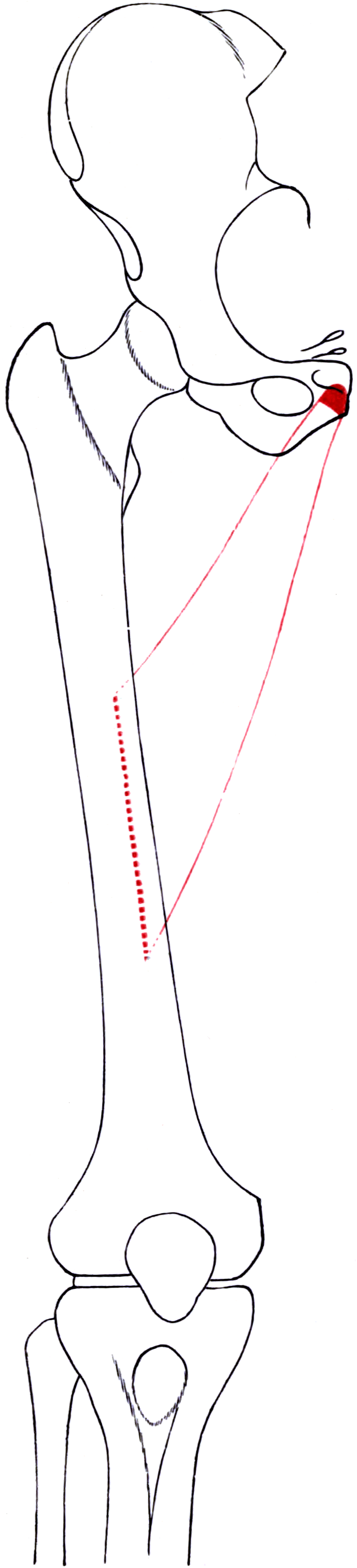
The aponeurotic plate. The tendon is continuous with the rectus abdominis aponeurosis across the front of the pubic body and the symphysis, forming a fibrocartilaginous aponeurotic plate anchored to the pubic bone and blending with the symphyseal disc and the superior and arcuate pubic ligaments. The anterior pubis is a shared tendon plate, not a set of separate insertions. The rectus abdominis pulls superiorly and posteriorly, the adductor longus inferiorly and laterally, and the symphysis is the fulcrum between the two opposing forces.
Why the plate matters. Load applied to one arm of the plate stresses the other. A kicking athlete generating hip extension and adduction loads the adductor enthesis while the trunk extends and rotates, loading the rectus arm.
- Adductor-related, pubic-related and inguinal-related groin pain frequently coexist, reported in a large proportion of athletes with longstanding groin pain, because they are mechanically coupled entities on one structure
- On MRI the secondary cleft sign, fluid or contrast tracking inferolaterally from the symphyseal primary cleft along the adductor enthesis, is a marker of enthesial injury on this plate rather than a separate diagnosis
- The examiner's point is to localise by entity, recognise the coupling, and explain why more than one entity is usually involved
Two layers at the enthesis. The origin has an anterior superficial tendinous portion and a deeper, broader aponeurotic portion continuous with the plate. That distinction is the anatomical basis of selective partial adductor release.
Insertion. The tendon expands into a broad fleshy belly and inserts by an aponeurosis into the middle third of the medial lip of the linea aspera on the posteromedial femur, between the vastus medialis anteriorly and laterally and the adductor brevis and magnus posteriorly. The insertion defines the depth of the posteromedial thigh compartment.
Shape and plane. Triangular, narrow proximally and broad distally, the opposite geometry to most muscles and a useful way to remember it. It lies anterior to the adductor brevis and medial to the pectineus, with the gracilis as the most medial and superficial strap of the compartment.
Innervation
Nerve and roots. The anterior division of the obturator nerve, root values L2, L3, L4, from the anterior divisions of the ventral rami of the lumbar plexus. Motor branches enter the posterior (deep) surface of the muscle in its proximal half.
Course of the obturator nerve. It descends on the medial border of the psoas, crosses the pelvic brim behind the common iliac vessels, runs on the lateral pelvic wall above the obturator vessels and exits through the obturator canal, approximately 2-3 cm inferolateral to the pubic tubercle, which is the point of nerve emergence, the block target and the obturator hernia site. It then divides into anterior and posterior divisions, separated first by the obturator externus and then by the adductor brevis.
The anterior division. It descends posterior to the pectineus and adductor longus and anterior to the adductor brevis, so it runs on the anterior surface of the brevis immediately deep to the adductor longus. It supplies adductor longus, adductor brevis, gracilis and pectineus, and gives the cutaneous branch to the distal medial thigh and an articular branch to the hip joint.
The compartment as a whole. Longus lies in front, brevis in the middle as the nerve landmark that separates the two obturator divisions, and magnus behind. Pectineus and magnus are the dually innervated pair: pectineus by the obturator anterior division and the femoral nerve, magnus by the obturator posterior division and the tibial division of the sciatic. Gracilis, supplied by the anterior division, is the only biarticular adductor.
What follows for the surgeon. A tenotomy performed close to the pubic origin is proximal to the motor entry, so it does not denervate the muscle: the muscle simply loses its proximal attachment and scars to the adjacent tissue at a slightly longer resting length, which is the mechanical basis of the operation. An obturator nerve block anaesthetises the adductor longus completely, and is used diagnostically in suspected obturator neuropathy, therapeutically for adductor spasticity, and to prevent the adductor jerk during transurethral bladder resection.
Localising a lesion. Hip adduction is L2/L3/L4. Sensation over the distal medial thigh is obturator, the anteromedial thigh is femoral, and the medial leg and medial border of the foot are saphenous. Weak adduction with medial thigh numbness therefore localises to the obturator nerve; add quadriceps weakness and the lesion is proximal, in the lumbar plexus or the L2-L4 roots, or from a retroperitoneal cause such as haematoma, abscess or tumour.
The denervation pattern. A complete obturator nerve lesion denervates adductor longus, brevis, gracilis and the adductor part of magnus, with relative sparing of the pectineus (femoral branch) and the ischiocondylar part of adductor magnus (tibial division of the sciatic). Adduction is therefore weakened, not abolished. On MRI this selective pattern of denervation oedema and later fatty atrophy is a reliable localiser for an obturator nerve lesion, and should prompt a search for a pelvic cause.
Blood Supply
Supply. Muscular branches of the profunda femoris are the dominant supply to the belly, with segmental contributions from its perforating branches as they pass posteriorly. The medial circumflex femoral artery supplies the proximal muscle and the enthesial region, and the anterior branch of the obturator artery contributes to the proximal muscle and the pubic enthesis. The external pudendal and inferior epigastric territories reach the enthesis through a fine periosteal plexus.
The enthesis is the poorly vascularised part. Compared with the muscle belly the pubic enthesis has a relatively poor blood supply, the typical pattern of a fibrocartilaginous insertion. That is one reason adductor-related groin pain is slow to resolve, and why enthesial injury behaves like tendinopathy rather than like a muscle strain. After a complete proximal avulsion the tendon retracts distally a short distance and heals by scar to the adjacent aponeurotic plate and periosteum, a functionally adequate result in most athletes and the reason non-operative management is the norm.
Vessels at risk.
- The profunda femoris and its perforating branches lie deep and posterior to the muscle; blind dissection posterior to the adductor longus in the proximal thigh risks them, so work under direct vision and control vessels deliberately
- The medial circumflex femoral artery lies deep to the pectineus and adductor brevis, more proximally and posteriorly, and is the vessel of concern in the medial approach to the hip
- The corona mortis, the anastomosis between the obturator and the external iliac or inferior epigastric systems, crosses the superior pubic ramus typically within 3-6 cm lateral to the symphysis, just deep to the adductor and pectineus origins; in anterior pelvic approaches it is inspected and ligated deliberately before plating
Relations, the Femoral Triangle and the Adductor Canal
Anterior. At its origin the tendon is essentially subcutaneous beneath the fascia lata, which is why it is so easily palpated and so precisely localised; the great saphenous vein and the superficial external pudendal artery lie in the subcutaneous plane proximally. The femoral vessels cross anterior to the muscle in the femoral triangle, and more distally the sartorius overlies the interval between adductor longus and vastus medialis, roofing the adductor canal.
Posterior. The anterior division of the obturator nerve on the anterior surface of the adductor brevis; the adductor brevis proximally and the adductor magnus distally; the profunda femoris artery and vein with their perforating branches; and, deeper still behind the brevis, the posterior division of the obturator nerve.
Lateral and medial. Laterally the pectineus proximally, then the femoral vessels in the femoral triangle, then the vastus medialis distally. Medially the gracilis.
The femoral triangle. Bounded by the inguinal ligament superiorly, the medial border of sartorius laterally and the medial border of adductor longus medially, with a floor of iliopsoas laterally, pectineus medially and adductor longus most medially, and a roof of fascia lata and cribriform fascia. Its apex is the point at which the medial border of sartorius crosses the medial border of adductor longus, and that is where the adductor canal begins. In an ultrasound study of 22 volunteers the apex lay a mean 27.4 cm from the anterior superior iliac spine, distal to the midpoint of the thigh (22.9 cm) in every one of them.
The adductor canal. The subsartorial (Hunter's) canal runs from the apex of the femoral triangle to the adductor hiatus in the adductor magnus, where the femoral vessels become the popliteal vessels.
- Anterolateral wall: vastus medialis
- Posteromedial wall: adductor longus proximally, adductor magnus distally
- Roof: the vastoadductor membrane, deep to the sartorius
- Contents: the femoral artery, the femoral vein posterior to it, the saphenous nerve anterolateral to it, the nerve to vastus medialis, and the descending genicular artery, which arises from the femoral artery within the canal just before the hiatus
- Exits: the saphenous nerve and the saphenous branch of the descending genicular artery pierce the vastoadductor membrane; the femoral artery and vein leave through the adductor hiatus
The saphenous nerve and the nerve to vastus medialis lie against the posteromedial wall, which is what makes them relevant to block technique and to distal medial dissection.
The adductor hiatus. An opening in the aponeurotic insertion of the adductor magnus at approximately the junction of the middle and distal thirds of the thigh. It is a fixed point of arterial tethering, which makes it a site of arterial injury in distal femoral fractures and knee dislocations, a site of entrapment in the rare popliteal artery entrapment variants, and the landmark in vascular surgical exposure of the distal femoral and proximal popliteal artery, where the adductor longus forms the posteromedial wall proximal to it.
Action and Biomechanics
- Action
- Hip adduction (a principal adductor)
- Position of greatest efficiency
- Hip abducted
- Note
- The dominant action; adductor longus is a major contributor
- Action
- Hip flexion or hip extension, depending on hip position
- Position of greatest efficiency
- Depends on hip position relative to the crossover point
- Note
- The position-dependent reversal is the exam point
- Action
- Contributes to rotation, direction depending on hip position
- Position of greatest efficiency
- Variable
- Note
- Described inconsistently in textbooks; state the mechanism, not a fixed direction
- Action
- Stabilises the pelvis in single-leg stance; decelerates hip abduction in change of direction
- Position of greatest efficiency
- Weight-bearing and cutting
- Note
- The functionally dominant role in sport
The flexion-extension reversal. The line of action crosses the hip's flexion-extension axis at a particular hip angle: below that angle the muscle flexes the hip, above it the muscle extends the hip. The commonly quoted crossover is around 50-70 degrees of hip flexion, varying with individual geometry. That is why the adductors are active in both the loading response and pre-swing phases of gait, why they are loaded in the kicking wind-up (hip extended and abducted, then explosive adduction and flexion), and why an isolated adductor test at a single hip angle can be misleading.
Force and architecture. The adductor group as a whole has a cross-sectional area comparable to the quadriceps in some estimates, and the adductor longus is a major contributor, with a large muscle volume relative to a small enthesial footprint. That mismatch is the injury mechanism: a big muscle transmitting force through a narrow fibrocartilaginous enthesis, on a bone that is also loaded by the rectus abdominis in the opposite direction, is a structurally demanding arrangement.
Eccentric demand in sport. The critical loading pattern is eccentric. The adductor decelerates hip abduction and external rotation during a change of direction, a sliding tackle, a reach for a ball, or a kick with the non-dominant limb, and peak strain falls on the enthesis with the hip abducted and extended. Adductor strength, particularly eccentric adduction strength and the adduction to abduction strength ratio, is a modifiable risk factor, which is the rationale for the Copenhagen adduction exercise.
Synergists and antagonists. Adduction synergists are the adductor brevis and magnus, pectineus, gracilis, the inferior fibres of gluteus maximus and quadratus femoris. The antagonists are gluteus medius and minimus and tensor fascia lata, and in the sagittal plane the antagonist changes with hip position. Across the symphysis the rectus abdominis is the functional antagonist, coupled to the adductor longus through the aponeurotic plate.
What happens when it fails.
- Acute strain or avulsion: sudden medial groin pain, often with an audible or palpable sensation, bruising in the medial thigh afterwards, and pain and weakness on resisted adduction. Function is surprisingly well preserved because of the other adductors
- Chronic adductor-related groin pain: exertional medial groin pain, tenderness at the enthesis, pain on resisted adduction and reduced adduction strength. The problem is enthesial
- Adductor contracture: fixed adduction deformity with reduced abduction, the picture in spastic hip disease and in long-standing hip arthritis
- After tenotomy: adduction strength recovers to a functionally adequate level in most patients because the remaining adductors compensate, though a measurable strength deficit may persist
Surface Anatomy and Examination
Finding the tendon. With the patient supine and the hip flexed, abducted and externally rotated (the frog-leg position), the adductor longus tendon stands out as a prominent cord running from just below the pubic tubercle distally and laterally. The muscle belly is the broad mass in the proximal medial thigh, and bruising after an avulsion tracks distally and medially with gravity.
The enthesis. Palpate immediately below and medial to the pubic tubercle. This is the point of maximal tenderness in adductor-related groin pain, and it must be distinguished from the pubic symphysis itself (pubic-related), the inguinal canal and conjoint tendon (inguinal-related), the iliopsoas (tenderness deep in the femoral triangle plus pain on resisted flexion) and the hip joint (FADIR, FABER, log roll).
- How to perform
- Supine; squeeze the examiner's fist or a dynamometer between the knees at 0, 45 and 90 degrees of hip flexion
- Positive finding
- Pain and/or reduced force compared with the uninjured side
- What it means
- Adductor-related groin pain; also a monitoring tool for rehabilitation progress
- False positives
- Pubic-related and inguinal-related groin pain, hip pathology, osteitis pubis
- How to perform
- Palpate immediately below and medial to the pubic tubercle
- Positive finding
- Localised tenderness at the enthesis
- What it means
- Required, together with a positive squeeze test, for a Doha diagnosis of adductor-related groin pain
- False positives
- Tenderness from the adjacent symphysis or from a coexisting entity
- How to perform
- Supine, pelvis square; abduct with the knee extended, then flexed
- Positive finding
- Pain or restriction; improvement with the knee flexed
- What it means
- Improvement with knee flexion implicates the biarticular gracilis; unchanged restriction implicates the monoarticular adductors including longus
- False positives
- Pelvic obliquity not controlled; hip joint restriction
- How to perform
- Abduct the hip passively and ask the patient to resist as you continue abducting
- Positive finding
- Pain at the enthesis
- What it means
- Sensitive for enthesial pathology because it loads the tendon eccentrically
- False positives
- Apprehension; pain from any adjacent structure
- How to perform
- Perform the squeeze bilaterally, then resist unilateral adduction
- Positive finding
- Asymmetry in force
- What it means
- Quantifies a strength deficit and is used as a return-to-play criterion
- False positives
- Effort dependence; pain inhibition
- How to perform
- Systematically palpate and resist for adductor, iliopsoas, inguinal, pubic and hip entities in turn
- Positive finding
- Findings in more than one entity
- What it means
- Coexisting groin pain entities, which is the rule rather than the exception
- False positives
- Overlapping referred pain; incomplete examination
- How to perform
- Standard hip provocation tests
- Positive finding
- Groin pain reproduced
- What it means
- Hip-related groin pain, which must be excluded before attributing symptoms to the adductors
- False positives
- Non-specific; FABER is positive in sacroiliac pathology
- How to perform
- Light touch over the distal medial thigh
- Positive finding
- Reduced sensation
- What it means
- Obturator nerve involvement, raising the possibility of obturator neuropathy rather than a muscle injury
- False positives
- Overlap with the medial cutaneous nerve of the thigh
Imaging. MRI is the investigation of choice. It shows:
- Enthesial oedema and tendon disruption at the adductor longus origin, graded by whether the tendon is partially or completely detached and by the degree of retraction
- The secondary cleft sign along the adductor enthesis
- Bone marrow oedema and subchondral change at the pubis (pubic-related involvement) and rectus abdominis aponeurotic change, the coupled entities
- Muscle belly oedema and haematoma, distinguishing a myofascial injury from an enthesial one
Ultrasound is useful for a dynamic assessment, for guiding an injection and in resource-limited settings, but it is inferior to MRI for the enthesis and the symphysis. Radiographs exclude an apophyseal avulsion in the adolescent, and show symphyseal changes and osteitis pubis in chronic cases.
Do not over-read imaging. Enthesial and symphyseal MRI changes are common in asymptomatic athletes. The diagnosis is clinical, and imaging refines it.
Pitfalls. Never diagnose "groin strain": use the Doha entities and expect more than one. Square the pelvis before measuring abduction. In an adolescent, think apophysis, because the pubic and ischial apophyses can avulse and the adductor origin is a recognised site. And consider the serious differentials before rehabilitating anything:
- Femoral neck or pubic ramus stress fracture (load-related pain, night pain, the endurance athlete)
- Slipped upper femoral epiphysis or Perthes disease in a child
- Apophyseal avulsion in an adolescent
- Inguinal or obturator hernia
- Testicular and urological causes
- Referred lumbar pain
- Rarely but importantly, a pelvic or proximal femoral tumour or infection
Adductor Tenotomy
In the athlete. The indication is refractory adductor-related groin pain in an athlete who has completed an adequate active rehabilitation programme, generally at least three to six months, without resolution, with clinical findings and imaging confirming an enthesial problem. It is not an early or first-line intervention, and not a substitute for rehabilitation.
Selective partial adductor release. This is the technique to describe. Dividing the superficial anterior tendinous portion selectively relieves the enthesial load while preserving the deep aponeurotic continuity, and therefore the stability of the pubic plate, which is why a complete tenotomy is not the default operation.
- Approach: a short transverse or longitudinal incision over the palpable adductor longus tendon, immediately below and medial to the pubic tubercle
- Steps: expose the tendon, identify the superficial anterior tendinous fibres, divide them selectively under direct vision preserving the deeper aponeurosis, and avoid dissection posteriorly, where the anterior division of the obturator nerve lies on the adductor brevis
- Rehabilitation: early mobilisation with graded loading, with reported return to sport typically in the region of a few months
- Outcomes: series of professional athletes undergoing selective partial release for chronic adductor-related groin pain report a high rate of return to play at the previous level
Complete adductor tenotomy. Divides the whole tendon at the pubis. It is effective for pain but carries a greater risk of measurable adduction weakness and, theoretically, of destabilising the aponeurotic plate, so it is reserved for cases where a selective release has failed, or where a complete release is being performed for a different indication (spasticity, contracture).
Adductor release for spastic hip displacement. In cerebral palsy the adductor longus tenotomy is the primary and first release, performed close to the pubic origin, followed if needed by gracilis, then adductor brevis, then pectineus.
- Indication: a young child with a migration percentage of roughly 30-50 percent, reduced passive abduction and a still-adequate acetabulum, detected through a hip surveillance programme because displacement is painless until late
- Target: approximately 40-45 degrees of abduction per side intraoperatively
- Combine with intramuscular iliopsoas lengthening at the pelvic brim rather than a tenotomy at the lesser trochanter, to preserve hip flexion power in an ambulant child
- Avoid complete anterior branch obturator neurectomy: it risks an abduction contracture, which is functionally worse, and it cannot abolish adduction anyway because the pectineus retains a femoral supply and the ischiocondylar adductor magnus a sciatic supply
- If the migration percentage exceeds roughly 50-60 percent, or the acetabulum is deficient, soft-tissue release alone will fail and the operation becomes a varus derotation femoral osteotomy with a pelvic osteotomy (Dega, San Diego, Pemberton) and capsulorrhaphy as required. Recurrent displacement after soft-tissue release alone follows a release performed too late, so adhere to the surveillance schedule
Before hip arthroplasty. A fixed adductor contracture in a long-standing arthritic or ankylosed hip prevents adequate positioning, dislocation and limb manipulation. Perform the adductor tenotomy first, before the arthrotomy, so that the limb can be moved freely for the rest of the procedure.
For perineal hygiene and catheterisation. In a non-ambulant adult with adductor spasticity the sequence is obturator nerve block (diagnostic and therapeutic), then botulinum toxin, then tenotomy with or without limited neurectomy for a fixed problem.
The Adductor Canal Block
What it blocks. The adductor canal block is widely used for analgesia after total knee arthroplasty and after knee arthroscopic surgery. It anaesthetises the saphenous nerve (medial knee and leg sensation), the nerve to vastus medialis and, variably, the articular branches to the knee. It is more quadriceps-sparing than a femoral nerve block but it is not motor-neutral, because the nerve to vastus medialis is a motor nerve within the canal.
Level matters more than the label. Ultrasound and anatomical studies show that a large proportion of injections labelled adductor canal blocks are placed proximal to the apex of the femoral triangle, in the distal femoral triangle. A more proximal injection reaches more motor branches and produces more quadriceps weakness, which is a genuine falls risk after knee arthroplasty. Identify the apex sonographically (the crossing of sartorius over adductor longus), know which block you are performing, and assess quadriceps power before the first mobilisation regardless of the block used, with a knee immobiliser or supervised ambulation as required.
The Medial Approach to the Hip and Other Pathology
The medial (Ludloff) approach. Used principally for open reduction of developmental dysplasia of the hip in the infant. The incision commences roughly 1-2 cm distal to the pubic tubercle over the adductor longus tendon, and the adductor longus is the first structure identified and tenotomised. The classic interval lies between adductor longus and gracilis superficially and adductor brevis and adductor magnus deeply; the Ferguson modification works anterior to the adductor brevis, which is the safer plane.
Why the plane matters. The anterior division of the obturator nerve lies anterior to the adductor brevis and the posterior division behind it, and the medial circumflex femoral artery, the dominant blood supply to the femoral head, lies posteriorly, deep to pectineus and adductor brevis. Working anterior to the brevis keeps you away from both, and keeping the anterior division in view confirms that you are in that plane. The femoral vessels lie anteriorly, so identify the adductor longus tendon first and work medially and posteriorly, never anteriorly.
Completing the reduction. The blocks to reduction released are the iliopsoas tendon at the lesser trochanter, the ligamentum teres, the pulvinar and the transverse acetabular ligament. Capsulorrhaphy is not possible, residual acetabular dysplasia is common, and avascular necrosis is the feared complication, from medial circumflex injury, excessive traction, forced reduction or extreme abduction in the spica. Immobilise in the human position, roughly 100 degrees of flexion and 45-55 degrees of abduction, not extreme abduction.
Adolescent apophyseal avulsion. The pubic and ischiopubic apophyses can avulse at the adductor origin in the skeletally immature athlete, presenting exactly like an adductor enthesial injury but with a bony fragment on radiograph or apophyseal oedema on MRI. Always radiograph an adolescent with adductor-origin pain rather than assuming a soft-tissue injury. Management is almost always non-operative, with protected weight-bearing and graded rehabilitation.
Osteitis pubis and pubic-related groin pain. Chronic overload of the symphysis and the aponeurotic plate, presenting with midline and paramedian pubic pain, tenderness over the symphysis, and radiographic and MRI changes (sclerosis, irregularity, marrow oedema, symphyseal widening). It very frequently coexists with adductor-related pain, for the mechanical reasons the plate explains. Treatment is load management and progressive strengthening; surgery (symphyseal debridement, curettage, arthrodesis) is a last resort with limited evidence.
Obturator neuropathy. Exertional medial thigh pain with adductor weakness and a small patch of medial thigh numbness, caused by fascial entrapment at the exit from the obturator canal in athletes, or by pelvic pathology. Post-exercise electromyography of the adductors is the most useful confirmatory test, MRI excludes a mass or hernia, and a diagnostic obturator nerve block supports the diagnosis. Fascial release at the obturator canal exit has been reported to allow return to sport.
Myositis ossificans and heterotopic ossification. Follows a direct blow, a large enthesial haematoma, extensive dissection, or head or spinal cord injury, and presents with a firm tender mass and progressive loss of abduction. Radiographs show centripetal (zonal) maturation, peripheral mature bone with an immature centre, the opposite of osteosarcoma. Gentle handling and haemostasis reduce the risk at surgery. Manage conservatively, avoid deep massage and early excision, and excise only a mature lesion causing mechanical restriction.
Surgical Relevance
The procedures in which the adductor longus is involved, and the structures at risk in each.
- How adductor longus is involved
- Superficial anterior tendinous fibres divided at the pubis; deep aponeurosis preserved
- Structures at risk with location
- Anterior division of the obturator nerve on adductor brevis behind it; aponeurotic plate stability if released completely
- How adductor longus is involved
- First and primary tenotomy, close to the pubic origin
- Structures at risk with location
- Obturator nerve anterior division deep to it; medial circumflex femoral artery deeper still behind pectineus and brevis
- How adductor longus is involved
- First structure identified and tenotomised; interval developed anterior to adductor brevis
- Structures at risk with location
- Medial circumflex femoral artery deep and posterior; femoral vessels anteriorly; obturator nerve divisions either side of adductor brevis
- How adductor longus is involved
- Forms the proximal posteromedial wall of the canal
- Structures at risk with location
- Femoral artery and vein; nerve to vastus medialis (quadriceps weakness); block level determines the effect
- How adductor longus is involved
- Divided to allow limb positioning and dislocation
- Structures at risk with location
- Obturator nerve; profunda femoris perforators posteriorly; excessive release causing weakness
- How adductor longus is involved
- Origin elevated from the anterior pubis
- Structures at risk with location
- Corona mortis within 3-6 cm of the symphysis; external iliac and obturator vessels; aponeurotic plate
- How adductor longus is involved
- The muscle is the posteromedial wall proximal to the hiatus
- Structures at risk with location
- Femoral vessels; saphenous nerve; adductor hiatus as the tethering point
- How adductor longus is involved
- The adductor longus is the adjacent landmark
- Structures at risk with location
- Anterior division of the obturator nerve; saphenous vein; adductor longus mistaken for gracilis
Complications
Of rehabilitation. The two failures are re-injury after a return by calendar rather than by restored strength, control and sport tolerance, and persistent groin pain after treatment of one entity only, when a coexisting pubic-related, inguinal-related or hip-related entity was never addressed. The avoidance is criteria-based progression with objective strength measurement and full symptom-free team training before match play, and a complete Doha screen that treats every entity present.
Of surgery.
- Adduction weakness after complete tenotomy: measure strength, rehabilitate, and counsel about a possible residual deficit
- Obturator nerve injury during release or medial approach: identify the anterior division before dividing the tendon
- Profunda femoris or perforator haemorrhage from blind dissection posterior to the muscle
- Avascular necrosis of the femoral head after medial open reduction
- Corona mortis haemorrhage during anterior pelvic exposure of the superior pubic ramus
- Abduction contracture after over-release combined with complete anterior branch obturator neurectomy
- Recurrent hip displacement after soft-tissue release alone
- Myositis ossificans or heterotopic ossification after extensive dissection or a large haematoma
- Wound problems in the groin, a moist contaminated area, particularly in a child in a spica or an incontinent adult: meticulous closure, careful cast padding, hygiene planning and early review
Of the block. Quadriceps weakness and falls after an adductor canal block, from the nerve to vastus medialis being blocked and from an injection placed proximal to the femoral triangle apex.
Of diagnosis. A missed stress fracture or other serious cause, when load-related groin pain is attributed to the adductors without imaging. Image when the pattern does not fit.
Guidelines, Registries & Global Practice
Variation and prevalence
- The relative contribution of the superficial tendinous and deep aponeurotic portions of the adductor longus enthesis varies between individuals, which is one reason a selective partial release requires direct visual identification rather than a fixed depth.
- Corona mortis prevalence and calibre vary between anatomical series, with venous connections more frequent than arterial. The practical instruction is always to inspect and ligate rather than to rely on an expected absence.
- The level at which the obturator nerve divides ranges from within the obturator canal to several centimetres distal, and an accessory obturator nerve is present in a minority — both relevant to the completeness of an obturator nerve block.
- Enthesial and symphyseal MRI changes are common in asymptomatic athletes, so prevalence data on imaging findings must not be read as prevalence of disease.
Differences in described technique and guidance
- Emphasis relating to adductor longus
- Requires anatomical classification of groin pain into defined clinical entities; adductor-related pain needs both enthesial tenderness and pain on resisted adduction; recognises frequent coexistence.
- Emphasis relating to adductor longus
- Emphasises active loading-based rehabilitation, objective strength measurement with a dynamometer, and criteria-based return to play; surgery only after an adequate programme fails.
- Emphasis relating to adductor longus
- Similar rehabilitation-first approach; greater use of imaging early in the professional athlete; selective partial adductor release described for the refractory case.
- Emphasis relating to adductor longus
- Explicit warning about the corona mortis crossing the superior pubic ramus in anterior pelvic approaches, immediately deep to the adductor and pectineus origins.
- Emphasis relating to adductor longus
- Adductor longus tenotomy as the primary release for spastic hip displacement, triggered by migration percentage rather than by symptoms, with stratified radiographic surveillance by GMFCS level.
- Emphasis relating to adductor longus
- Adductor canal block preferred over femoral nerve block for knee arthroplasty; explicit acknowledgement that it is not motor-neutral and that many blocks are placed proximal to the true canal.
Registry and outcome signals
- Elite football injury surveillance programmes (the UEFA Elite Club Injury Study and comparable national programmes) consistently identify the adductors as one of the four dominant muscle-injury groups, with a substantial recurrence rate — the epidemiological basis for prevention programmes and for criteria-based return.
- Randomised prevention trials in football have shown that adductor strengthening programmes centred on the Copenhagen adduction exercise reduce the prevalence of groin problems, which has driven their inclusion in team injury prevention protocols internationally.
- Hip surveillance programmes for cerebral palsy have demonstrated a measurable reduction in hip dislocation rates where implemented systematically, supporting programme-based rather than opportunistic radiographic review.
- There is no registry of adductor surgery, and published series of selective partial release are single-centre cohorts in professional athletes — an honest statement of the evidence base is expected.
High- versus limited-resource practice
- Well-resourced settings: MRI for the enthesis, symphysis and rectus arm of the plate; handheld dynamometry for objective adduction strength and adduction to abduction ratio; ultrasound-guided injections and blocks with sonographic identification of the femoral triangle apex; post-exercise electromyography for suspected obturator neuropathy.
- Limited-resource settings: the diagnosis of adductor-related groin pain is entirely clinical under the Doha framework and needs no imaging — enthesial tenderness plus a painful squeeze test. The treatment with the best evidence, an active progressive strengthening programme including the Copenhagen adduction exercise, needs no equipment at all. Radiographs remain essential in the adolescent to exclude apophyseal avulsion, and in chronic cases to assess the symphysis. Hip surveillance in cerebral palsy needs only a correctly positioned AP pelvis and a ruler.
- Universal principles: localise by entity and expect coexistence; load rather than rest; return by criteria rather than calendar; treat a complete proximal avulsion non-operatively; stay anterior to adductor brevis in a medial hip approach; and inspect for a corona mortis before plating the superior pubic ramus.
Related pages: Adductor Groin Strain is the injury this enthesis produces and the page for its acute management; Athletic Groin Pain and Athletic Pubalgia are the neighbouring Doha entities that coexist with it so often that finding one is a reason to look for the others, and Osteitis Pubis is the pubic-related entity at the other end of the shared aponeurotic plate. Obturator Nerve Anatomy carries the anterior division that supplies this muscle and runs between it and adductor brevis. Adductor Brevis, Adductor Magnus, Gracilis and Pectineus complete the medial compartment, with adductor longus the most anterior and the one forming the medial border of the femoral triangle. Femoroacetabular Impingement is the hip-related cause that must be excluded before groin pain is attributed to the adductor alone, and Hamstring Injuries is the muscle group that outranks the adductors in the same surveillance data and shares the criteria-based return principle.
MCQ Practice Points
Q: Give the origin and insertion of the adductor longus. A: Origin — a narrow strong tendon from the anterior surface of the body of the pubis, in the angle between the pubic crest and the symphysis, immediately below the pubic tubercle. Insertion — the middle third of the medial lip of the linea aspera.
Q: What is the nerve supply of the adductor longus and where does that nerve lie? A: The anterior division of the obturator nerve (L2, L3, L4). It lies posterior to the pectineus and adductor longus and anterior to the adductor brevis.
Q: Why do adductor-related and pubic-related groin pain coexist? A: The adductor longus enthesis is continuous with the rectus abdominis aponeurosis across the anterior pubis, forming a single aponeurotic plate with the symphysis as the fulcrum. Load on one arm stresses the other.
Q: What is required to diagnose adductor-related groin pain under the Doha framework? A: Adductor tenderness AND pain on resisted adduction. Both are needed, and multiple entities coexist in a large proportion of athletes.
Q: How is a complete proximal adductor longus avulsion in a professional athlete managed? A: Non-operatively. Return-to-play times and outcomes after criteria-based rehabilitation are comparable with surgical repair, so surgery is not routinely indicated.
Q: What are the boundaries and floor of the femoral triangle? A: Inguinal ligament superiorly, medial border of sartorius laterally, medial border of adductor longus medially. Floor: iliopsoas laterally, pectineus medially, adductor longus most medially.
Q: Name the walls, roof and contents of the adductor canal. A: Anterolateral wall vastus medialis; posteromedial wall adductor longus proximally and adductor magnus distally; roof the vastoadductor membrane deep to sartorius. Contents: femoral artery and vein, saphenous nerve, nerve to vastus medialis, and the descending genicular artery arising within it.
Q: Where does the adductor canal begin? A: At the apex of the femoral triangle, where the medial border of the sartorius crosses the medial border of the adductor longus. A block proximal to this point is a femoral triangle block, not an adductor canal block, and causes more quadriceps weakness.
Q: What is the anatomical rationale for a selective partial adductor release? A: The enthesis has an anterior superficial tendinous portion and a deeper aponeurotic portion continuous with the pubic plate. Dividing the superficial anterior fibres unloads the enthesis while preserving plate stability and adduction strength.
Q: What is the evidence-based prevention programme for groin problems in football? A: An adductor strengthening programme centred on the Copenhagen adduction exercise, which increases eccentric adduction strength and reduced groin problem prevalence in a cluster-randomised trial.
Q: In the medial approach to the hip, what plane keeps you safest and why? A: Anterior to the adductor brevis (the Ferguson modification). The posterior division of the obturator nerve and, critically, the medial circumflex femoral artery — the dominant blood supply to the femoral head — lie posterior to the adductor brevis.
Q: What is the adductor hiatus and why does it matter? A: The opening in the aponeurotic insertion of the adductor magnus through which the femoral vessels pass to become the popliteal vessels. It is a fixed point of arterial tethering, and therefore a site of arterial injury in distal femoral fractures and knee dislocations.
Exam Viva Scenarios
Practise clinical reasoning and management decisions out loud
“A 27-year-old professional footballer felt a sudden tearing sensation in the right groin while stretching for a ball. MRI shows a complete avulsion of the adductor longus from the pubis with retraction and a large haematoma. His club want to know whether he needs an operation. What do you advise?”
“A 30-year-old semi-professional footballer has had medial groin pain for eight months. He has completed six months of physiotherapy including progressive adductor strengthening. He has tenderness at the adductor origin, a painful squeeze test at all three angles, no symphyseal tenderness, a normal hip examination and MRI showing adductor longus enthesopathy with a secondary cleft sign but no other abnormality. What now?”
“A 68-year-old woman has had a total knee arthroplasty with what the anaesthetic chart records as an adductor canal block. On the ward she nearly falls on first mobilisation because her quadriceps gives way. The physiotherapist asks whether the block was performed incorrectly. How do you answer, using the anatomy?”
Anatomy
- Origin: narrow tendon, anterior body of pubis below the pubic tubercle
- Insert: middle third of the medial lip of the linea aspera
- Triangular: narrow proximally, broad distally
- Nerve: obturator anterior division, L2-L4
- Enthesis continuous with rectus abdominis aponeurosis (aponeurotic plate)
Femoral Triangle and Canal
- Triangle: inguinal ligament, sartorius, adductor longus
- Floor: iliopsoas, pectineus, adductor longus
- Apex: sartorius crosses adductor longus — canal begins here
- Canal walls: vastus medialis anterolateral, adductor longus then magnus posteromedial
- Roof: vastoadductor membrane deep to sartorius
- Contents: femoral artery and vein, saphenous nerve, nerve to vastus medialis, descending genicular artery
Groin Pain
- Doha entities: adductor, iliopsoas, inguinal, pubic, hip, other
- Adductor-related needs tenderness PLUS painful resisted adduction
- Coexistence is the rule
- Secondary cleft sign on MRI
- Active training beats passive modalities (months, not weeks)
- Copenhagen adduction exercise for prevention
Return to Play
- Pain-free full range, symmetrical
- No enthesial or symphyseal tenderness
- Pain-free squeeze at 0, 45 and 90 degrees
- Strength and adduction to abduction ratio restored
- Graded sport-specific progression completed
- Full symptom-free team training before match play
Surgery
- Complete avulsion: non-operative default
- Selective partial release: superficial anterior fibres, preserve deep aponeurosis
- No posterior dissection — obturator anterior division on adductor brevis
- Ludloff: adductor longus first, but work ANTERIOR to adductor brevis
- MCFA deep and posterior — avascular necrosis risk
- CP release order: longus, gracilis, brevis, pectineus; target 40-45 degrees
Evidence Base
Doha Agreement Meeting on Terminology and Definitions in Groin Pain in Athletes
- International consensus establishing a clinical classification of groin pain in athletes
- Three categories: defined clinical entities (adductor-related, iliopsoas-related, inguinal-related, pubic-related), hip-related, and other causes
- Adductor-related groin pain requires adductor tenderness AND pain on resisted adduction
- Recognised that multiple entities frequently coexist in the same athlete
Effectiveness of Active Physical Training as Treatment for Long-Standing Adductor-Related Groin Pain in Athletes: Randomised Trial
- Randomised trial comparing an active physical training programme with conventional physiotherapy without active strengthening
- The active training group achieved substantially higher rates of return to sport without groin pain
- The programme emphasised progressive adductor and abdominal strengthening and coordination
- Benefits were maintained at follow-up
Effectiveness of a Selective Partial Adductor Release for Chronic Adductor-Related Groin Pain in Professional Athletes
- Series of professional athletes with chronic adductor-related groin pain refractory to conservative treatment
- Selective partial release of the superficial anterior adductor longus tendon fibres preserving the deeper aponeurosis
- A high proportion returned to their previous level of professional sport
- Adduction strength was largely preserved because the deeper aponeurotic attachment was retained
Return to Sport After Criteria-Based Rehabilitation of Acute Adductor Injuries in Male Athletes: A Prospective Cohort Study
- 81 male athletes with acute adductor injury, graded on MRI from 0 (no finding) to 3 (complete tear or avulsion), managed by a standardised criteria-based exercise programme
- Return to sport was assessed against three milestones: clinically pain-free, completion of controlled sports training, and return to full team training
- GRADE 0-2 injuries: pain-free at a median 13 days and back to full team training at 18 days
- GRADE 3 injuries (complete tear or avulsion): pain-free at a median 55 days and back to full team training at 78 days - FOUR TIMES LONGER, not comparable
- Milestone attainment fell in the middle: 75% became clinically pain-free, only 62% completed controlled sports training, yet 93% returned to full team training
- Overall one-year reinjury 8%; athletes who met the clinically pain-free criterion reinjured at 5% versus 21% in those who did not (p=0.048)
Defining the Location of the Adductor Canal Using Ultrasound
- Ultrasound study of 22 volunteers locating the proximal end of the adductor canal, defined where the medial border of SARTORIUS crosses the medial border of ADDUCTOR LONGUS
- Mean distance from the anterior superior iliac spine to the midpoint of the thigh was 22.9 cm; to the proximal end of the adductor canal, 27.4 cm
- The midpoint of the thigh - the conventional needle insertion point for an 'adductor canal block' - lay PROXIMAL to the adductor canal in ALL 22 volunteers, by a mean of 4.6 cm
- The authors conclude that the standard 'adductor canal block' is a misnomer and is in fact a FEMORAL TRIANGLE block
- This matters because a more proximal injection reaches more motor branches, with correspondingly greater quadriceps weakness
The Adductor Strengthening Programme Prevents Groin Problems Among Male Football Players: A Cluster-Randomised Controlled Trial
- Cluster-randomised trial in 35 SEMIPROFESSIONAL Norwegian football teams - 18 teams (339 players) intervention, 17 teams (313 players) control
- A single exercise with three progression levels, based on the Copenhagen Adduction exercise: three times weekly in preseason, once weekly through 28 competitive weeks
- Average prevalence of groin problems 13.5% in the intervention group against 21.3% in controls
- Risk of reporting a groin problem 41% lower: OR 0.59 (95% CI 0.40 to 0.86, p=0.008)
- Note the outcome is SELF-REPORTED groin PROBLEMS on the OSTRC Overuse Injury Questionnaire, not clinician-diagnosed injury or time loss
Epidemiology of Muscle Injuries in Professional Football (Soccer)
- Prospective cohort of European professional football clubs across multiple seasons
- Muscle injuries accounted for roughly one third of all time-loss injuries
- Four muscle groups dominated: hamstrings, adductors, quadriceps and calf
- Adductor injuries recurred at a substantial rate