The Expendable Muscle That Rebuilds a Limb and a Smile
- Gracilis is supplied by the anterior division of the obturator nerve, root values L2, L3, L4.
- It is the only biarticular adductor, crossing both the hip and the knee.
- Its dominant pedicle is the adductor branch of the medial circumflex femoral artery, entering the deep surface approximately 8-10 cm distal to the pubic tubercle.
- It is a Mathes-Nahai type II muscle: one dominant proximal pedicle plus one or two minor distal pedicles, which is what makes free transfer reliable.
- It is the middle tendon of the pes anserinus, lying between the sartorius fascia superficially and the semitendinosus deep.
- “Three pes tendons, three nerves: sartorius from the femoral, gracilis from the obturator, semitendinosus from the tibial division of the sciatic.
- “The obturator nerve enters the muscle proximal to and slightly deep to the vascular pedicle — take a long nerve for a free transfer.
- “As a free functioning muscle it is neurotised by the spinal accessory, intercostals, or contralateral facial nerve via a cross-face graft.
- “Regeneration of the harvested tendon cannot be assumed: one two-year MRI study found both semitendinosus and gracilis had regenerated in only 35 per cent of patients.
Overview
Gracilis is the most superficial and most medial of the adductor group: a long, flat, strap-like muscle running the whole length of the medial thigh from the pubis to the tibia. It is the only adductor that crosses two joints, and this biarticular course explains both its knee flexion and tibial internal rotation function and its position as the middle tendon of the pes anserinus.
Its clinical importance has almost nothing to do with what it does in the leg. Gracilis is functionally expendable — its loss produces no measurable adduction deficit because adductor longus, brevis and magnus dwarf it — and it has a single dominant proximal neurovascular pedicle with a long, accessible motor nerve. Those three facts have made it the most-transferred muscle in reconstructive surgery: as a free tendon graft in the knee, hand and ankle; as a pedicled flap for perineal, vaginal and ischial pressure-sore reconstruction; and as a functioning free muscle transfer to restore elbow flexion after brachial plexus avulsion and to restore a smile in facial paralysis.
Examiners want the criteria, not just the name. A muscle is a good functioning free transfer donor when it satisfies all of the following, and gracilis satisfies every one:
- Expendable. Its function is fully covered by synergists (the other adductors), so donor morbidity is negligible.
- Single dominant pedicle (Mathes-Nahai type II). The muscle survives on one artery and vein, so one anastomosis restores the whole muscle.
- Long, single, accessible motor nerve. The anterior division of the obturator nerve enters as a single trunk and can be dissected proximally to gain 6-10 cm of nerve length, allowing a tension-free coaptation away from the zone of injury.
- Long parallel fibre architecture with adequate excursion. Gracilis has long, parallel fibres and an excursion of roughly 10-12 cm, enough to flex an elbow through a functional arc or to pull a commissure.
- Adequate but not excessive bulk. Thin enough to be split for facial reanimation without creating a bulky cheek; strong enough, when whole, to lift a forearm against gravity.
- Reliable, constant anatomy. The pedicle position varies little, which matters enormously when the same operation must be reproducible.
Say Grace before TeaPes Anserinus Order
Hook:Superficial to deep and anterior to posterior are the same order. Femoral, obturator, tibial — three tendons, three nerves.
GALA-BOXObturator Nerve Supply
Hook:The adductor magnus has dual innervation — obturator for the adductor part, tibial division of sciatic for the hamstring part.
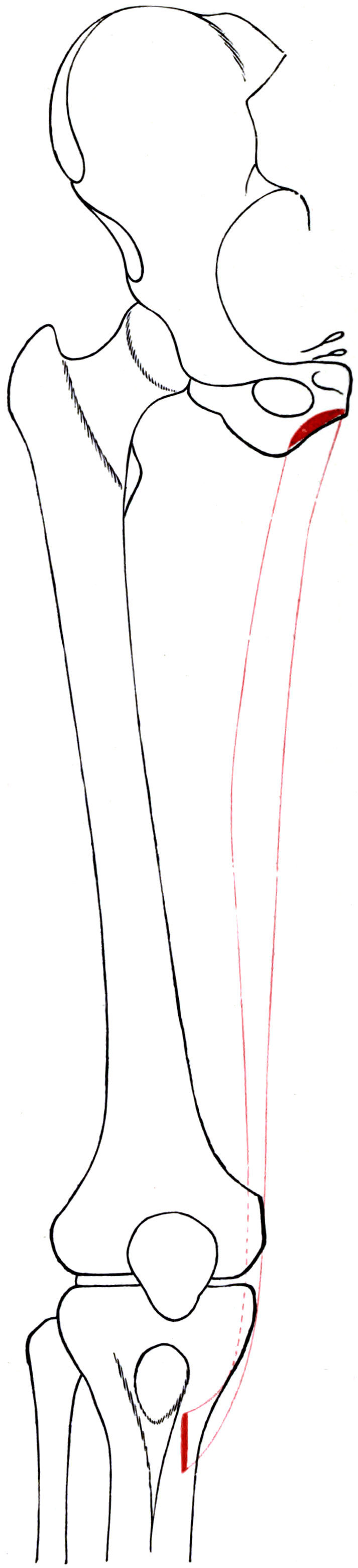
Attachments, Innervation and Relations
Proximal Origin
- Site: a thin, flat aponeurosis from the body and inferior ramus of the pubis and the adjacent ischiopubic ramus, immediately lateral to the symphysis.
- Character: the origin is broad and membranous, only about 2-3 cm wide, and it lies superficial to adductor brevis and adductor magnus.
- Palpable landmark: the pubic tubercle is the reference point for all gracilis measurements. The muscle's leading edge is roughly a fingerbreadth lateral to it.
Course
- Descends vertically along the medial thigh, narrowing progressively — it is broad proximally and becomes a narrow strap by the mid-thigh.
- The muscle belly ends at approximately the junction of the middle and distal thirds of the thigh, giving way to a long, thin, round-to-flat tendon.
- The tendon passes behind the medial femoral condyle, then turns anteriorly around it to reach the tibia.
Distal Insertion
- Site: the superomedial surface of the tibia, as the middle tendon of the pes anserinus, roughly 2-3 cm distal to the medial joint line and medial to the tibial tuberosity.
- Layers at the pes (superficial to deep): sartorius fascia, gracilis, semitendinosus, then the pes anserine bursa, then the superficial MCL, then bone.
- Order at the tibia: gracilis lies proximal and anterior to semitendinosus at the insertion, which is why the gracilis is the first tendon encountered when the sartorial fascia is opened.
Harvestable Dimensions
- Tendon length: approximately 20-26 cm of usable tendon, shorter than semitendinosus.
- Muscle length: typically 35-45 cm of total muscle length in an adult; for free transfer a segment of 12-18 cm is usually taken, tailored to the excursion required.
- Doubled gracilis diameter: roughly 5-6 mm — insufficient alone for an ACL, which is why it is combined with semitendinosus.
Get these three relationships right and the harvest is straightforward; get them wrong and you lose the flap.
- The dominant pedicle enters the deep surface about 8-10 cm distal to the pubic tubercle. Mark the pubic tubercle before you drape, and expect the pedicle in the proximal third.
- The obturator nerve branch enters slightly proximal to and deep to the pedicle. Find the nerve first, on the anterior surface of adductor brevis, by retracting adductor longus anteriorly.
- Take the nerve long. Dissect it proximally toward the obturator canal to gain 6-10 cm, so the coaptation sits outside the zone of injury and under no tension.
- Skin paddle only over the proximal third. Perforators to the distal skin are unreliable; an over-long paddle is the classic cause of distal skin necrosis.
- Ligate the minor distal pedicles deliberately — do not avulse them, as the resulting haematoma obscures the dissection.
Action and Biomechanics
Actions by Plane
- Hip: adduction, with a small contribution to flexion. Its contribution to total adduction torque is small, roughly a tenth of the adductor group's capacity.
- Knee: flexion, and internal rotation of the flexed tibia.
- As a pes tendon: contributes with sartorius and semitendinosus to the medial dynamic sling, a secondary restraint to valgus and to tibial external rotation.
Architecture
- Long, parallel fibres running almost the whole length of the muscle, with a small physiological cross-sectional area but a large excursion of roughly 10-12 cm.
- The trade-off: excursion, not power. A muscle with long parallel fibres moves a long way but generates modest force — which is exactly the profile required for a functioning free transfer that must pull a tendon or a commissure through a distance.
- Contrast with a pennate muscle such as adductor magnus: high force, short excursion, and useless as a functioning transfer for elbow flexion.
Biarticular Consequences
- Gracilis cannot shorten maximally at both hip and knee simultaneously. Hip abduction with knee extension places it on maximal stretch — the position that provokes pain in adductor-related groin pain and the position in which its tendon is most easily palpated.
- Its knee flexion moment arm is small compared with the hamstrings, so isolated gracilis loss is functionally silent at the knee.
What Happens When It Is Removed
- No measurable adduction deficit after unilateral harvest in the great majority of patients; the other adductors compensate completely.
- No measurable hip flexion or knee flexion deficit attributable to the gracilis alone.
- Sensory disturbance over the medial thigh may occur if the cutaneous branches of the obturator nerve are injured during proximal dissection.
- The reproducible deficit after a combined semitendinosus and gracilis harvest is the deep-flexion knee flexion weakness, and that is largely attributable to the semitendinosus.
The commonest technical failure in a functioning free muscle transfer is not thrombosis — it is setting the wrong resting length.
- A muscle produces maximal active force at its optimal sarcomere length. Set it too short and it has no force reserve; set it too long and it cannot shorten enough to complete the arc.
- The standard technique: before dividing the pedicle, place marking sutures at fixed intervals (commonly 5 cm) along the muscle in situ, at its natural resting length. After inset, re-tension the transferred muscle until those marks are the same distance apart. This restores the in-vivo sarcomere length reliably.
- For elbow flexion, the muscle is inset with the elbow in a defined position (commonly around 90-100 degrees of flexion) and tensioned so the marks are restored; too little tension gives a weak flexion, too much gives a flexion contracture.
- For facial reanimation, the vector and tension are set against the contralateral resting commissure position, with deliberate slight over-correction because the transferred muscle will relax.
- Reinnervation timing: a transferred muscle takes 3-6 months to show first contraction and 12-24 months to reach plateau function. Counsel the patient with those numbers.
Surface Anatomy and Examination
Palpation
- Proximal: with the hip abducted and the knee extended, the gracilis stands out as the most posterior and most superficial cord at the medial thigh root, immediately posterior to adductor longus. Adductor longus is the more prominent, more anterior cord at the groin — the two are easily distinguished by resisted adduction.
- Distal: with the patient supine in the figure-of-four position (hip flexed, abducted and externally rotated, knee flexed) and the knee flexed against resistance, two cords appear beneath the sartorial fascia at the posteromedial knee. The anterior, thinner, flatter cord is the gracilis; the posterior, thicker, rounder cord is the semitendinosus.
- At the pes: roughly 2-3 cm distal to the medial joint line and 1-2 cm medial to the tibial tuberosity.
Named Clinical Tests
- How to perform
- Supine, knees flexed 45 degrees, patient squeezes the examiner's fist between the knees
- Positive finding
- Pain at the pubic origin or medial thigh
- What it means
- Adductor-related groin pain, including gracilis origin tendinopathy
- False positives
- Pubic-related groin pain, osteitis pubis, inguinal-related groin pain
- How to perform
- Supine, hip abducted with the knee extended, then knee flexed
- Positive finding
- Pain relieved by knee flexion
- What it means
- Localises to a biarticular muscle — gracilis rather than a uniarticular adductor
- False positives
- Hamstring or medial capsular pain
- How to perform
- History and examination in a patient with an obturator hernia
- Positive finding
- Medial thigh and knee pain relieved by hip flexion, provoked by extension and internal rotation
- What it means
- Obturator nerve compression by an obturator hernia
- False positives
- Hip osteoarthritis; lumbar radiculopathy
- How to perform
- Prone, knee at 90 degrees, foot turned in
- Positive finding
- Weakness or pain
- What it means
- Medial dynamic sling involvement (gracilis, semitendinosus, semimembranosus)
- False positives
- Posterior horn medial meniscal tear
- How to perform
- Squeeze test repeated at three hip flexion angles, measured with a sphygmomanometer cuff
- Positive finding
- Reduced force compared with the contralateral side or with normative values
- What it means
- Objective monitoring of adductor-related groin pain and return to play
- False positives
- Pain inhibition rather than true weakness
- How to perform
- After free gracilis transfer, ask for the donor nerve's natural action
- Positive finding
- Visible or palpable contraction of the transferred muscle
- What it means
- Successful reinnervation; expect first contraction at 3-6 months
- False positives
- Tendon tenodesis effect mimicking active motion
Grading and Interpretation
- Adduction strength is graded 0-5 but is insensitive to isolated gracilis loss.
- After a functioning free transfer, grading uses the British Medical Research Council motor scale (M0-M5) applied to the new function: for elbow flexion, M3 (movement against gravity) is the minimum useful result and M4 (against resistance) the goal.
- For facial reanimation, outcome is graded by symmetry of the smile at rest and on animation, commissure excursion in millimetres, and validated patient-reported measures.
Pitfalls
- Do not mistake adductor longus for gracilis at the groin — adductor longus is anterior and much more prominent.
- Medial thigh numbness after harvest is from the obturator cutaneous branches, not the saphenous nerve.
- A palpable gap at the pes after harvest is expected and does not indicate a complication.
Complications
Donor-Site Morbidity
- Adduction weakness is minimal and rarely measurable after unilateral harvest, because the remaining adductors are far larger.
- Medial thigh numbness from injury to the cutaneous branches of the obturator nerve.
- Seroma and haematoma in the medial thigh dead space — the commonest donor-site problem after a muscle harvest. Reduced by careful haemostasis of the minor pedicles, quilting sutures and a drain.
- Wound problems in the groin — a moist, mobile, contaminated area; infection and dehiscence rates are higher than at most donor sites.
- Lymphocele if the dissection strays into the femoral triangle.
- Scar along the medial thigh; a two-incision technique with tunnelling gives a better cosmetic result.
Neurological
- Obturator nerve injury during proximal dissection — with all four adductors at risk if the main trunk rather than the gracilis branch is damaged.
- Infrapatellar branch of the saphenous nerve injury at the pes during a tendon harvest — the same problem as with semitendinosus, prevented by an oblique incision.
Free-Transfer Specific
- Flap failure from arterial or venous thrombosis — the standard microvascular risk, mitigated by careful anastomosis, appropriate recipient vessel choice and vigilant monitoring.
- Failure of reinnervation — from a tense coaptation, a coaptation inside the zone of injury, an inadequate axon source, or too long a delay before transfer.
- Incorrect tension — a flexion contracture if over-tensioned, a weak or absent excursion if under-tensioned. Prevented by the in-situ marking sutures.
- Bulkiness at the recipient site — particularly in facial reanimation, where a full-width gracilis produces an obviously fat cheek. Take a narrow slip and consider secondary debulking.
- Distal skin paddle necrosis — from an over-long skin island.
- Synkinesis or unnatural movement — the intercostal-neurotised muscle contracts with respiration and the masseteric-neurotised smile requires biting; both need dedicated motor retraining.
Preventing Each
- Mechanism
- Blind dissection of the deep surface in the proximal third
- Prevention
- Expose the pedicle under direct vision before mobilising the muscle
- Mechanism
- Short nerve, tense coaptation, or coaptation in scar
- Prevention
- Dissect the obturator branch proximally for 6-10 cm; coapt outside the zone of injury
- Mechanism
- Guessing resting length after the muscle is free
- Prevention
- Place 5 cm marking sutures in situ before dividing the pedicle
- Mechanism
- Skin paddle beyond the reliable perforator zone
- Prevention
- Confine the paddle to the proximal third
- Mechanism
- Large dead space after muscle removal
- Prevention
- Quilting sutures, meticulous haemostasis, closed suction drain
- Mechanism
- Vertical incision at the pes
- Prevention
- Oblique incision parallel to the nerve
Clinical Relevance
As a Free Tendon Graft
- ACL reconstruction: combined with semitendinosus as the classic four-strand hamstring graft. Doubled gracilis alone is only 5-6 mm and is never adequate on its own.
- MPFL reconstruction: the standard graft in most units. A doubled gracilis is more than strong enough — the native MPFL fails at around 200 N, whereas a doubled gracilis is several times stronger, so graft strength is never the limiting factor. Tunnel position and tension are.
- MCL and posteromedial corner, posterolateral corner, PCL augmentation and multiligament reconstruction.
- Hand and wrist: ligament reconstruction where a longer graft than the palmaris longus is required.
- Ankle and foot: lateral ligament, spring ligament and deltoid reconstruction; peroneal and tibialis posterior tendon reconstruction.
- Acromioclavicular joint reconstruction and coracoclavicular ligament reconstruction.
Harvest Technique
- Position: supine, hip flexed, abducted and externally rotated, knee flexed 90 degrees (figure-of-four).
- Incision: a 2-3 cm oblique incision over the pes, roughly 2-3 fingerbreadths distal to the medial joint line, oriented parallel to the infrapatellar branch of the saphenous nerve.
- Identify the sartorial fascia; the gracilis is the more proximal and thinner cord palpable through it, semitendinosus the more distal and thicker.
- Incise the fascia along the superior border of the gracilis, deliver the tendon with a right-angled clamp, and whip-stitch it.
- Release the fascial expansions and the dense investing fascia on the deep surface with firm distal traction, sharply and under direct vision. The gracilis has fewer and less substantial accessory attachments than the semitendinosus, whose accessory insertion was found in 77 per cent of cadaveric knees, but they still misdirect a stripper.
- Detach the tibial insertion subperiosteally, then advance the closed-loop stripper coaxially with steady traction. Never force it.
- Prepare and measure the graft; close the sartorial fascia.
Practical Notes
- The gracilis strips more easily than the semitendinosus because it has fewer accessory bands — but the same discipline applies.
- Sparing the gracilis is increasingly favoured in young patients, using a quadrupled or quintupled semitendinosus alone, to preserve a future graft source and to reduce donor deficit.
- Regeneration cannot be assumed. In a two-year MRI study, both the semitendinosus and gracilis had regenerated in only 35 per cent of patients, and donor muscle loss was greater where the tendon had not regenerated — another argument for sparing the gracilis where the semitendinosus alone will suffice.
Distal skin necrosis.
- Reliable musculocutaneous perforators are confined to the proximal third, within roughly 8-12 cm of the pubic tubercle.
- A skin paddle designed over the distal muscle looks generous on the table and dies at 72 hours.
- Keep the paddle proximal and small, or take the muscle alone with a split-thickness graft.
Failure to reinnervate.
- Dividing the obturator branch at the muscle leaves 2-3 cm of nerve and forces a coaptation inside the zone of injury.
- Dissect the branch proximally toward the obturator canal to gain 6-10 cm.
- A tension-free coaptation, outside scarred tissue, is worth more than any suture technique.
Surgical Relevance
Structures at Risk with Distances
- Location relative to a landmark
- Enters the deep surface approximately 8-10 cm distal to the pubic tubercle
- How to protect it
- Mark the tubercle before draping; expose the proximal third first
- Location relative to a landmark
- On the anterior surface of adductor brevis, entering gracilis 8-12 cm distal to the pubic tubercle
- How to protect it
- Retract adductor longus anteriorly to expose it; take it long
- Location relative to a landmark
- Subcutaneous, immediately anterior to gracilis in the distal medial thigh
- How to protect it
- Identify at the distal incision; retract rather than divide
- Location relative to a landmark
- Crosses the pes transversely, 1-4 cm distal to the medial joint line
- How to protect it
- Oblique incision parallel to the nerve; blunt subcutaneous dissection
- Location relative to a landmark
- Deep to adductor brevis at the level of the pedicle origin
- How to protect it
- Dissect the pedicle under vision; avoid blind clamping
- Location relative to a landmark
- Deep and anterior, beneath sartorius in the middle third of the thigh
- How to protect it
- Stay in the plane superficial to adductor longus and magnus
- Location relative to a landmark
- Deep to the pes tendons, separated by the pes anserine bursa
- How to protect it
- Preserve its tibial insertion when raising a periosteal cuff
The Medial Approach to the Thigh
- Incision: longitudinal along the medial thigh, one fingerbreadth posterior to the palpable adductor longus — or, for a cosmetically superior free harvest, two short transverse incisions (proximal for the pedicle and nerve, distal for the tendon) with subcutaneous tunnelling between them.
- Superficial dissection: incise deep fascia to expose gracilis, the most superficial and posterior muscle in the field.
- Key interval: develop the plane between adductor longus (anterior) and gracilis (posterior); retracting adductor longus anteriorly exposes adductor brevis and, on its anterior surface, the anterior division of the obturator nerve with the branch to gracilis.
- Pedicle: found on the deep surface of gracilis in the proximal third, entering from the direction of the profunda.
- Uses of this approach: free and pedicled gracilis harvest, adductor tenotomy, obturator neurectomy, and access to the medial femur and the lesser trochanter.
Sequence of a Free Functioning Gracilis Harvest
- Mark the pubic tubercle, the medial femoral condyle and the line between them — the muscle lies along this line.
- Proximal incision; expose gracilis and confirm identity by tracing it distally (adductor longus is anterior and does not cross the knee).
- Identify the pedicle on the deep surface in the proximal third.
- Identify the obturator branch just proximal and deep to the pedicle; dissect it proximally to maximum length.
- Distal incision at the pes; detach the tendon subperiosteally and deliver the muscle proximally.
- Place marking sutures at 5 cm intervals along the muscle in situ, at resting length, before dividing anything.
- Ligate the minor pedicles; divide the muscle to the required length.
- Divide the pedicle last, immediately before transfer, to minimise ischaemia time.
- Inset at the recipient site with the marking sutures restored to their original intervals.
- Anastomose artery and vein, then perform the nerve coaptation without tension.
Contraindications and Cautions
- Prior groin surgery or radiotherapy compromising the pedicle.
- Significant peripheral vascular disease — a gracilis harvest is not dangerous to the limb, but the microvascular anastomosis may fail.
- Bilateral harvest is possible and is done in the Doi double free muscle technique, but should not be undertaken casually.
- Lymphoedema risk if the groin dissection strays into the femoral triangle lymphatics.
Guidelines, Registries & Global Practice
Anatomical Variation Across Populations
- The gracilis pedicle position is remarkably constant across cadaveric series from European, North American, South Asian and East Asian populations, entering the proximal third of the muscle. Reported distances from the pubic tubercle vary with limb length rather than with population, which is why measuring from a bony landmark rather than in absolute centimetres is the safer habit.
- Muscle length and width scale with stature, and reported gracilis dimensions are consistently smaller in South and East Asian series. For facial reanimation this is an advantage — less debulking is required.
- Accessory obturator nerve, present in roughly one in ten to one in five people, supplies pectineus and occasionally contributes to the hip joint; it does not alter gracilis innervation but matters in obturator neurectomy.
- Pedicle origin variation: the adductor branch arises from the medial circumflex femoral artery in most limbs, but directly from the profunda femoris or the superficial femoral artery in a minority. Expect the variant rather than being surprised by it.
Differences in Described Technique
- Position on gracilis use
- Gracilis is the reference functioning free muscle transfer for both brachial plexus and facial reanimation; donor morbidity is accepted as negligible.
- Position on gracilis use
- Double free gracilis transfer to restore prehension after complete brachial plexus avulsion, using spinal accessory and intercostal donors.
- Position on gracilis use
- Two-stage cross-face nerve graft followed by free gracilis, prioritising a spontaneous, emotionally driven smile.
- Position on gracilis use
- Single-stage masseteric neurotisation for a stronger, faster, more reliable smile, accepting the need for retraining; dual innervation as a hybrid.
- Position on gracilis use
- Gracilis with semitendinosus as the standard four-strand hamstring graft; gracilis alone as the standard MPFL graft.
- Position on gracilis use
- Quadrupled or quintupled semitendinosus alone for ACL, preserving the gracilis as a future graft source and reducing donor deficit.
Practice Signals
- National ligament registries report hamstring autograft (semitendinosus with or without gracilis) as the most-used ACL graft worldwide, and demonstrate that graft diameter influences revision risk — the practical reason the gracilis is added to the semitendinosus.
- There is no international registry for functioning free muscle transfer; the evidence base is single-centre series and systematic reviews of them. The consistent signals are that younger patients, shorter denervation intervals and stronger motor donors give better results.
- For facial reanimation, comparative series consistently show that masseteric neurotisation gives greater commissure excursion while cross-face grafting gives more spontaneous, emotionally driven movement, which is why dual innervation has gained ground.
High- and Limited-Resource Practice
- Well-resourced settings: microvascular free transfer with intraoperative nerve stimulation, dual innervation techniques, and formal motor retraining programmes.
- Limited-resource settings: the pedicled gracilis flap requires no microsurgery and remains a highly effective option for perineal and ischial reconstruction. For the knee, the gracilis tendon graft needs only a stripper and sutures. Where microsurgical facilities are unavailable, regional muscle transfers (such as a Steindler flexorplasty or a pedicled latissimus transfer) remain the alternative for elbow flexion, and static slings remain an option for facial symmetry at rest.
- The universal point: gracilis is the donor of choice precisely because its anatomy is constant and its loss costs the patient nothing.
MCQ Practice Points
Q: What innervates gracilis? A: The anterior division of the obturator nerve, L2-L4. It is the only pes tendon supplied by the obturator nerve.
Q: Which adductor crosses two joints? A: Gracilis. That is why it flexes the knee and internally rotates the tibia, and why its stretch pain is relieved by knee flexion.
Q: Where does the dominant pedicle enter gracilis? A: The deep surface, approximately 8-10 cm distal to the pubic tubercle, from the adductor branch of the medial circumflex femoral artery.
Q: What Mathes-Nahai type is gracilis? A: Type II — one dominant pedicle plus minor pedicles. This is why the whole muscle survives on the proximal pedicle alone.
Q: What is the relationship of the obturator branch to the vascular pedicle? A: The nerve enters slightly proximal to and deep to the pedicle. Find the nerve on adductor brevis by retracting adductor longus anteriorly.
Q: Where can a reliable skin paddle be raised on a gracilis flap? A: Over the proximal third only, within roughly 8-12 cm of the pubic tubercle. Distal paddles necrose.
Q: How is resting length restored in a functioning free muscle transfer? A: Marking sutures placed at fixed intervals (usually 5 cm) along the muscle in situ, before the pedicle is divided, and restored at inset.
Q: Why can a nerve transfer not be used for elbow flexion 16 months after a plexus avulsion? A: Motor endplates degenerate irreversibly after about 12 months of denervation. The target muscle is fibrotic; a functioning free muscle transfer supplies a new one.
Q: Would you use a pedicled gracilis for proximal tibial cover? A: No. Its arc reaches the perineum, ischium and groin, not the knee. The medial gastrocnemius is the flap for the proximal third of the tibia.
Q: Does gracilis regenerate after tendon harvest? A: Not reliably. A two-year MRI study found both the semitendinosus and gracilis had regenerated in only 35 per cent of patients, and donor muscle loss was greater where regeneration failed. This is one argument for sparing the gracilis and using a quadrupled semitendinosus alone in a young patient.
Exam Viva Scenarios
Practise clinical reasoning and management decisions out loud
“A 26-year-old motorcyclist presents 16 months after a complete C5-T1 brachial plexus avulsion. He has a flail, insensate arm with a Horner sign. He wants his elbow to bend. What can you offer and why?”
“A 28-year-old woman has had a complete left facial palsy for three years after acoustic neuroma surgery. The nerve was sacrificed. She wants to smile. What are the anatomical and technical steps?”
“You are reconstructing the MPFL in a 17-year-old with recurrent lateral patellar dislocation. Why gracilis, and what will determine your result?”
Core Anatomy
- Origin: body and inferior ramus of pubis, thin aponeurosis
- Insertion: middle tendon of the pes anserinus
- Nerve: anterior division of obturator, L2-L4
- Only biarticular adductor; most superficial and medial
Flap Numbers
- Dominant pedicle 8-10 cm distal to the pubic tubercle, deep surface
- Pedicle length 6-8 cm; artery 1.5-2.5 mm; two venae comitantes
- Obturator branch enters just proximal and deep to the pedicle
- Mathes-Nahai type II; skin paddle over the proximal third only
Free Transfer
- Brachial plexus: spinal accessory or intercostals; goal M3-M4 elbow flexion
- Facial: cross-face sural graft then gracilis at 9-12 months, or masseteric single stage
- Tension set by 5 cm marking sutures placed in situ
- First contraction 3-6 months; plateau 18-24 months
Tendon Graft
- Length 20-26 cm; doubled diameter only 5-6 mm
- ACL: with semitendinosus as a four-strand graft
- MPFL: the standard graft; tunnel position beats graft strength
- Regeneration is not reliable — both tendons regenerated in only 35 per cent at 2 years
Evidence Base
Free Gracilis Muscle Transplantation, with Microneurovascular Anastomoses, for the Treatment of Facial Paralysis
- First clinical description of free muscle transplantation by microneurovascular anastomosis
- Two cases of free gracilis transfer for dynamic reconstruction of facial paralysis are reported
- Follow-up included electromyography, light microscopy and electron microscopy
- Established that a transferred muscle can be reinnervated and produce voluntary movement
Restoration of Prehension with the Double Free Muscle Technique Following Complete Avulsion of the Brachial Plexus
- 32 patients with complete brachial plexus avulsion underwent double free gracilis transfer; 26 were followed for at least 24 months
- The first muscle, neurotised by the spinal accessory nerve, restored elbow flexion and finger extension
- The second muscle, neurotised by the fifth and sixth intercostal nerves, restored finger flexion
- Satisfactory elbow flexion was restored in 25 of 26 patients (96 per cent) and satisfactory prehension in 17 (65 per cent)
- 14 patients (54 per cent) could position the hand in space and use it for two-handed tasks
Analysis of 100 Cases of Free-Muscle Transplantation for Facial Paralysis
- 100 free muscle transplantations to the face in 93 patients by a single surgeon between 1981 and 1993
- The gracilis was used in 63 cases and the pectoralis minor in 34
- Onset of muscle function averaged 21.6 plus or minus 9.1 weeks postoperatively, with a range of 6 to 48 weeks
- 80 per cent of patients achieved a moderate or better result on independent rating, and 94 per cent improved
- There was a trend toward earlier onset of function and higher aesthetic rating in young female patients
Classification of the Vascular Anatomy of Muscles: Experimental and Clinical Correlation
- Described five patterns of muscle circulation based on studies of the vascular anatomy of muscle
- Correlated each vascular pattern with arc of rotation, skin territory and the feasibility of distally based flaps
- Also correlated the patterns with suitability for microvascular composite tissue transplantation and with muscle-delay experimental models
- The classification was designed to guide both the choice and the design of muscle and musculocutaneous flaps
Anatomic Considerations in Harvesting the Semitendinosus and Gracilis Tendons and a Technique of Harvest
- Cadaveric dissection of fresh-frozen adult knees to define the anatomy relevant to hamstring tendon harvest
- An accessory insertion of the semitendinosus tendon was present in 77 per cent of knees and should be identified and transected to avoid tendon damage at harvest
- The tendons are ensheathed in a dense fascial layer that may impede tendon stripping
- The superficial medial collateral ligament lies deep to the tendons and should not be disturbed
- Knee flexion may reduce the risk of injury to the saphenous nerve as it crosses the gracilis tendon
Graft Size and Patient Age Are Predictors of Early Revision After ACL Reconstruction with Hamstring Autograft
- 256 of 338 consecutive primary hamstring autograft ACL reconstructions evaluated
- Revision was performed in 1.7 per cent of grafts greater than 8 mm, 6.5 per cent of 7.5 to 8 mm grafts and 13.6 per cent of grafts 7 mm or less
- Revision rate was 0.7 per cent in patients aged 20 years or over versus 14.3 per cent in those under 20
- 16 of 18 revisions occurred in patients under 20 years with grafts 8 mm or less, a revision rate of 16.4 per cent in that group
- Age under 20 years and decreased graft size were independent predictors of early revision