Two Muscles in One: The Adductor and the Hamstring
- Adductor magnus has two functionally and neurologically distinct parts: the adductor (pubofemoral) part supplied by the posterior division of the obturator nerve, and the hamstring (ischiocondylar) part supplied by the tibial division of the sciatic nerve.
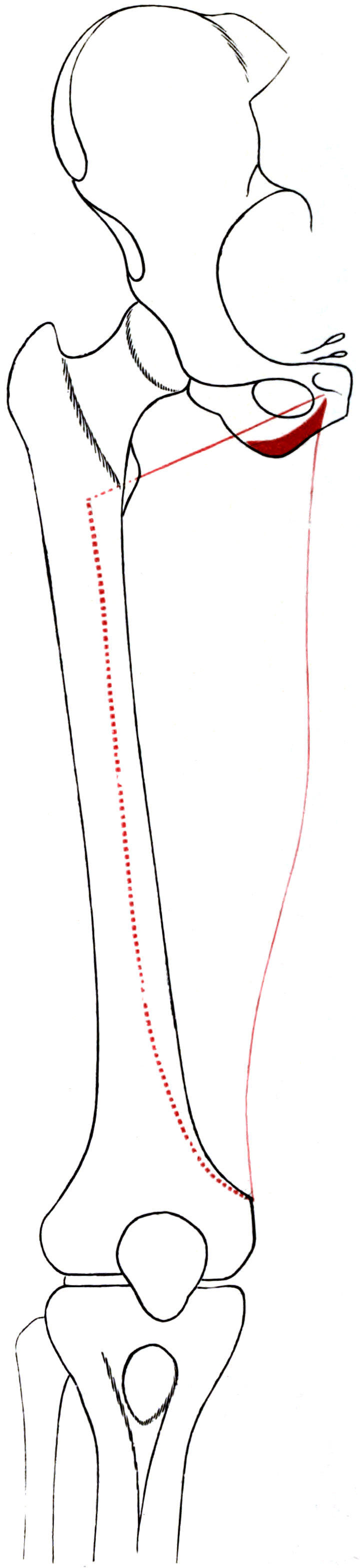
- Origin is the inferior pubic ramus, the ischial ramus and the inferolateral ischial tuberosity; insertion sweeps along the gluteal tuberosity, the whole medial lip of the linea aspera and the medial supracondylar line, ending as a rounded tendon on the adductor tubercle.
- It is the largest muscle of the adductor group and, through its ischiocondylar part, a genuine hip extensor whose extension moment arm increases as the hip flexes.
- The adductor hiatus is the opening in its aponeurotic insertion through which the femoral artery and vein pass to become the popliteal vessels — a fixed tethering point that makes the artery vulnerable in knee dislocation and displaced distal femoral fractures.
- The adductor tubercle, its distal tendinous insertion, is the landmark for the femoral origin of the medial patellofemoral ligament, which lies between the adductor tubercle and the medial epicondyle.
- “The dual innervation of adductor magnus is the classic exam point: it explains why hip adduction is weakened but never abolished by an obturator nerve lesion, and why the ischiocondylar part behaves like a hamstring.
- “The ischiocondylar part is the hamstring that does not cross the knee — it arises from the ischial tuberosity, is supplied by the tibial division of the sciatic nerve, and extends the hip, but has no attachment below the knee.
- “The popliteal artery is tethered at the adductor hiatus proximally and the fibrous arch of the soleus distally, which is why it is stretched or torn in knee dislocation — and why a palpable pulse never excludes an intimal injury.
- “Trace the adductor magnus tendon to the adductor tubercle to find the MPFL femoral origin between the tubercle and the medial epicondyle, then confirm the position fluoroscopically rather than trusting palpation.
Overview
The adductor magnus is the largest muscle of the adductor compartment and one of the largest muscles in the body. It is a triangular fan whose apex is at the pubis and ischium and whose base sweeps almost the entire length of the medial femur, from the gluteal tuberosity proximally to the adductor tubercle distally.
Two muscles in one. It is best understood not as one muscle but as two, and this is the point examiners return to. The anterior, more horizontally orientated adductor (pubofemoral) part arises from the pubic and ischial rami and inserts into the linea aspera; the posterior, vertically orientated hamstring (ischiocondylar) part arises from the ischial tuberosity and inserts by a rounded tendon on the adductor tubercle. The two differ in origin, orientation, action, innervation and embryological derivation, so the muscle is a hybrid of the adductor and hamstring groups, and the adductor hiatus sits in the aponeurosis where the two parts meet.
- Origin
- Inferior pubic ramus, ischial ramus
- Orientation
- Oblique to horizontal
- Insertion
- Gluteal tuberosity, medial lip of linea aspera, medial supracondylar line
- Nerve
- Posterior division of the obturator nerve (L2, L3, L4)
- Origin
- Inferolateral ischial tuberosity
- Orientation
- Vertical
- Insertion
- Rounded tendon on the adductor tubercle
- Nerve
- Tibial division of the sciatic nerve (L4, L5, S1)
Why there are two. The ischiocondylar part shares its ischial tuberosity origin and its innervation with the hamstrings and derives from the same flexor (posterior) muscle mass; the adductor part derives from the adductor (medial) mass. The muscle is a fusion of two developmental units, which is why one half of it is supplied exactly like the true hamstrings.
Attachments
Origin. The adductor part arises from a small area on the inferior (ischiopubic) ramus of the pubis and the ramus of the ischium, deep to the origins of adductor brevis and gracilis. The hamstring part arises from the inferolateral aspect of the ischial tuberosity, immediately adjacent to and partly blended with the origins of the semimembranosus and the conjoint tendon of biceps femoris and semitendinosus.
Why the ischial origin matters. Because the ischiocondylar origin blends with the hamstring origins, the adductor magnus is involved in proximal hamstring pathology: an ischial tuberosity avulsion or apophyseal injury frequently involves it, and in any posterior approach to the ischium its origin is encountered alongside the hamstring origins and must be recognised.
Insertion. A continuous sweep down the medial femur:
- Gluteal tuberosity, the rough area on the posterior proximal femur medial to the vastus lateralis origin, and a landmark in posterior proximal femoral exposure
- The entire length of the medial lip of the linea aspera
- Medial supracondylar line
- A rounded tendon onto the adductor tubercle, the distal ischiocondylar portion
This is the longest femoral insertion of any muscle, spanning almost the whole shaft.
Innervation
Two nerves, two parts. The adductor part is supplied by the posterior division of the obturator nerve, root values L2, L3, L4. The hamstring part is supplied by the tibial division of the sciatic nerve, root values commonly given as L4, L5, S1, the same territory that supplies the true hamstrings. Adductor magnus is, with the pectineus, one of the two dually innervated muscles of the adductor group, and its dual supply comes from two different nerves entirely.
The obturator nerve. It arises from the anterior divisions of the ventral rami of L2-L4, descends on the medial border of the psoas, crosses the pelvic brim behind the common iliac vessels, runs along the lateral pelvic wall above the obturator vessels, and exits through the obturator canal approximately 2-3 cm inferolateral to the pubic tubercle. Its divisions are separated by the obturator externus in the pelvis and canal, and by the adductor brevis in the thigh, so the posterior division lies posterior to the adductor brevis on the anterior surface of the adductor magnus.
What the posterior division supplies. The adductor part of adductor magnus, the obturator externus and variably the adductor brevis, and an articular branch to the knee that descends through the adductor hiatus with the popliteal artery to the posterior capsule. That knee branch is the anatomical basis of referred medial knee pain in hip pathology, and the reason a child with a slipped upper femoral epiphysis, Perthes disease or a septic hip presents complaining of the knee. Never accept medial knee pain at face value in a child.
The sciatic branch. The branch to the ischiocondylar part arises from the tibial division of the sciatic nerve in the posterior thigh, alongside the branches to semimembranosus, semitendinosus and the long head of biceps femoris, and enters the posterior surface of the ischiocondylar portion.
Why adduction survives an obturator lesion. A complete obturator nerve lesion denervates adductor longus, brevis, gracilis and the adductor part of magnus, but the ischiocondylar part retains its sciatic supply and the pectineus retains its femoral branch. Adduction is therefore weakened, not abolished, which is the answer examiners want. An obturator nerve block or neurectomy cannot abolish adduction for the same reason: state this explicitly when consenting for neurectomy, and to the urologist or anaesthetist relying on a block to abolish the adductor jerk during transurethral bladder resection, where a residual adductor twitch may persist despite an apparently successful block.
The MRI denervation pattern. In an obturator lesion, oedema and later fatty atrophy affect adductor longus, brevis, gracilis and the adductor part of magnus, with relative sparing of the ischiocondylar part and of the pectineus. In a tibial division sciatic lesion the pattern for adductor magnus is the reverse, alongside the true hamstrings. The selective pattern is a reliable localiser of an obturator lesion and should prompt a search for a pelvic cause.
- Denervated
- Adductor longus, brevis, gracilis, adductor part of magnus
- Spared
- Pectineus (femoral branch), ischiocondylar part of magnus (sciatic)
- Clinical picture
- Weakened but not abolished adduction; small patch of distal medial thigh numbness
- Denervated
- Adductor part of magnus, obturator externus, variably adductor brevis
- Spared
- Adductor longus, gracilis, pectineus, ischiocondylar magnus
- Clinical picture
- Subtle adduction weakness; loss of the knee articular branch
- Denervated
- Ischiocondylar part of magnus plus the true hamstrings
- Spared
- Adductor part of magnus, adductor longus, brevis, gracilis, pectineus
- Clinical picture
- Weak hip extension AND weak knee flexion
- Denervated
- All obturator-supplied muscles plus the quadriceps
- Spared
- Nothing in the obturator territory
- Clinical picture
- Weak adduction AND weak knee extension — localises proximally; search for a retroperitoneal cause
Localising a lesion by root value. Hip adduction is L2/L3/L4 (obturator) with an L4/L5/S1 (sciatic) contribution through the ischiocondylar part; hip extension is L5/S1 predominantly (gluteus maximus, hamstrings, ischiocondylar adductor magnus); distal medial thigh sensation is the obturator cutaneous branch, from the anterior division. Weak adduction with a medial thigh sensory patch localises to the obturator nerve; add quadriceps weakness and the lesion is proximal, in the lumbar plexus, the L2-L4 roots, or a retroperitoneal haematoma, abscess or tumour.
The other dually innervated muscles. Pair adductor magnus with the pectineus (obturator anterior division plus femoral), the biceps femoris (long head tibial division, short head common peroneal division) and, in the upper limb, flexor digitorum profundus (median and ulnar).
Blood Supply
Sources. The profunda femoris dominates; the others contribute near the origin, the ischial tuberosity and the hiatus.
- Profunda femoris artery: muscular branches, the dominant supply
- Perforating branches of the profunda femoris: typically three to four, which pierce the adductor magnus on their way from the anterior to the posterior compartment, supply the muscle and give nutrient branches to the femur
- Obturator artery, posterior branch: the proximal muscle near its pubic and ischial origin
- Medial circumflex femoral artery: proximal supply, and the vessel of concern in the medial approach to the hip
- Inferior gluteal artery: the ischiocondylar part near the ischial tuberosity, alongside the hamstring origins
- Femoral artery and, distally, the descending genicular artery: near the hiatus and the adductor tubercle
The perforators. Because they pass through the muscle to reach the posterior compartment, dividing the adductor magnus blindly bleeds from a vessel that then retracts into muscle, or into the posterior compartment where the sciatic nerve lies.
The descending genicular artery. It arises from the femoral artery within the adductor canal, just proximal to the adductor hiatus, and gives muscular, saphenous and articular branches. It is the pedicle of the medial femoral condyle vascularised corticoperiosteal flap, so in a subvastus exposure it is identified and ligated deliberately, and preserved if such a flap may be needed.
Relations and the Adductor Canal
Anterior. Adductor brevis proximally and adductor longus distally; the posterior division of the obturator nerve on its anterior surface; the profunda femoris artery and vein with their perforating branches, which pierce the muscle; and distally the adductor canal contents against its posteromedial surface, where the muscle forms the distal posteromedial wall of the canal.
Posterior. The true hamstrings, semimembranosus, semitendinosus and biceps femoris, and the sciatic nerve in the posterior compartment. The muscle forms the anterior wall of the posterior compartment and the floor on which the hamstrings and the sciatic nerve lie.
Medial and distal. Medially the gracilis, the most medial strap and the only biarticular adductor, and the great saphenous vein in the subcutaneous plane. Distally the adductor tubercle, with the MPFL femoral origin between it and the medial epicondyle; the medial epicondyle and superficial MCL origin distal and anterior; the medial head of gastrocnemius arising just posterior to the adductor tubercle region; and the popliteal fossa contents beyond the hiatus.
The adductor canal. It runs from the apex of the femoral triangle, where the medial border of sartorius crosses the medial border of adductor longus, to the adductor hiatus, which is its distal boundary.
- Anterolateral wall: vastus medialis
- Posteromedial wall: adductor longus proximally, adductor magnus distally
- Roof: the vastoadductor membrane, deep to the sartorius
- Contents: femoral artery, femoral vein, saphenous nerve, nerve to vastus medialis, and the descending genicular artery arising within it
Action and Biomechanics
- Principal action
- Hip adduction; hip flexion in extension, extension in deep flexion
- Line of pull
- Oblique to horizontal, pubic and ischial rami to linea aspera
- Nerve
- Obturator, posterior division (L2-L4)
- Principal action
- Hip extension, with adduction
- Line of pull
- Vertical, ischial tuberosity to adductor tubercle
- Nerve
- Tibial division of sciatic (L4-S1)
- Principal action
- The largest adductor; stabilises the pelvis in single-leg stance
- Line of pull
- Fan-shaped, spanning the whole medial femur
- Nerve
- Dual
- Principal action
- Contributes to rotation, direction depending on hip position
- Line of pull
- Variable
- Nerve
- Dual
The hamstring that does not cross the knee. The ischiocondylar part has a vertical line of pull from the ischial tuberosity to the adductor tubercle, which is a hip extension vector, and it shares its origin, its innervation and its embryological derivation with the true hamstrings. It extends the hip but has no attachment below the knee, and so has no knee flexion action.
Be precise about magnitude. In vivo measurement of hip extensor moment arms found that in the anatomical position the adductor magnus moment arm averaged about 15 mm, against roughly 61 mm for the hamstrings and 79 mm for the gluteus maximus. Its moment arm then increased with hip flexion up to about 75 degrees before declining, whereas the gluteus maximus moment arm decreased as the hip flexed and the hamstrings peaked at around 35 degrees.
The clinical corollary. The adductor magnus contributes relatively more to hip extension out of a flexed hip, in the drive phase of a deep squat, rising from a low seat or accelerating from a crouched start, precisely where the gluteus maximus is at its mechanical worst. Combined with its large cross-sectional area, that makes it functionally relevant in exactly those tasks. Do not overstate it as the second or third strongest hip extensor in the anatomical position; its advantage is positional.
Architecture and moment arms. The muscle is fan-shaped and multipennate, and its portions have different moment arms: the more horizontal proximal fibres favour adduction, the vertical distal fibres favour extension. The adduction moment arm increases with hip abduction, which is why the muscle is loaded eccentrically at the extremes of abduction in change-of-direction sport.
Gait. The adductor group is active in loading response and pre-swing, contributing to pelvic stabilisation and to the stance-to-swing transition. The ischiocondylar part is additionally active in terminal swing and early stance, working with the hamstrings to decelerate the limb and extend the hip.
Synergists and antagonists.
- Adduction synergists: adductor longus and brevis, pectineus, gracilis, inferior gluteus maximus fibres, quadratus femoris
- Hip extension synergists: gluteus maximus, semimembranosus, semitendinosus, long head of biceps femoris
- Antagonists: gluteus medius and minimus and tensor fascia lata (abduction); iliopsoas, rectus femoris and sartorius (flexion)
Surface Anatomy and Examination
Finding the adductor tubercle. Palpate the medial epicondyle, then move proximally and slightly posteriorly to the small prominence at the distal end of the medial supracondylar line, where the rounded adductor magnus tendon inserts. It is best appreciated with the knee slightly flexed and the muscle relaxed. The tendon itself is palpable as a firm cord running to the tubercle in the distal medial thigh, particularly with resisted hip extension in slight abduction.
The rest of the muscle. The bulk is the deep posteromedial mass of the thigh, palpable between the hamstrings posteriorly and the adductor longus and gracilis anteriorly and medially. The ischial tuberosity is palpable with the hip flexed, and the ischiocondylar origin blends with the hamstring origins there. The adductor hiatus is not directly palpable, but its level should be known.
- How to perform
- Prone or supine with the hip slightly abducted; resist hip extension with the knee extended
- Positive finding
- Pain or weakness in the posteromedial thigh
- What it means
- Loads the ischiocondylar (hamstring) part of adductor magnus preferentially
- False positives
- Hamstring pathology; ischial tuberosity enthesopathy; lumbar referred pain
- How to perform
- Supine; squeeze the examiner's fist or a dynamometer between the knees at 0, 45 and 90 degrees of hip flexion
- Positive finding
- Pain and/or reduced force
- What it means
- Adductor-related groin pain or adductor weakness
- False positives
- Pubic-related, inguinal-related and hip-related groin pain
- How to perform
- Supine, pelvis squared; abduct with the knee extended, then flexed
- Positive finding
- Abduction improves with the knee flexed
- What it means
- The limiting structure crosses the knee, that is the gracilis; unchanged restriction implicates the monoarticular adductors including magnus
- False positives
- Pelvic obliquity not controlled; hip joint restriction
- How to perform
- Doppler systolic pressure at the ankle divided by the brachial systolic pressure, both limbs
- Positive finding
- Less than 0.9
- What it means
- Arterial injury requiring further imaging — mandatory in every knee dislocation because the artery is tethered at the hiatus
- False positives
- Calcified vessels in diabetes or renal disease giving falsely high values; operator error
- How to perform
- Assess and DOCUMENT at both time points in a dislocated knee
- Positive finding
- Absent pulses, delayed refill, cool pale limb
- What it means
- Arterial injury — hard signs mandate immediate exploration
- False positives
- A palpable pulse does NOT exclude an intimal tear with delayed thrombosis
- How to perform
- Test ankle and great toe dorsiflexion, eversion, and sensation over the dorsum of the foot and first web space
- Positive finding
- Weakness or numbness
- What it means
- Common peroneal nerve injury, which accompanies a substantial proportion of knee dislocations
- False positives
- Compartment syndrome causing a similar picture; L5 radiculopathy
- How to perform
- Palpate the medial epicondyle then move proximally and slightly posteriorly
- Positive finding
- A discrete prominence with a firm tendon inserting
- What it means
- Confirms the landmark for the MPFL femoral origin
- False positives
- Obesity; a prominent medial epicondyle mistaken for the tubercle
- How to perform
- In a patient with medial knee pain, examine the hip fully (log roll, FADIR, FABER)
- Positive finding
- Hip signs with a normal knee examination
- What it means
- Hip pathology referred via the obturator posterior division articular branch through the adductor hiatus
- False positives
- Concurrent hip and knee degeneration in the older patient
Measuring abduction. Always square the pelvis before measuring abduction, and test with the knee both flexed and extended.
Imaging. MRI distinguishes an adductor magnus strain from a hamstring or adductor longus injury, and the selective denervation pattern, adductor part against ischiocondylar part, localises an obturator versus a tibial division sciatic lesion. CT angiography is the investigation of choice for suspected arterial injury at the hiatus. Radiographs of the pelvis in an adolescent with posterior thigh pain exclude an ischial tuberosity apophyseal avulsion, which involves the blended hamstring and adductor magnus origin.
The Adductor Hiatus and Popliteal Artery Injury
The anatomy. The adductor hiatus is an opening (an osseoaponeurotic hiatus) in the aponeurotic distal insertion of the adductor magnus, between the insertion into the medial supracondylar line and the rounded tendon to the adductor tubercle, at roughly the junction of the middle and distal thirds of the thigh on the posteromedial aspect. The femoral artery and vein pass through it from the anterior compartment to the popliteal fossa and become, by definition, the popliteal artery and vein; the articular branch of the posterior division of the obturator nerve passes through with the artery to the knee. The descending genicular artery arises just proximal to it.
Why the artery is vulnerable. The vessel is relatively tethered at the hiatus by its aponeurotic margins, and tethered again distally by the fibrous arch of the soleus. Between these two fixed points the popliteal artery has limited capacity to accommodate displacement, so in knee dislocation and in displaced distal femoral and proximal tibial fractures it is stretched or torn between two tethered points. Injury ranges from an intimal tear with delayed thrombosis to complete disruption, and a palpable pulse does not exclude an intimal injury: that is the clinical trap.
- Mechanism
- High- or low-energy tibiofemoral dislocation, particularly anterior (traction) and posterior (direct) mechanisms
- Vascular consequence
- Popliteal artery stretch, intimal tear with delayed thrombosis, or complete disruption; also common peroneal nerve injury in a substantial proportion
- Mechanism
- Metaphyseal fragment displacing posteriorly toward the popliteal fossa
- Vascular consequence
- Direct laceration or intimal injury at or just distal to the hiatus
- Mechanism
- Varus force with medial plateau displacement, effectively a knee dislocation equivalent
- Vascular consequence
- Popliteal artery injury with a high rate of associated nerve injury
- Mechanism
- Dissection at or through the hiatus, or retractor placement in the popliteal fossa
- Vascular consequence
- Direct arterial or venous injury; the vessels lie immediately deep to the muscle here
- Mechanism
- Anomalous relationship of the artery to the medial head of gastrocnemius or a fibrous band
- Vascular consequence
- Exertional claudication in a young athlete; the hiatus region is one described level
The protocol for a knee dislocation. Any knee dislocation or grossly displaced distal femoral fracture mandates a documented vascular assessment, and delay to diagnosis and revascularisation markedly increases the amputation rate, which is what makes the assessment time-critical.
- Reduce urgently and immobilise in about 20-30 degrees of flexion
- Document a full vascular examination before and after reduction: pulses, capillary refill, colour, temperature, and a Doppler ankle-brachial pressure index (ABPI)
- ABPI of 0.9 or greater with a normal examination: serial documented examinations for at least 24-48 hours, because thrombosis can be delayed. Prospective data support a selective rather than a routine arteriography policy on this basis
- ABPI less than 0.9, an abnormal examination, or an expanding haematoma or bruit: urgent CT angiography or on-table angiography and vascular surgical involvement. Hard signs of ischaemia mandate immediate exploration without waiting for imaging
- Examine the common peroneal nerve, injured in a substantial proportion of knee dislocations, and anticipate compartment syndrome after revascularisation
Intimal injury at the adductor hiatus or in the popliteal segment can present with a palpable pulse that later disappears as thrombus propagates. Documenting a pulse once and moving on is the error that costs limbs: measure and record the ABPI, repeat the examination serially, and escalate if the index is below 0.9 or the examination changes.
The Adductor Tubercle and MPFL Reconstruction
Three landmarks a few millimetres apart. The medial distal femur has three closely spaced landmarks that are routinely confused, and getting them right is what separates an accurate MPFL reconstruction from a stiff or unstable knee.
- Adductor tubercle: the rounded tendinous insertion of the adductor magnus at the distal end of the medial supracondylar line, proximal and slightly posterior to the medial epicondyle
- Medial epicondyle: distal and anterior to the tubercle; the origin of the superficial medial collateral ligament
- MPFL femoral origin: between the adductor tubercle and the medial epicondyle, slightly distal and anterior to the tubercle (the quantitative cadaveric mapping places it 1.9 mm anterior and 3.8 mm distal)
Why the tendon is the practical landmark. The tubercle can be difficult to identify by palpation alone in a thick knee, but the adductor magnus tendon is a firm, easily traced cord leading directly to it. Following the tendon distally reliably finds the tubercle, and from there the MPFL origin. Quantitative cadaveric mapping of the medial knee has described the MPFL femoral attachment in relation to the adductor tubercle and the medial epicondyle, and has defined the superficial MCL femoral attachment as a distinct site lying distal and anterior to the adductor tubercle.
Then confirm it on the screen. Fluoroscopic confirmation on a true lateral, using a radiographic point localised in relation to the posterior cortical extension line and the level of the posterior origin of the medial femoral condyle, has been proposed as more reliable than palpation of the bony landmarks alone. Femoral tunnel malposition is a well-recognised technical error in MPFL reconstruction, and the table gives what each error costs.
- Biomechanical effect
- Graft tightens progressively with knee flexion (non-anatomic length change)
- Clinical result
- Loss of flexion, medial patellofemoral overload, graft failure
- Biomechanical effect
- Graft slackens in flexion
- Clinical result
- Persistent instability
- Biomechanical effect
- Increased graft tension through flexion
- Clinical result
- Medial overload and cartilage damage
- Biomechanical effect
- Reduced isometry and an altered tension pattern
- Clinical result
- Instability or stiffness depending on graft tension
- Biomechanical effect
- Excessive medial restraint
- Clinical result
- Medial patellar facet overload, loss of lateral glide, iatrogenic medial instability
The tendon as a construct. Adductor magnus tendon sling techniques leave the distal tendon attached to the adductor tubercle and use it as a pulley or fixation point for a gracilis or semitendinosus graft, or as the graft itself, which avoids drilling a femoral tunnel altogether. That is particularly attractive in children, where a femoral tunnel adjacent to the distal femoral physis risks growth disturbance; physeal-sparing techniques built around the adductor magnus tendon are a recognised option for paediatric patellar instability. In posterior cruciate and multiligament surgery the tendon and the tubercle also serve as reference points for medial-side reconstruction tunnels.
Injury and Neuropathy
Adductor magnus strain. The mechanism is high-velocity eccentric loading during sprint acceleration, hurdling or a change of direction, with the hip abducted and extending. It presents as deep posteromedial thigh pain, often described by the athlete as neither a hamstring nor a groin injury, with impaired acceleration as the functional hallmark, because the ischiocondylar part is a hip extensor whose moment arm is greatest with the hip flexed. It is frequently mislabelled as a hamstring strain, because of the posterior location and the ischial origin, or as an adductor strain, because of the medial location; MRI localises it precisely.
Rehabilitation. Criteria-based, as for other muscle injuries, with a deliberate emphasis on hip extension and eccentric adduction loading rather than knee flexion work, because the muscle does not cross the knee. Return to play requires:
- Pain-free full range and no tenderness
- Pain-free maximal resisted adduction and resisted hip extension
- Restored strength on dynamometry
- A completed sport-specific progression through acceleration and change of direction before match play
Ischial tuberosity avulsion. In the adolescent athlete an ischial apophyseal avulsion is managed almost always non-operatively, with radiographs mandatory to make the diagnosis. In the adult a proximal hamstring avulsion involves the same blended origin, and the extent of tendon involvement determines whether surgical repair is offered.
Obturator neuropathy. The causes are pelvic and acetabular surgery, pelvic fracture, obstetric compression, gynaecological and urological surgery, tumour or lymphadenopathy, pelvic haematoma, obturator hernia, and in athletes fascial entrapment at the exit from the obturator canal over the short adductor. Investigate with MRI of the pelvis to exclude a mass or hernia, electromyography of the adductors, ideally after exercise provocation, and a diagnostic obturator nerve block.
Adductor compartment syndrome. Rare but recognised, most often after crush injury, a large adductor haematoma (particularly with anticoagulation), reperfusion, or prolonged compression. It presents with a tense tender medial thigh, pain out of proportion, and pain on passive hip abduction, which stretches the adductors. Treatment is a medial thigh fasciotomy through a longitudinal medial incision, combined with a lateral incision when the anterior compartment also requires decompression.
Adductor Release and the Medial Approach to the Hip
Contracture. Adductor contracture gives fixed adduction with reduced abduction, apparent limb shortening, and difficulty with hygiene and positioning. In spastic hip disease the magnus is a deep contributor but is rarely released.
The release sequence. In an adductor release for spastic hip displacement the sequence is adductor longus, then gracilis, then adductor brevis (partial), then pectineus if abduction remains restricted, to a target of approximately 40-45 degrees of abduction per side. The adductor magnus is rarely released, for three anatomical reasons:
- The perforating branches of the profunda femoris pass through it, so blind division bleeds
- The sciatic nerve and the true hamstrings lie immediately behind it in the posterior compartment
- Its ischiocondylar part is a hip extensor whose loss would compromise standing and transfers in a child whose extension power is already limited
Where a deeper release is genuinely required it is performed under direct vision with deliberate control of perforators, addressing only the anterior adductor portion. Avoid a complete anterior branch obturator neurectomy: it risks an abduction contracture, and adduction cannot be abolished anyway.
Selection matters more than technique. Age at surgery and the preoperative migration percentage are the variables most often quoted as selection criteria, alongside the degree of acetabular deficiency, but in the long-term series cited in the evidence base neither age at surgery nor preoperative migration percentage significantly affected outcome; the ability to walk preoperatively and a spastic diplegic pattern predicted success, and the migration percentage at one year after surgery was the strongest predictor of the final result. A proportion of hips displace further despite release and require later reconstruction.
The medial (Ludloff) approach. The original Ludloff interval passed between adductor brevis and adductor magnus, behind the brevis and in front of the magnus, which is where the posterior division of the obturator nerve lies and which brings the surgeon closer to the medial circumflex femoral artery, lying proximally deep to pectineus and adductor brevis. The Ferguson modification works anterior to the adductor brevis, avoiding both, and is the safer plane; limit posterior dissection.
What is released and what follows. The blocks to reduction are the iliopsoas tendon at the lesser trochanter, the ligamentum teres, the pulvinar and the transverse acetabular ligament. Capsulorrhaphy is not possible, residual dysplasia is common, and avascular necrosis is the feared complication; immobilise in the human position, roughly 100 degrees of flexion and 45-55 degrees of abduction.
Medial and Posteromedial Approaches to the Femur
At every level. Because the insertion runs the whole length of the medial lip of the linea aspera, the muscle is encountered at every level of a medial or posteromedial femoral exposure. The rule is the same at each: the perforating branches of the profunda femoris pass through the muscle and the sciatic nerve lies immediately behind it, so divide it only under direct vision, control perforators deliberately, never chase a retracted vessel posteriorly, and never dissect blindly behind it; pack and gain vision instead.
Distally. At and beyond the adductor hiatus the popliteal vessels lie immediately deep to the muscle, which is why retractors here must be seated on bone and placed under vision, and why flexing the knee relaxes the vessels. The medial approach to the distal femur uses the interval between the vastus medialis anteriorly and the adductor magnus posteriorly, with the adductor magnus tendon as the posterior landmark and the vessels at the hiatus as the posterior danger.
Two operations in that field. For a medial closing-wedge or opening-wedge distal femoral osteotomy the adductor tubercle and the adductor magnus insertion mark the proximal extent, and the muscle must be elevated to expose the medial cortex. The medial femoral condyle vascularised corticoperiosteal flap is raised on the descending genicular artery in the field that the adductor magnus and its tendon define.
Surgical Relevance
- How adductor magnus is involved
- The adductor hiatus in its aponeurosis tethers the artery proximally
- Structures at risk with location
- Popliteal artery and vein (ABPI mandatory); common peroneal nerve; compartment syndrome after revascularisation
- How adductor magnus is involved
- Its tendon on the adductor tubercle locates the femoral origin, and can serve as a physeal-sparing pulley or fixation point
- Structures at risk with location
- Femoral tunnel malposition; distal femoral physis in children; saphenous nerve branches
- How adductor magnus is involved
- The original interval passed between adductor brevis and magnus; the safer Ferguson plane is anterior to the brevis
- Structures at risk with location
- Medial circumflex femoral artery; posterior division of the obturator nerve on the anterior surface of magnus
- How adductor magnus is involved
- Rarely released; only under direct vision if a deeper release is needed
- Structures at risk with location
- Perforating branches of profunda femoris passing through it; sciatic nerve and hamstrings behind it; loss of hip extension power
- How adductor magnus is involved
- Encountered at every level because of its long insertion along the medial linea aspera
- Structures at risk with location
- Perforating branches through the muscle; sciatic nerve behind it; popliteal vessels distally at the hiatus
- How adductor magnus is involved
- Elevated from the medial cortex; the adductor tubercle marks the proximal extent
- Structures at risk with location
- Popliteal vessels at the hiatus; a posterior retractor must be seated on bone
- How adductor magnus is involved
- The muscle defines the field; the descending genicular artery arises just proximal to the hiatus
- Structures at risk with location
- Descending genicular artery must be preserved; joint line and MCL distally
- How adductor magnus is involved
- Forms the distal posteromedial wall of the canal; the hiatus is the canal's distal boundary
- Structures at risk with location
- Femoral artery and vein; nerve to vastus medialis (quadriceps weakness); block level determines the effect
- How adductor magnus is involved
- Its ischiocondylar origin blends with the hamstring origins
- Structures at risk with location
- Sciatic nerve lateral to the tuberosity; posterior femoral cutaneous nerve; inferior gluteal vessels
- How adductor magnus is involved
- The compartment containing it is decompressed
- Structures at risk with location
- Perforating branches through the muscle; obturator nerve; saphenous vein
Complications
- Mechanism
- Intimal tear at the tethered adductor hiatus with delayed thrombosis; a palpable pulse falsely reassures
- Avoidance / management
- Document ABPI in every knee dislocation and displaced distal femoral or proximal tibial fracture; serial examination; angiography if ABPI is under 0.9
- Mechanism
- Delay between injury and restoration of flow
- Avoidance / management
- Time-critical assessment; immediate exploration for hard signs without waiting for imaging
- Mechanism
- Reperfusion injury following prolonged ischaemia
- Avoidance / management
- Anticipate it; consider prophylactic fasciotomy after prolonged ischaemia; monitor closely
- Mechanism
- Perforating branches of profunda femoris divided blindly as they pass through the muscle
- Avoidance / management
- Divide the muscle under direct vision; control perforators deliberately; do not chase a retracted vessel posteriorly
- Mechanism
- Blind dissection posterior to the adductor magnus into the posterior compartment
- Avoidance / management
- Never dissect blindly behind the muscle; pack and gain vision
- Mechanism
- Reliance on palpation alone; confusing the adductor tubercle with the medial epicondyle
- Avoidance / management
- Trace the adductor magnus tendon to the tubercle; confirm the position fluoroscopically on a true lateral
- Mechanism
- Femoral tunnel adjacent to the distal femoral physis
- Avoidance / management
- Use a physeal-sparing technique, including adductor magnus tendon sling or pulley methods
- Mechanism
- Over-tensioned MPFL graft, often combined with a lateral release
- Avoidance / management
- Tension the graft at the recommended flexion angle and confirm lateral glide intraoperatively
- Mechanism
- Release of the ischiocondylar part, which is a hip extensor
- Avoidance / management
- Avoid releasing the adductor magnus in a child dependent on extension for standing and transfers
- Mechanism
- Medial circumflex femoral artery injury; the original Ludloff interval passes closer to it
- Avoidance / management
- Use the Ferguson modification anterior to adductor brevis; limit posterior dissection; human position spica
- Mechanism
- Posterior retractor not seated on bone at the level of the hiatus
- Avoidance / management
- Seat retractors directly on bone; recognise the hiatus level; flex the knee to relax the vessels
- Mechanism
- Crush, haematoma with anticoagulation, reperfusion, prolonged compression
- Avoidance / management
- High suspicion; pain on passive hip abduction; medial thigh fasciotomy
- Mechanism
- Posterior thigh pain attributed to a hamstring strain without radiographs
- Avoidance / management
- Radiograph the pelvis in any adolescent with posterior thigh or ischial pain
- Mechanism
- Acetabular fracture surgery, hip arthroplasty, head or spinal cord injury
- Avoidance / management
- Gentle handling, haemostasis, prophylaxis in high-risk patients; excise only when mature
Guidelines, Registries & Global Practice
Variation and prevalence
- The degree of separation between the adductor and ischiocondylar parts varies: in some individuals the two are clearly distinct with a definable fascial plane, in others they merge gradually. The innervation split is consistent, which is why it is a more reliable teaching point than the morphology.
- The size and shape of the adductor hiatus vary, as does the exact level at which it lies. The clinical point — that the vessel is tethered there — is invariant.
- The reported prevalence of popliteal artery injury in knee dislocation varies widely between series depending on case mix, energy of injury and whether low-energy dislocations are included. The consistent finding across series is that delay to revascularisation, not the reported incidence, determines the amputation rate.
Differences in described technique and guidance
- Emphasis relating to adductor magnus
- Selective rather than routine arteriography in knee dislocation, guided by physical examination and ankle-brachial index with a threshold of 0.9; immediate exploration for hard signs.
- Emphasis relating to adductor magnus
- Similar selective imaging policy with strong emphasis on documented serial examination and early vascular surgical involvement, and an explicit warning that a palpable pulse does not exclude injury.
- Emphasis relating to adductor magnus
- Warns that the popliteal vessels lie immediately deep to the adductor magnus at the hiatus during medial and posteromedial distal femoral exposure, and that posterior retractors must be seated on bone.
- Emphasis relating to adductor magnus
- MPFL reconstruction as the cornerstone for recurrent instability, with radiographic confirmation of the femoral origin, and physeal-sparing techniques including adductor magnus tendon methods in the skeletally immature.
- Emphasis relating to adductor magnus
- Adductor release sequence begins with adductor longus and gracilis; the adductor magnus is generally spared, both to protect the perforators and the sciatic nerve and to preserve hip extension power.
- Emphasis relating to adductor magnus
- Recognition of adductor magnus strain as a distinct sprinting injury separate from hamstring and adductor longus injury, with rehabilitation emphasising hip extension loading.
Registry and outcome signals
- National trauma registries and audit programmes capture amputation and revascularisation after knee dislocation and displaced periarticular fracture; the consistent signal is that time to revascularisation is the dominant modifiable determinant of limb salvage, which is the basis for a mandatory documented vascular assessment.
- Patellofemoral instability cohorts consistently show lower recurrence after MPFL reconstruction than after isolated soft-tissue medial reefing, and identify femoral tunnel malposition as a leading cause of failure — reinforcing the importance of the adductor tubercle landmark and of radiographic confirmation.
- Hip surveillance programmes for cerebral palsy have reduced dislocation rates where implemented systematically, and the standard release sequence deliberately spares the adductor magnus.
High- versus limited-resource practice
- Well-resourced settings: bedside Doppler ABPI, immediate CT angiography, on-table angiography, vascular surgical cover, intraoperative fluoroscopy for the MPFL femoral point, MRI for muscle injury localisation and denervation pattern, and physeal-sparing MPFL techniques with modern fixation.
- Limited-resource settings: the assessment that matters most is nearly free — repeated documented clinical examination of the reduced knee, and a handheld Doppler for an ABPI, which is inexpensive and decisive. Where angiography is unavailable, hard signs of ischaemia mandate exploration, and the surgeon should be prepared to expose the popliteal artery through a posteromedial approach with the adductor magnus and the hiatus as the proximal landmark. MPFL reconstruction using the adductor magnus tendon as a pulley or anchor is attractive where implants are scarce and in children, because it needs no femoral tunnel and no interference fixation.
- Universal principles: state the dual innervation and what it explains; measure and document an ABPI in every knee dislocation, because an intimal flap can preserve a palpable pulse and declare itself later; trace the adductor magnus tendon to the adductor tubercle and confirm the MPFL origin radiographically; divide the adductor magnus only under direct vision because the perforators pass through it and the sciatic nerve lies behind it; and spare the muscle in adductor release because it is a hip extensor.
Related pages: Knee Dislocation Management is where the adductor hiatus stops being anatomy and becomes an emergency, and Vascular Injury in Fracture-Dislocation covers the assessment pathway in full. MPFL Injury and Patellofemoral Instability are where the adductor tubercle earns its keep as the landmark for the femoral origin, and where the adductor magnus tendon is itself used as a pulley or anchor. Adductor Longus, Adductor Brevis, Gracilis and Pectineus complete the medial compartment, with adductor magnus the deepest and the only one to cross into the hamstring group. Obturator Nerve Anatomy supplies the adductor part while the tibial division of the sciatic supplies the ischiocondylar part - the dual innervation this page turns on. Hamstring Injuries and Proximal Hamstring Avulsion share the ischial tuberosity origin and the sprint-acceleration mechanism. Popliteal Artery Entrapment Syndrome is the chronic counterpart of the same tethering problem at the other end of the vessel's course.
MCQ Practice Points
Q: What is the nerve supply of the adductor magnus? A: Two nerves. The adductor (pubofemoral) part by the posterior division of the obturator nerve (L2, L3, L4); the hamstring (ischiocondylar) part by the tibial division of the sciatic nerve (L4, L5, S1).
Q: Why is adduction weakened but not abolished by a complete obturator nerve lesion? A: The ischiocondylar part of adductor magnus retains its tibial (sciatic) supply and the pectineus retains a femoral nerve branch.
Q: Why is the adductor magnus described as a hamstring that does not cross the knee? A: Its ischiocondylar part arises from the ischial tuberosity, is supplied by the tibial division of the sciatic nerve, and has a vertical line of pull to the adductor tubercle, so it extends the hip — but it has no attachment below the knee and therefore no knee flexion action.
Q: How does the adductor magnus hip extension moment arm compare with the gluteus maximus, and how does it change with hip flexion? A: It is much smaller in the anatomical position — about 15 mm against roughly 79 mm for gluteus maximus and 61 mm for the hamstrings — but it increases up to about 75 degrees of hip flexion while the gluteus maximus moment arm decreases. Its extension contribution is therefore positional, mattering most out of a flexed hip.
Q: What is the adductor hiatus and what passes through it? A: An opening in the aponeurotic distal insertion of the adductor magnus, at about the junction of the middle and distal thirds of the thigh. The femoral artery and vein pass through it to become the popliteal artery and vein, along with the articular branch of the posterior division of the obturator nerve to the knee.
Q: What ankle-brachial pressure index mandates further vascular imaging after a knee dislocation? A: Less than 0.9. An index of 0.9 or greater with a normal examination permits serial documented observation. A palpable pulse alone does not exclude an intimal injury.
Q: Where is the MPFL femoral origin in relation to the adductor tubercle? A: Between the adductor tubercle and the medial epicondyle, slightly distal and anterior to the tubercle — confirmed radiographically on a true lateral against the posterior cortical extension line.
Q: Why is the adductor magnus generally not released in an adductor release for spastic hip displacement? A: The profunda femoris perforators pass through it, the sciatic nerve and hamstrings lie immediately behind it, and its ischiocondylar part is a hip extensor whose loss compromises standing and transfers.
Q: Which nerve branch explains referred medial knee pain in hip pathology, and how does it reach the knee? A: The articular branch of the posterior division of the obturator nerve, which descends with the popliteal artery through the adductor hiatus to the posterior knee capsule.
Exam Viva Scenarios
Practise clinical reasoning and management decisions out loud
“A 32-year-old man sustained a tibiofemoral dislocation in a road traffic collision. It was reduced in the emergency department and the dorsalis pedis pulse is palpable. The registrar wants to splint him and review in the morning. Using the anatomy, explain why you disagree and what you would do.”
“A 54-year-old woman has weak hip adduction and a numb patch on the distal medial thigh six weeks after a complex pelvic operation. Adduction is weak but she can still adduct against gravity. Explain the anatomy of what you would expect to be denervated, and why adduction is not abolished.”
“A 12-year-old girl has had three lateral patellar dislocations. Her trochlea shows mild dysplasia, TT-TG is 14 mm, patellar height is normal, and her distal femoral physis is widely open. How does the adductor magnus help you here, and what are the landmark pitfalls?”
Two Muscles, Two Nerves
- Adductor (pubofemoral) part: inferior pubic and ischial rami to linea aspera; OBTURATOR posterior division, L2-L4
- Hamstring (ischiocondylar) part: ischial tuberosity to adductor tubercle; TIBIAL division of SCIATIC, L4-S1
- Largest adductor; also a hip EXTENSOR
- The hamstring that does not cross the knee
- Obturator lesion weakens but never abolishes adduction
Attachments
- Origin: inferior pubic ramus, ischial ramus, inferolateral ischial tuberosity
- Insert: gluteal tuberosity, whole medial lip of linea aspera, medial supracondylar line, adductor tubercle
- Longest femoral insertion of any muscle
- Ischial origin blends with hamstring origins
Adductor Hiatus
- Opening in the aponeurotic distal insertion
- Junction of middle and distal thirds of thigh, posteromedial
- Femoral artery and vein become POPLITEAL here
- Also transmits the obturator posterior division knee articular branch
- Descending genicular artery arises just PROXIMAL to it
- Distal boundary of the adductor canal
Vascular Emergency
- Artery tethered at the hiatus and at the soleal arch
- Knee dislocation stretches or tears it between the two
- A palpable pulse does NOT exclude an intimal tear
- ABPI in every case; under 0.9 mandates angiography
- Hard signs: explore immediately, do not wait for imaging
- Delay to revascularisation markedly raises the amputation rate
- Check the common peroneal nerve; anticipate compartment syndrome
Medial Knee Landmarks
- Adductor tubercle: adductor magnus tendon, proximal and slightly posterior
- Medial epicondyle: superficial MCL origin, distal and anterior
- MPFL origin: between the tubercle and the epicondyle
- Confirm fluoroscopically on a true lateral
- Too proximal equals stiffness; too distal equals instability
- Adductor magnus tendon sling is a physeal-sparing option in children
Surgical Cautions
- Profunda femoris perforators pass THROUGH the muscle
- Sciatic nerve lies immediately behind it
- Popliteal vessels immediately deep at the hiatus
- Rarely released in CP adductor release — it is a hip extensor
- Original Ludloff interval ran between brevis and magnus; use Ferguson anterior to brevis instead
Evidence Base
In Vivo Moment Arm Lengths for Hip Extensor Muscles at Different Angles of Hip Flexion
- Moment arm lengths of three hip extensors — the gluteus maximus, the hamstrings and the adductor magnus — determined from 0 to 90 degrees of hip flexion, combining data from ten autopsy specimens with twenty patients examined by computed tomography
- In the anatomical position the mean moment arm to the bilateral motion axis was 79 mm for the gluteus maximus, 61 mm for the hamstrings and 15 mm for the adductor magnus
- The gluteus maximus moment arm decreased as the hip flexion angle increased
- The adductor magnus moment arm increased up to about 75 degrees of hip flexion and then decreased; the hamstrings increased up to about 35 degrees and then decreased
- Statistically significant differences in moment arm length were found between men and women
Vascular Injuries Associated with Dislocation of the Knee
- 245 knee dislocations analysed, including 41 new cases - the series that established the scale of the problem
- Popliteal artery injury in 32% of dislocations
- Vascular repair must be completed within six, at most eight, hours of injury to avoid amputation
- Of patients not revascularised within that window, 86% underwent AMPUTATION, and two-thirds of the remaining 14% had ischaemic changes
- The artery is vulnerable because it is tethered proximally at the ADDUCTOR HIATUS - the outlet of the adductor magnus - and distally at the soleal arch, so it cannot displace with the knee
The Value of the Ankle-Brachial Index for Diagnosing Arterial Injury After Knee Dislocation: A Prospective Study
- 38 patients with knee dislocation assessed prospectively by pulse examination and ankle-brachial index
- 11 of 38 (29%) had an ABI below 0.90 - and ALL 11 had an arterial injury requiring surgical treatment
- All 27 patients with an ABI of 0.90 or above had no vascular injury on serial examination or duplex ultrasonography
- Sensitivity, specificity and positive predictive value of an ABI below 0.90 were each 100%, as was the negative predictive value of an ABI at or above 0.90
- The authors conclude that routine arteriography for every knee dislocation is not supported
The Anatomy of the Medial Part of the Knee
- Eight non-paired fresh-frozen cadaveric knees mapped with an electromagnetic three-dimensional tracking system - the quantitative study the qualitative literature had lacked
- The MEDIAL PATELLOFEMORAL LIGAMENT attaches 1.9 mm ANTERIOR and 3.8 mm DISTAL to the adductor tubercle, so the adductor magnus insertion is the reference point for the MPFL femoral origin
- The POSTERIOR OBLIQUE LIGAMENT attaches 7.7 mm distal and 6.4 mm posterior to the adductor tubercle
- A third osseous prominence was identified between the two familiar ones - the GASTROCNEMIUS TUBERCLE, marking the medial gastrocnemius tendon attachment
- The superficial MCL femoral attachment is 3.2 mm proximal and 4.8 mm posterior to the medial epicondyle, is 94.8 mm long, and has TWO separate tibial attachments, the distal one 61.2 mm below the joint line
Radiographic Landmarks for Femoral Tunnel Placement in Medial Patellofemoral Ligament Reconstruction
- Eight fresh-frozen cadaveric knees; the MPFL femoral insertion centre was marked with a 2 mm lead ball and localised on straight lateral radiographs
- The resulting point: 1 mm ANTERIOR to the posterior cortex extension line, 2.5 mm DISTAL to the posterior origin of the medial femoral condyle, and PROXIMAL to the level of the posterior point of Blumensaat's line
- Six of the eight insertion points lay anterior to the posterior cortex extension line, one touched it and one lay posterior - so the point is a mean, not an invariant
- The lateral radiograph must have both posterior condyles superimposed for the landmark to be valid
- Proposed as an intraoperative and postoperative check where palpation of the adductor tubercle and medial epicondyle is unreliable
Anatomy of the Medial Femoral Circumflex Artery and Its Surgical Implications
- 24 cadaver hips injected with neoprene-latex to define the course of the medial femoral circumflex artery and its deep branch
- The deep branch is the primary blood supply to the femoral head and its extracapsular course was CONSTANT in every specimen
- It is crossed posteriorly by the tendon of obturator externus, then passes anterior to the conjoint tendon before perforating the capsule at the level of gemellus superior
- OBTURATOR EXTERNUS PROTECTS the deep branch: after serial release of every other soft-tissue attachment including a complete circumferential capsulotomy, the vessel was neither disrupted nor stretched during dislocation in any direction
- Intracapsularly it divides into two to four subsynovial retinacular vessels along the posterosuperior neck
Soft-Tissue Releases to Treat Spastic Hip Subluxation in Children with Cerebral Palsy
- 65 children (129 hips) treated by open adductor tenotomy with psoas recession or iliopsoas tenotomy, followed for a mean of 10.8 years
- 49% good, 17% fair, 4% poor and 30% OUTRIGHT FAILURE; release prevented dislocation long term in 67%
- NEITHER preoperative migration percentage NOR age at surgery significantly affected outcome - the two variables most often quoted as the selection criteria
- What did predict success: ability to WALK preoperatively and a spastic diplegic pattern (p=0.01)
- The migration percentage at ONE YEAR AFTER SURGERY was the strongest predictor of final outcome (p=0.001)