Post-Fusion Degeneration | Radiographic vs Symptomatic | Motion Preservation vs Fusion Extension
- Define your terms - radiographic degeneration (ASDeg) far exceeds symptomatic disease (ASDis); only about a quarter to a third of radiographic change becomes symptomatic
- Symptomatic ASD is roughly 2.5-3% per year - cervical 2.9%/yr (Hilibrand), lumbar reaching ~36% needing surgery by 10 years (Ghiselli)
- PI-LL mismatch and loss of lumbar lordosis are the key MODIFIABLE risk factors; pre-existing adjacent degeneration is the key non-modifiable one
- Facet joint violation during screw placement and adjacent-level laminectomy are technique-dependent risks the surgeon controls
- Motion preservation (TDR) has NOT clearly reduced adjacent-level reoperation versus fusion in RCTs/reviews - it is not a guaranteed solution
- “Hilibrand 1999 (cervical): single-level fusion had HIGHER adjacent-level risk than multilevel - supports natural-history contribution
- “Ghiselli 2004 (lumbar): found NO correlation between fusion length and ASD - a classic exam trap
- “PI-LL mismatch of 10 degrees or more and SVA of 50 mm or more are associated with ASD even after single-level PLIF
- “Radcliff RCT: most 'extra' reoperations after fusion were instrumentation removal, not adjacent-level surgery
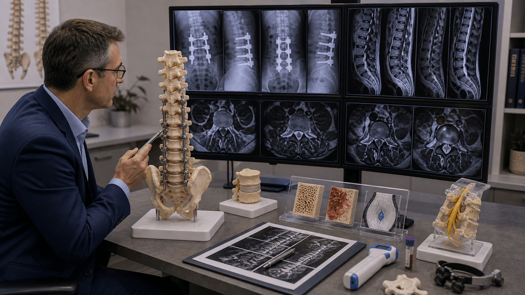
Clinical Imaging
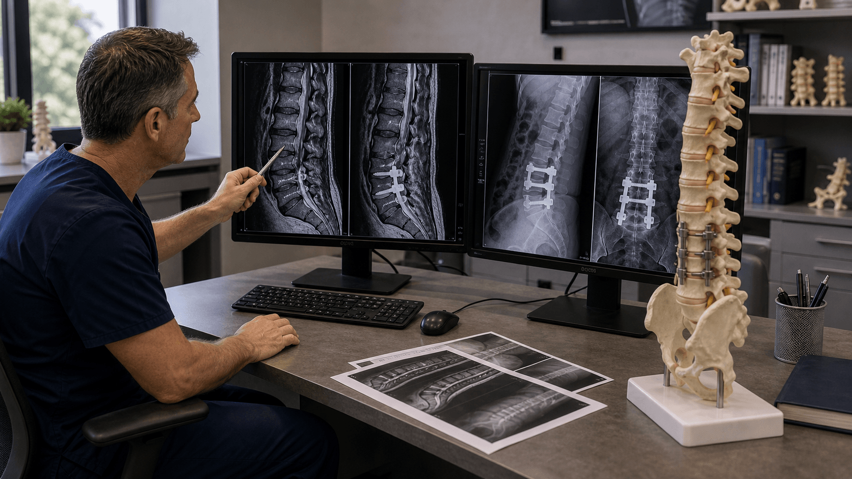
Imaging Atlas
Radiographic ASD (imaging changes) occurs in up to 30% at 10 years. Symptomatic ASD (causing clinical problems) is much less common at 2.5% per year. Not all radiographic changes require intervention.
Symptomatic ASD rate of 2.5% per year is consistent. Risk factors include long fusions, sagittal imbalance, fusion ending at L5 (vs S1), stiff adjacent segments, and pre-existing degeneration.
Restoring sagittal balance at index surgery is protective against ASD. Lumbar lordosis should match pelvic incidence (PI - LL mismatch less than 10 degrees). Fusion in kyphosis accelerates ASD.
Treatment for symptomatic ASD: extension of fusion to include affected level(s), or targeted decompression if stenosis without instability. Motion preservation at adjacent level remains investigational.
| Parameter | Radiographic ASD | Symptomatic ASD |
|---|---|---|
| Definition | Imaging changes at adjacent level | Clinical symptoms from adjacent degeneration |
| Incidence | Up to 30% at 10 years | 2.5% per year (10-15% at 10 years) |
| Clinical significance | May not progress | Requires treatment consideration |
| Management | Observation, optimize factors | Extension of fusion or decompression |
| Reoperation rate | N/A | 10-15% at 10 years |
ASDASD - Risk Factors
Hook:ASD risk is higher with Age, Sagittal imbalance, and Destabilization
FUSIONFUSION - Factors Increasing ASD Risk
Hook:FUSION technique and patient factors drive ASD - sagittal balance and facet preservation matter most
PROTECTPROTECT - Reducing ASD Risk
Hook:PROTECT adjacent segments by optimizing fusion parameters
L4L4-L5 - Common ASD Level
Hook:L4-L5 is the most common ASD level when fusion ends at L5
Overview and Epidemiology
Adjacent segment disease (ASD) refers to the development of degenerative changes at spinal levels immediately adjacent to a previous fusion. It remains one of the most debated topics in spine surgery.
Key concepts:
- Radiographic ASD: Imaging evidence of degeneration (disc space narrowing, osteophytes, facet hypertrophy) at adjacent levels
- Symptomatic ASD: Clinical presentation with pain or neurological symptoms from adjacent level degeneration
- Adjacent segment degeneration: Used interchangeably with radiographic ASD
- Adjacent segment pathology: Symptomatic disease requiring treatment
Epidemiology:
- Radiographic degeneration (ASDeg): pooled ~26.6% lumbar and ~32.8% cervical (Hashimoto 2018 systematic review)
- Symptomatic disease (ASDis): roughly 2.5-3% per year - cervical 2.9%/yr (Hilibrand 1999); lumbar disease-free survival 83.5% at 5yr and 63.9% at 10yr, with up to ~36% needing surgery by 10 years (Ghiselli 2004)
- Only about one-quarter to one-third of radiographic degeneration becomes symptomatic
- More common cephalad to fusion than caudad; L4-5 is the most common lumbar level, C5-6/C6-7 the most common cervical levels
Is ASD a consequence of altered biomechanics from fusion (iatrogenic) or natural progression of degenerative disease that would have occurred anyway? Evidence suggests both contribute. The high rate of imaging changes with much lower clinical disease rate supports natural history playing a major role.
Pathophysiology and Biomechanics
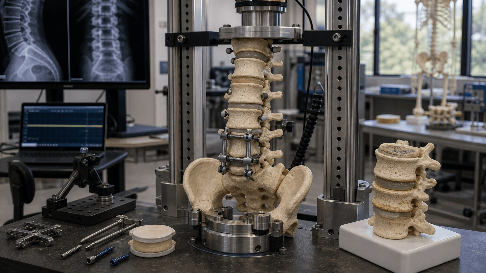
Biomechanical basis for ASD:
Load transfer hypothesis:
- Fusion eliminates motion at treated segments
- Load and motion transfer to adjacent levels
- Increased stress on adjacent disc and facets
- Accelerated degeneration at these levels
Cadaveric studies show:
- Increased intradiscal pressure at adjacent levels after fusion
- Increased facet joint loading above fusion
- Greater motion at adjacent segments to compensate for fused levels
Cadaveric and finite-element models consistently show fusion increases intradiscal pressure, facet load and compensatory motion at adjacent levels, and that longer constructs raise stress at transition zones. However, this biomechanical signal does NOT translate cleanly into clinical ASD: Ghiselli (2004) found no correlation between lumbar fusion length and reoperation, and Hilibrand (1999) found single-level cervical fusion carried higher adjacent-level risk. Biomechanics is necessary but not sufficient to explain ASD.
Factors affecting load transfer:
- Fusion length: Longer fusion = greater stress on adjacent levels
- Fusion position: Sagittal balance affects load distribution
- Adjacent segment quality: Pre-existing degeneration is vulnerable
- Rigid vs less rigid constructs: Pedicle screws vs cables/wires
Natural history argument:
- Patients requiring fusion already have degenerative disease
- Adjacent segments may have subclinical degeneration at surgery
- Some ASD represents natural disease progression
- Age-matched controls also develop degeneration
The truth likely involves both biomechanical factors and natural history.
Classification Systems
Patient and surgical factors affecting ASD
| Category | Factor | Impact |
|---|---|---|
| Patient | Age over 60 | Higher baseline degeneration |
| Patient | Pre-existing adjacent degeneration | Most significant risk factor |
| Patient | Obesity | Increased spinal loading |
| Patient | Osteoporosis | Altered load transfer |
| Surgical | PI-LL mismatch / lost lordosis | Strongest modifiable risk (Lau 2021) |
| Surgical | Sagittal imbalance (SVA over 50 mm) | Abnormal load distribution |
| Surgical | Facet joint violation | Iatrogenic destabilization of adjacent level |
| Surgical | Adjacent-level laminectomy | Destabilizes the next segment |
| Surgical | Fusion length | Disputed - Ghiselli found NO correlation |
| Surgical | Stiff instrumentation | Greater load transfer (biomechanical) |
Pre-existing degeneration at the adjacent level is the strongest non-modifiable predictor of ASD; PI-LL mismatch / loss of lordosis and facet joint violation are the most important modifiable, technique-dependent factors.
It is intuitive that longer fusions cause more ASD, but the landmark studies are nuanced. Ghiselli (2004) found NO correlation between lumbar fusion length and ASD, and Hilibrand (1999) found single-level cervical fusion carried a HIGHER adjacent-level risk than multilevel fusion. These findings are frequently used to argue that natural-history progression of spondylosis - not just biomechanical stress - drives ASD.

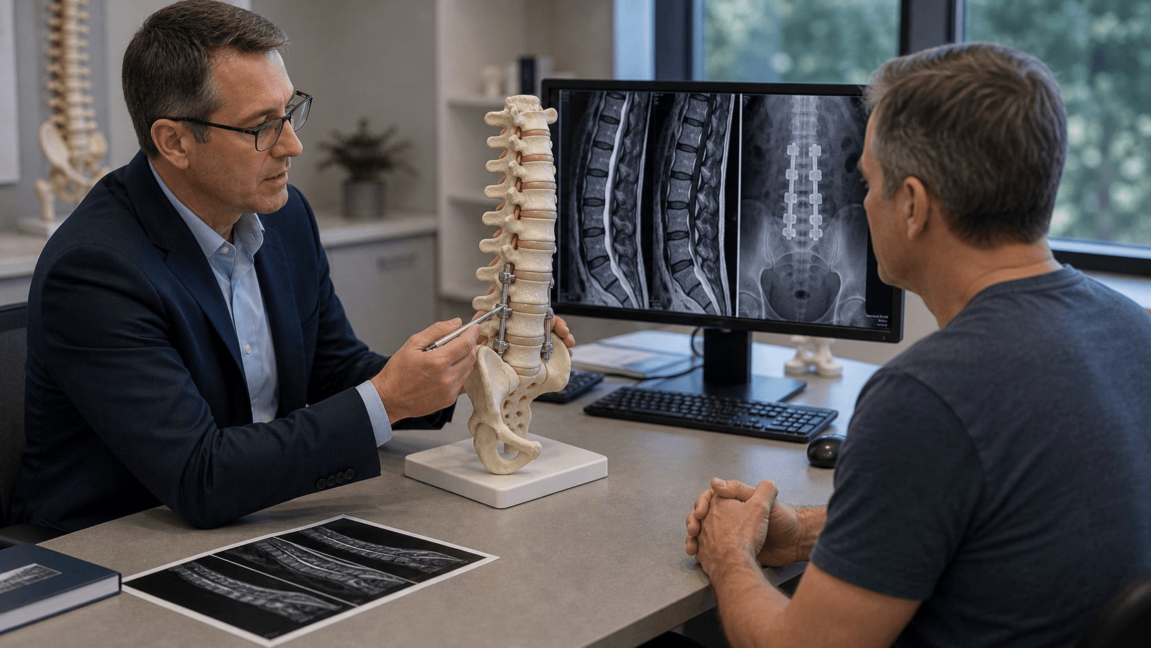
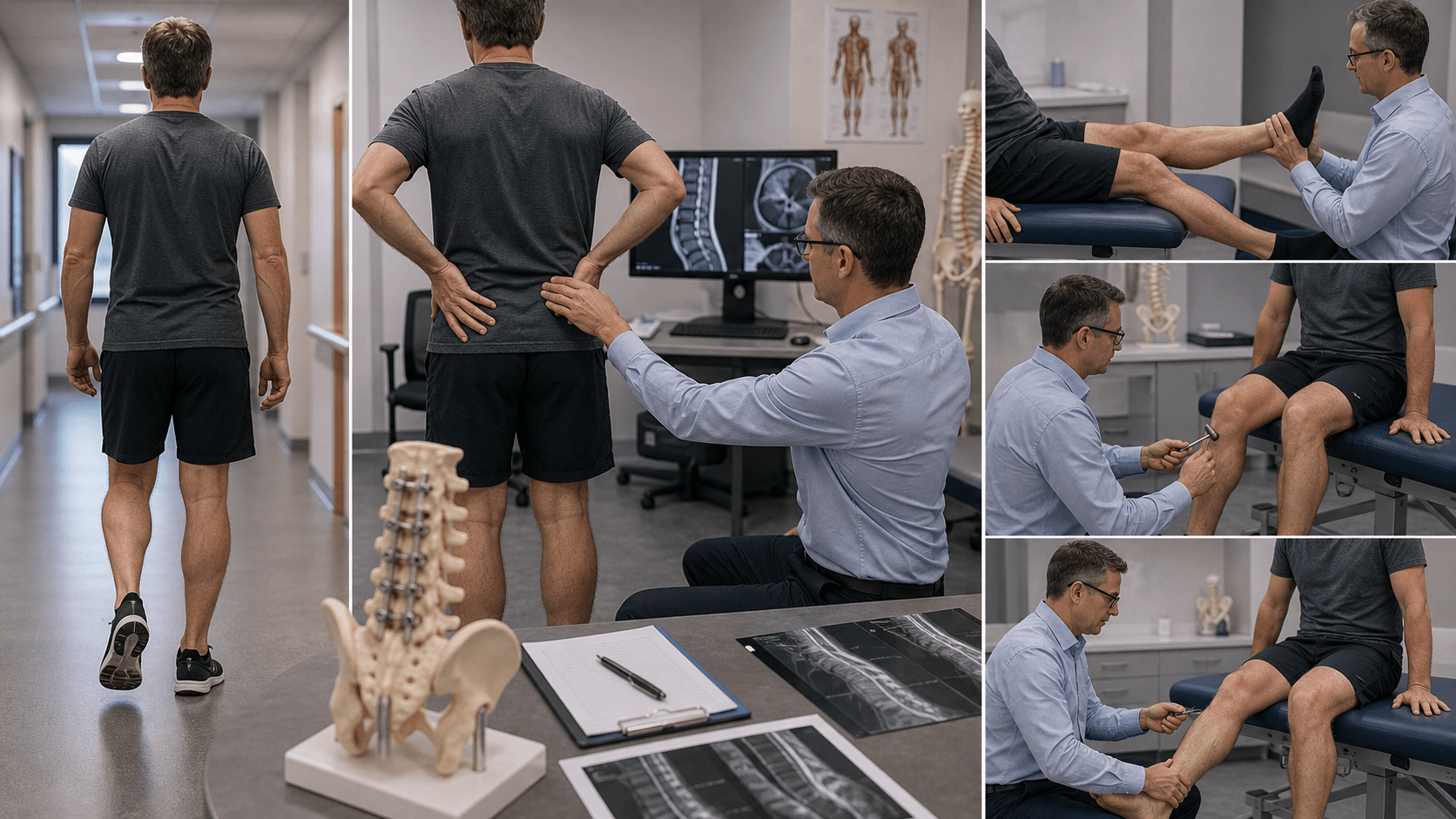
Clinical Assessment
History:
- Timing: Symptom-free interval after index surgery (months to years)
- Location: New or different pain pattern from original surgery
- Character: Axial back pain, radicular symptoms, or both
- Relationship: May mimic original presentation
- Functional impact: Walking distance, activities of daily living
- Previous surgery details: Levels fused, approach, complications
Clinical presentation patterns:
- Stenosis: Neurogenic claudication, radiculopathy
- Disc herniation: Radicular pain, dermatomal sensory changes
- Instability: Mechanical back pain, worse with motion
- Spondylolisthesis: Back pain, radicular symptoms, gait changes
Examination:
| Finding | Suggests | Management Implication |
|---|---|---|
| New radiculopathy | Nerve root compression at adjacent level | May need decompression |
| Neurogenic claudication | Central stenosis above fusion | Extension of fusion likely |
| Sagittal imbalance | Failed to restore lordosis at index | Major reconstruction needed |
| Positive extension pain | Instability at adjacent level | Fusion extension required |
| Normal neurological exam | May be discogenic/facetogenic | Consider less invasive options |
Progressive neurological deficit, cauda equina symptoms, or severe instability require urgent assessment and likely intervention. Most ASD presents insidiously with gradual symptom development.
Differential diagnosis:
New or recurrent symptoms after a fusion are NOT automatically ASD. Systematically exclude the mimics below before attributing symptoms to the adjacent level - several require entirely different management.
| Diagnosis | Distinguishing Features | Key Investigation | Management Difference |
|---|---|---|---|
| Adjacent segment disease | Symptom-free interval then new pain at NEW level/dermatome | MRI shows degeneration at level next to fusion | Decompression or fusion extension |
| Pseudarthrosis | Persistent/recurrent axial pain at SAME level, no symptom-free interval | CT (lucency, hardware halo), flexion-extension motion | Revision of original fusion, not extension |
| Hardware failure/loosening | Mechanical pain, screw haloing or rod fracture | CT and radiographs | Hardware revision |
| Recurrent stenosis at fused level | Symptoms localize to operated level | MRI/CT at index level | Targeted re-decompression |
| Infection (delayed/low-grade) | Constitutional symptoms, raised CRP/ESR | Inflammatory markers, MRI, aspiration | Debridement, antibiotics |
| Insufficiency fracture | Acute pain, osteoporosis, often at UIV | CT/MRI (STIR oedema) | Bracing, cement, or extension |
| Hip / SI joint pathology | Groin or buttock pain, positive provocation tests | Hip/SI exam, diagnostic injection | Hip or SI-directed treatment |
The single most important distinction is ASD versus pseudarthrosis. ASD produces new symptoms at a different level after a symptom-free interval; pseudarthrosis produces persistent or recurrent axial pain at the original level with no pain-free window. A solid fusion must be confirmed (ideally on CT) before considering fusion extension - extending a fusion onto a non-united segment will fail.
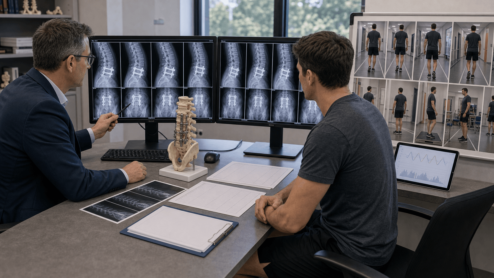
Investigations
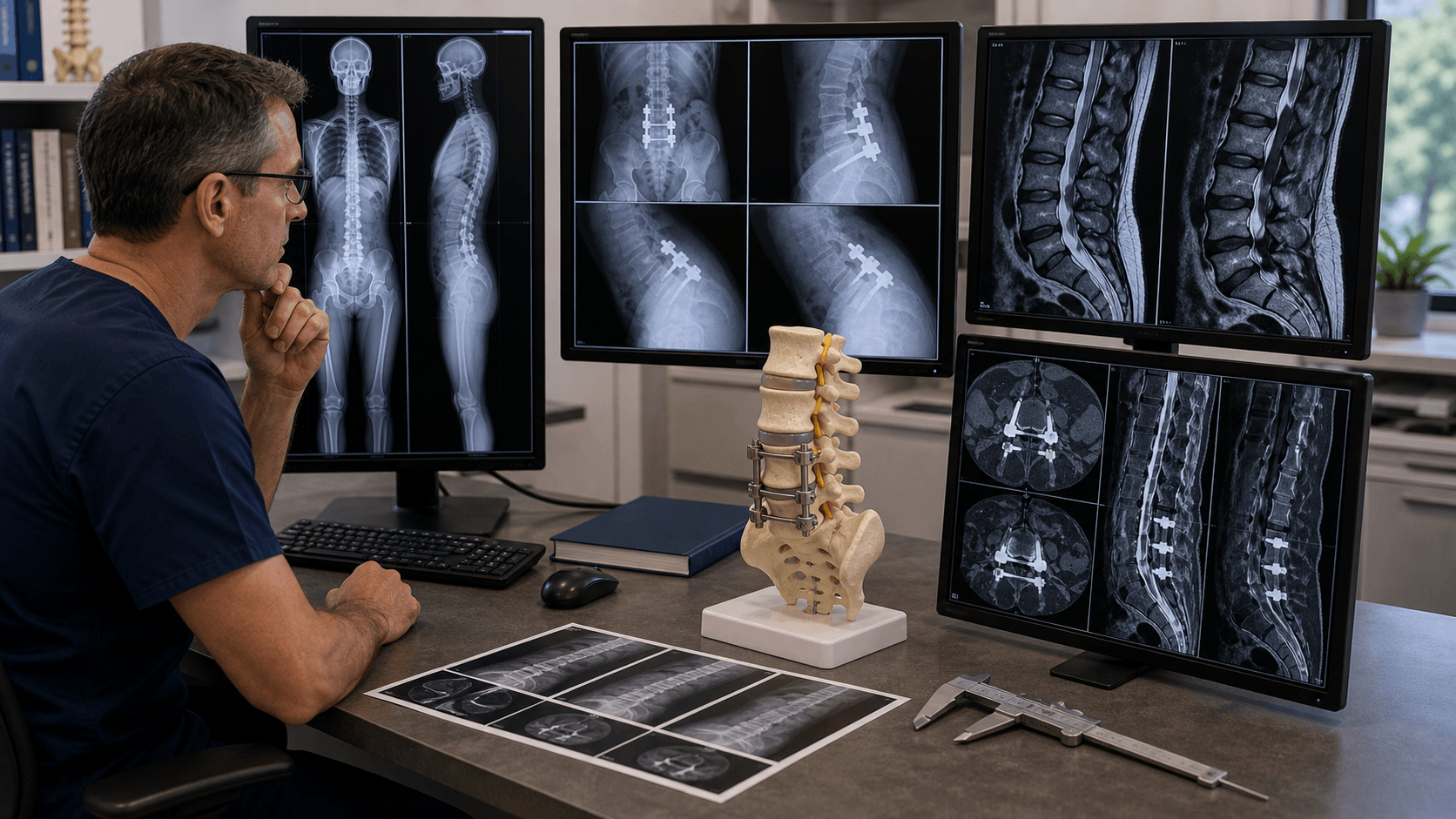

First-line imaging
Standing views essential:
- AP and lateral lumbar spine
- Flexion-extension views (dynamic instability)
- Long cassette scoliosis views if deformity
Key findings:
- Disc space narrowing at adjacent levels
- Osteophyte formation
- Endplate sclerosis
- Spondylolisthesis development
- Loss of disc height compared to pre-op
- Sagittal alignment changes
Dynamic instability:
- Greater than 4mm translation or greater than 10 degrees angulation on flexion-extension
Standing radiographs reveal functional alignment and dynamic changes.
Management Algorithm
Initial management for symptomatic ASD
Physical therapy:
- Core strengthening
- Flexibility exercises
- Posture optimization
- Activity modification
Medications:
- NSAIDs
- Neuropathic pain agents (gabapentin, pregabalin)
- Short-term oral steroids for acute exacerbation
- Muscle relaxants
Injections:
- Epidural steroid injections
- Facet joint injections
- Medial branch blocks
- Selective nerve root blocks
Bracing:
- Limited role in lumbar spine
- May provide short-term relief
Approximately 50% of patients with symptomatic ASD may improve with conservative treatment.
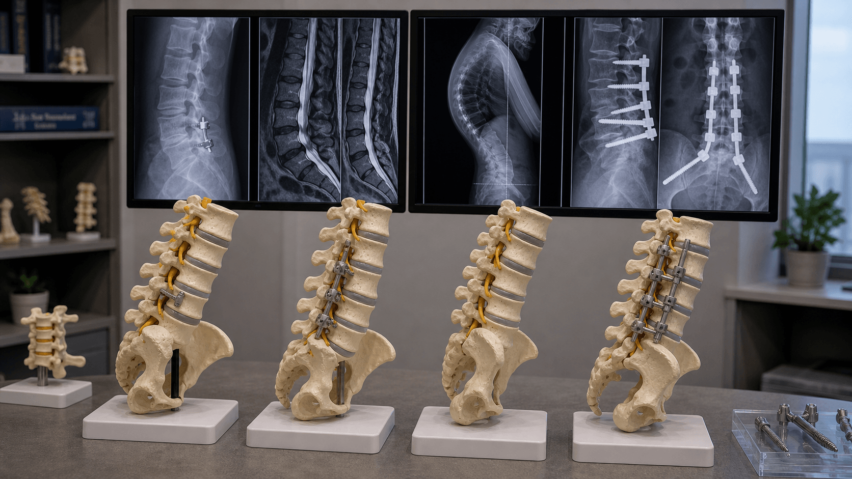
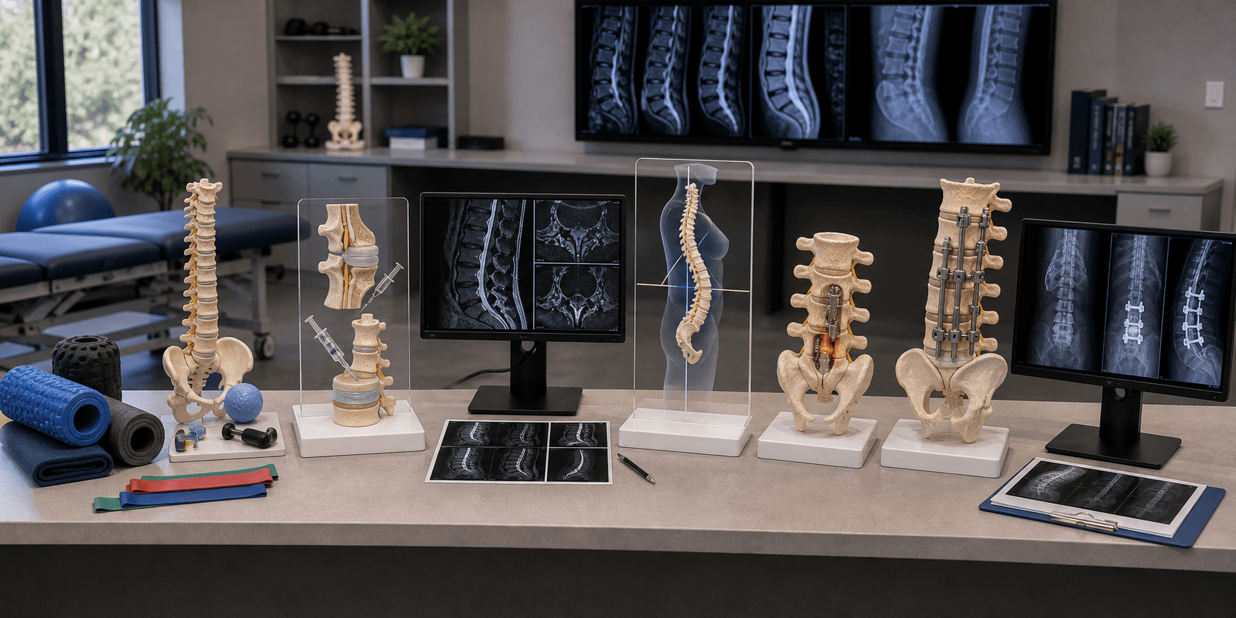
Surgical Technique
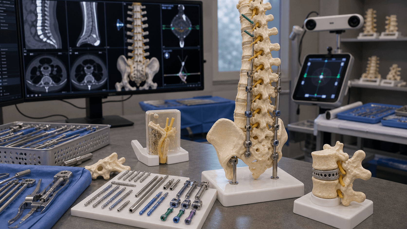
Extending the fusion cephalad or caudad
Preoperative planning:
- Assess number of levels to add
- Consider sagittal balance correction
- Plan upper instrumented vertebra (UIV)
- Evaluate bone quality
Fusion Extension Steps
Posterior approach extending above or below previous incision. Identify previous hardware. Assess for loosening or pseudarthrosis at original levels.
Evaluate existing instrumentation stability. May need to revise if loose. Connect new segments to existing construct if stable.
Decompress neural elements at affected level(s). Perform adequate foraminotomy if radicular symptoms present.
Place pedicle screws at new levels. Connect to existing construct with rods or rod-to-rod connectors. Consider interbody fusion for stability.
Address sagittal balance if needed. Restore appropriate lordosis. Confirm alignment on imaging.
Extending to sacrum and pelvis may be needed if fusing below L4.
Complications
Complications of revision surgery for ASD
Intraoperative:
- Dural tear (more common in revision)
- Nerve root injury
- Vascular injury (if anterior approach)
- Hardware malposition
Early postoperative:
- Wound infection (higher in revision surgery)
- Hardware failure
- Adjacent level injury from retraction
- Medical complications
Late complications:
- Pseudarthrosis at new fusion
- Hardware loosening
- Recurrent ASD at next level
- Persistent symptoms
Revision surgery for ASD has higher complication rates than primary surgery.
Postoperative Care
After fusion extension surgery
Recovery Phases
Mobilization with PT. DVT prophylaxis. Wound monitoring. Pain management.
Limited bending, twisting, lifting. Walking program. Wound healing. Brace if prescribed.
Gradual activity increase. PT for core strengthening. Fusion assessment on X-ray.
Progressive return to normal activities. Impact activities when fusion solid. Long-term follow-up for next adjacent level.
Bone healing assessment at 3-6 months with standing radiographs.
Outcomes and Prognosis
| Treatment | Success Rate | Recurrent ASD Risk | Notes |
|---|---|---|---|
| Conservative | 50% | N/A | Initial trial for all |
| Decompression alone | 60-70% | 10-20% need later fusion | Select patients only |
| Fusion extension | 70-80% | Continues at 2.5%/yr | Most common approach |
| Motion preservation | Variable | May be reduced | Limited long-term data |
Prognostic factors:
| Factor | Better Prognosis | Worse Prognosis |
|---|---|---|
| Symptom duration | Short (less than 6 months) | Prolonged (greater than 2 years) |
| Number of levels | Single level | Multiple levels |
| Sagittal balance | Balanced | Fixed imbalance |
| Bone quality | Normal | Osteoporotic |
| Smoking status | Non-smoker | Current smoker |
| Prior revisions | None | Multiple prior surgeries |
Surgical treatment of ASD is generally effective but carries risk of further ASD at next levels.
Controversies and Areas of Uncertainty
ASD is one of the most debated topics in spine surgery, and examiners use it to test whether a candidate can reason with uncertain evidence rather than recite dogma.
The central debate: is ASD caused by fusion-altered biomechanics or simply progression of the patient's underlying spondylosis? The biomechanical case is strong in the lab, but Hilibrand (higher risk after single-level fusion) and Ghiselli (no fusion-length effect) argue for a large natural-history component. The honest answer is both contribute.
Total disc replacement reduces overall reoperation in some trials but has not consistently reduced adjacent-level surgery versus fusion (Radcliff lumbar RCT; Harrod/Hilibrand cervical review). Whether arthroplasty truly prevents ASD - rather than just avoiding fusion-specific reoperations - remains unproven at mid-term.
Should a mildly degenerate adjacent level be included in the index fusion? Hilibrand argued symptomatic degenerate segments should be fused, but prophylactically extending onto an asymptomatic level lengthens the construct, creates a NEW transition zone, and is not evidence-supported. Most surgeons fuse only symptomatic levels.
Dynamic stabilization or an interspinous device above a fusion ("topping-off") is theoretically attractive but has mixed, low-quality evidence and device-specific complications. It remains investigational and should not be presented as standard care.
Do not commit dogmatically to "fusion causes ASD." A strong answer states that ASD is multifactorial - part iatrogenic (sagittal malalignment, facet violation), part natural history (pre-existing spondylosis) - cites Hilibrand and Ghiselli, and emphasises that the surgeon's controllable levers are restoring lordosis, preserving facets, and selecting the shortest appropriate construct.
Evidence Base
Landmark Cervical ASD Natural History (Hilibrand)
- 374 patients, 409 anterior cervical arthrodeses, followed up to 21 years
- Symptomatic adjacent-segment disease occurred at a relatively constant 2.9% per year over the first 10 years
- Survivorship predicted 25.6% of patients (95% CI 20-32%) would develop new adjacent-level disease within 10 years
- Highest-risk levels were C5-6 and C6-7; risk was HIGHER after single-level than multilevel arthrodesis
- Authors concluded all symptomatic degenerated segments should be included in the index fusion
Lumbar Adjacent-Segment Survivorship (Ghiselli)
- 215 patients after posterior lumbar arthrodesis, mean follow-up 6.7 years
- Symptomatic degeneration warranting decompression or fusion predicted at 16.5% at 5 years and 36.1% at 10 years
- Disease-free survival 83.5% at 5 years and 63.9% at 10 years (Kaplan-Meier)
- No significant correlation between fusion length and adjacent-segment disease
- No significant correlation between preoperative adjacent-level arthritic grade and need for further surgery
Pooled Incidence of ASDeg and ASDis (Systematic Review)
- Pooled incidence of radiographic adjacent-segment degeneration: 26.6% (lumbar) and 32.8% (cervical)
- Approximately one-quarter to one-third of radiographic degeneration progresses to symptomatic disease
- Lumbar risk factors: age, high BMI, pre-existing adjacent degeneration, adjacent-level laminectomy, insufficient lumbar lordosis, multilevel fixation, osteoporosis
- Cervical risk factors: young age, pre-existing degeneration, short fusion, high T1 slope, plate placed close to adjacent disc
- Motion-preserving surgery appeared to carry lower ASD risk than conventional fusion
Risk Factors for Symptomatic Lumbar ASD (Meta-analysis)
- 16 studies, 3,553 patients pooled for symptomatic ASD after lumbar fusion
- High BMI and facet joint violation during screw insertion were significant surgical/patient risk factors
- Decreased pre- and post-operative lumbar lordosis and increased postoperative PI-LL mismatch significantly increased ASD risk
- Pre-existing adjacent disc degeneration and decreased adjacent disc height predicted ASD
- Anterior shift of the sagittal plumb line and higher postoperative pelvic tilt were also associated with ASD
Spinopelvic Imbalance and ASD After PLIF (Matsumoto/Okuda)
- 1:5 matched case-control study: 20 revision-for-ASD patients vs 100 controls after L4-5 single-level PLIF
- PI-LL mismatch of 10 degrees or more was present in 75% of ASD patients vs 43% of controls (p less than 0.01)
- Sagittal vertical axis of 50 mm or more was seen in 50% of ASD patients vs 21% of controls (p less than 0.01)
- ASD patients had significantly lower lumbar lordosis and higher pelvic tilt
- Even after single-level fusion, restoring segmental and lumbar lordosis appeared protective against ASD
Sagittal Alignment as an ASD Risk Factor (Djurasovic)
- Case-control study: 51 patients with adjacent-segment degeneration matched to controls
- ASD patients had significantly less lordosis through the fusion and across the lumbar spine after the index operation
- No significant difference in pre-existing adjacent-level degeneration between ASD and control groups
- No significant difference in smoking rates between groups
- Concluded fusion in abnormal sagittal alignment - not merely progression of pre-existing degeneration - predisposes to ASD
Lumbar TDR vs Fusion - 2-Level Reoperation (Radcliff RCT)
- Prospective RCT: 229 patients, 2-level degenerative disc disease, TDR (n=161) vs circumferential fusion (n=68)
- Overall adjacent-segment disease rate was only 3.5% (8/229) at 5 years
- Adjacent-level reoperation did not differ significantly between TDR (2.5%) and fusion (5.9%)
- Overall secondary surgery was lower with TDR (5.6%) than fusion (19.1%), but most fusion reoperations were instrumentation removal
- Excluding instrumentation removal, reoperation rates were similar - TDR was non-inferior to fusion
Cervical Motion Preservation vs Fusion for ASD (Systematic Review)
- Systematic review of 14 studies comparing cervical TDR with anterior cervical decompression and fusion (ACDF)
- Reoperation for clinical adjacent-segment pathology ranged 1.0% to 4.8%, with no statistically significant difference between TDR and ACDF
- No significant difference in radiographic or clinical ASP between cervical TDR and ACDF at short- to mid-term follow-up
- Insufficient evidence to recommend non-arthroplasty motion-preserving devices over fusion
- Highlighted lack of consistent ASD definitions across the literature
Exam Viva Scenarios
Practise clinical reasoning and management decisions out loud
“A 60-year-old woman who had L4-S1 fusion 5 years ago presents with new-onset bilateral leg pain and back pain. She has difficulty walking more than 100 meters. Examination reveals reduced ankle reflexes and sensory changes in the L3 distribution. Her previous surgery was uneventful with good relief of symptoms for the first 4 years.”
“You are planning a L4-L5 fusion for a 55-year-old man with spondylolisthesis. He asks about the risk of needing more surgery in the future because of problems at other levels. How would you counsel him?”
“A 58-year-old woman had L5-S1 fusion 8 years ago and now has symptomatic stenosis at L4-5. MRI shows severe stenosis with a 5mm spondylolisthesis at L4-5. Her standing X-rays show positive sagittal balance with loss of lumbar lordosis. How would you approach this?”
MCQ Practice Points
Key facts for MCQs:
- Symptomatic ASD rate: 2.5% per year
- Radiographic ASD at 10 years: approximately 30%
- Reoperation rate: 10-15% at 10 years
- L4-5 is most common ASD level (when fusion ends at L5)
- PI-LL mismatch increases ASD risk
- Pre-existing adjacent degeneration is strongest risk factor
Common MCQ topics:
- Radiographic vs symptomatic ASD distinction
- Risk factors for ASD
- Role of sagittal balance
- Treatment options for symptomatic ASD
- When to decompress alone vs extend fusion
- Prevention strategies
- Fusion length effect on ASD
Key concepts:
- Long fusions have higher ASD rate
- Fusion to L5 (vs S1) has higher ASD at L4-5
- Motion preservation may reduce but not eliminate ASD
- Both iatrogenic and natural history factors contribute
Q: What is the symptomatic ASD rate after lumbar fusion? A: Approximately 2.5% per year. Radiographic ASD is much more common (~30% at 10 years), but only a fraction become clinically symptomatic.
Q: What is the single strongest risk factor for developing ASD? A: Pre-existing degeneration at adjacent levels at the time of index surgery is the strongest predictor.
Q: What sagittal parameter mismatch increases ASD risk? A: PI-LL mismatch greater than 10 degrees. Restoring appropriate lumbar lordosis is protective against ASD.
Q: How does fusion length affect ASD risk? A: Longer fusions have higher ASD rates. Each additional fused level increases stress concentration at transition zones. Use shortest necessary fusion.
Q: When can decompression alone be performed for ASD? A: When there is stable stenosis without spondylolisthesis and preserved disc height. Instability requires fusion extension.
Guidelines, Registries & Global Practice
Global epidemiology
- Symptomatic ASD requiring surgery is a leading cause of lumbar fusion revision worldwide; pooled radiographic degeneration is ~26.6% (lumbar) and ~32.8% (cervical) (Hashimoto 2018).
- A systematic review reported a higher prevalence of lumbar ASDeg in Western than in Eastern populations (Cannizzaro 2022), likely reflecting differences in surgical indications, BMI distribution and follow-up imaging practice rather than a single biological cause.
- As global life expectancy and fusion volumes rise, the absolute burden of ASD is increasing, making prevention at the index operation a worldwide priority.
Side-by-side guidance (recommendations converge more than they differ)
| Body / Source | Region | Position relevant to ASD |
|---|---|---|
| NASS (degenerative spondylolisthesis / stenosis guidelines) | US / international | Decompression with fusion for instability; emphasises restoring alignment and shortest appropriate construct |
| NICE NG59 (low back pain and sciatica) | UK | Exhaust non-surgical care before revision; no routine prophylactic extension of fusion |
| AOSpine knowledge forum | International (AO Foundation) | Standardising ASD definitions (ASDeg vs ASDis); restore sagittal balance and avoid facet violation |
| EUROSPINE / EFORT consensus | Europe | Spinopelvic alignment central to deformity and ASD prevention; PI-LL matching emphasised |
| SRS / ISSG sagittal alignment targets | International | Age-adjusted PI-LL and SVA targets to reduce mechanical complications and ASD |
All major societies agree on three points: (1) confirm the diagnosis and exclude pseudarthrosis/infection before surgery, (2) treat conservatively first, and (3) restore sagittal alignment and avoid iatrogenic destabilization at the index operation. Genuine differences are minor and largely about thresholds rather than principles.
Registry evidence
- Large national spine registries (e.g. the Swedish Spine Register (Swespine), the British Spine Registry, and Norwegian (NORspine) datasets) capture reoperation as a proxy for symptomatic ASD and consistently identify revision for adjacent-level pathology as a major late reoperation cause.
- Registry reoperation data underpin patient counselling on long-term revision risk and are increasingly used to benchmark alignment-restoring techniques.
High- vs limited-resource practice variation
- Well-resourced settings: routine long-cassette standing radiographs for spinopelvic parameters, MRI with metal-artifact-reduction sequences, navigation/robotics to reduce facet violation, and access to revision deformity surgery and arthroplasty.
- Limited-resource settings: sagittal alignment may be judged on standard radiographs without full spinopelvic measurement; metal-artifact MRI and revision deformity capacity may be scarce, so prevention at the index operation and prolonged non-operative management carry even greater weight.
- Bone-health optimization (vitamin D, osteoporosis treatment) and smoking cessation are universally relevant, low-cost, modifiable measures that improve both prevention and revision outcomes regardless of setting.
Exam Cheat Sheet
Key Numbers
- Symptomatic ASD: 2.5% per year
- Radiographic ASD at 10yr: 30%
- Reoperation rate: 10-15% at 10yr
- PI-LL mismatch: less than 10 degrees target
Risk Factors
- Pre-existing adjacent degeneration (strongest)
- Long fusion (more levels = more risk)
- Sagittal imbalance/loss of lordosis
- Fusion ending at L5 (vs S1)
- Wide decompression destabilizing facets
Treatment Approach
- Conservative first: PT, NSAIDs, injections
- Decompression alone: stable, preserved disc
- Fusion extension: instability, spondylolisthesis
- Address sagittal imbalance when present
Prevention Strategies
- Shortest necessary fusion length
- Restore appropriate sagittal balance
- Preserve adjacent facet joints
- Smoking cessation, weight management
Exam Traps
- Confusing radiographic and symptomatic ASD
- Decompression alone with instability
- Ignoring sagittal balance
- Not discussing ASD with fusion patients