Doha Classification | Adductor-Related Most Common | Multidisciplinary Management
- Doha classification: four defined clinical entities (adductor-, iliopsoas-, inguinal-, pubic-related) plus hip-related and other causes
- Adductor squeeze test is key diagnostic manoeuvre for adductor-related pain
- Copenhagen adductor protocol is evidence-based rehab for adductor injuries
- Differential diagnosis includes FAI - always examine hip ROM and impingement
- Multidisciplinary approach essential: physio, sports medicine, surgery last resort
- “Athletic pubalgia = chronic groin pain in athletes without inguinal hernia
- “Soccer/football players most affected - kicking and change of direction
- “MRI shows pubic symphysis oedema, secondary cleft sign, adductor pathology
- “Conservative management succeeds in 70-80% with proper rehabilitation
Overview and Epidemiology
Athletic groin pain (also termed athletic pubalgia or sports hernia) is chronic groin pain in athletes that interferes with sport participation. The term "sports hernia" is a misnomer: most cases do not have a true hernia but rather musculotendinous injury or pubic symphysis pathology.
Burden. In prospective surveillance, hip/groin injuries are 12-16% of all time-loss injuries in men's professional football, with a consistent incidence over time (UEFA injury studies: Werner 2009, PMID 19945984; Werner 2018, PMID 29691289). Adductor-related injury is the commonest entity, about 63% of hip/groin injuries in elite football (Werner 2018, PMID 29691289). Onset is often insidious and the problem is chronic by the time of presentation; mean absence is about 15 days per injury, with about 15% re-injury in elite football (Werner 2009, PMID 19945984).
Who and how. Soccer/football, ice hockey, rugby codes, Australian Rules Football and other change-of-direction sports. The mechanism is kicking, rapid acceleration and deceleration, and change of direction: chronic repetitive microtrauma, with the pubic symphysis and adductor insertion subjected to repeated eccentric loads during deceleration and change of direction. Unlike an acute adductor strain, this is a chronic overuse syndrome.
Terminology follows the Doha agreement (2014), set out under Classification Systems.
Pathophysiology and Mechanisms
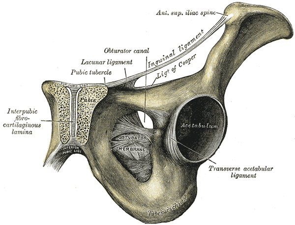
The pubic symphysis is the fibrocartilaginous joint connecting the left and right pubic bones, and it experiences significant shear and compression forces during athletic movements. The structures inserting around it:
- Adductor longus - the primary pain generator, inserting on the anterior pubic body
- Gracilis - medial aspect of the groin
- Rectus abdominis - superior pubic insertion
- External oblique - forms the anterior inguinal canal wall
- Transversalis fascia - posterior inguinal canal wall
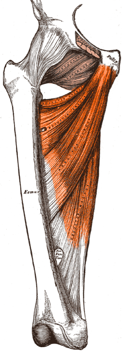
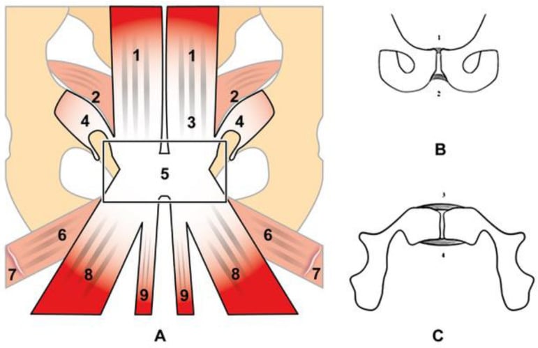
Adductor-related. Repeated eccentric loading of adductor longus at its enthesis, the pubic insertion. Microtrauma leads to tendinopathy, partial tears and chronic inflammation.
Inguinal-related. Weakness or disruption of the posterior inguinal wall (transversalis fascia, conjoint tendon), which creates a "dilated superficial inguinal ring" without a frank hernia sac. The underlying imbalance is between strong hip flexors and adductors and a weak abdominal wall.
Pubic-related. Pubic symphysis stress reaction, or osteitis pubis. Repeated shear forces cause bone marrow oedema, secondary cleft formation (a parasymphyseal cyst) and eventual sclerosis, and MRI shows a characteristic oedema pattern.
Hip-related. Femoroacetabular impingement (FAI) or a labral tear presenting as groin pain. Cam or pincer morphology causes anterior impingement. It often coexists with adductor pathology: the hip is the hidden driver.
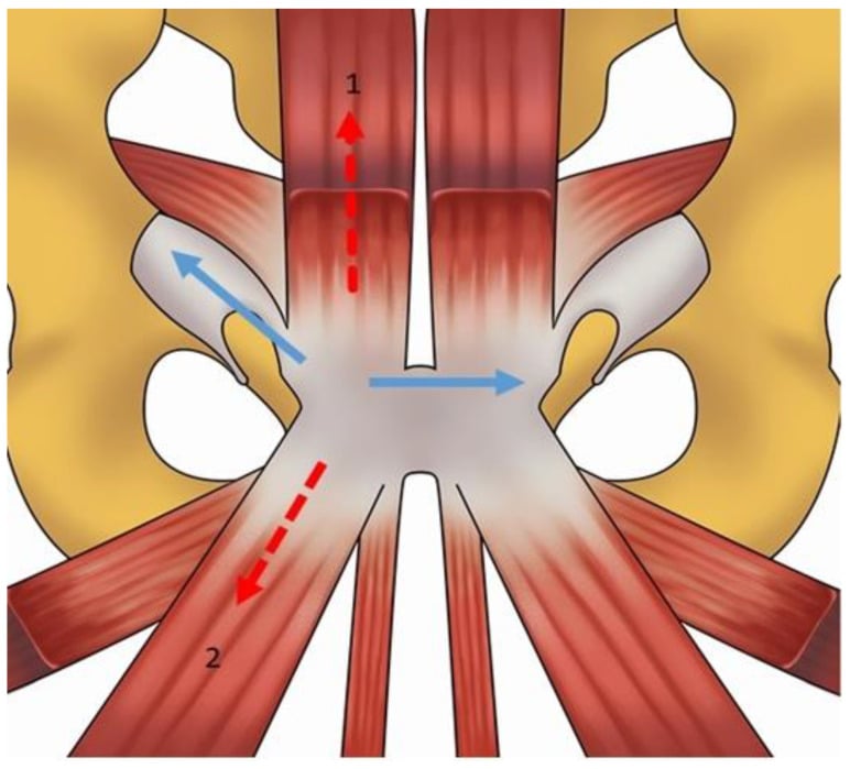
Always evaluate the hip first. Up to 50% of athletes with "groin pain" have underlying hip pathology (FAI, labral tear), which is common in athletes and presents as groin pain. Hip pathology can cause secondary adductor overload as compensation: the adductor compensates for hip dysfunction, and treating the groin without addressing the hip leads to failure and recurrent symptoms. Perform the FADIR test and assess hip internal rotation; if FADIR is positive or internal rotation is limited, obtain hip MRI.
Biomechanics of kicking. During the soccer kick:
- Wind-up phase: hip extension, adductors eccentrically control abduction
- Acceleration phase: rapid hip flexion, adductors co-contract
- Deceleration phase: peak adductor load, eccentric lengthening to control the leg
- Follow-through: continued eccentric adductor demand
The deceleration phase generates the highest forces on the adductor longus enthesis.
Classification Systems
The Doha agreement (2014) established a clinical entity-based classification for groin pain in athletes, diagnosed by history and examination, in three categories: the defined clinical entities (adductor-, iliopsoas-, inguinal- and pubic-related groin pain), hip-related groin pain, and other causes. It replaced the confusing historical terminology (Gilmore's groin, sportsman's hernia, hockey groin syndrome and the like) because those eponymous and pathoanatomical labels assert a pathology that is often unproven, so quote the Doha term and give the old name ("sports hernia", "osteitis pubis") only as a gloss. The entities coexist, so finding one does not exclude the others. Two are developed further in adductor-related groin strain and pubic-related groin pain.
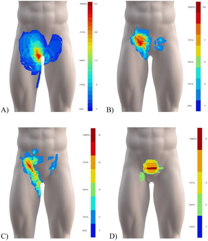
Adductor-related groin pain is the most common entity. It presents as pain with resisted adduction (a positive squeeze test), on palpation of the adductor tendons (especially longus), on stretching the adductors, and with kicking or change of direction.
Examination. The adductor squeeze test is performed at three hip flexion angles, 0, 45 and 90 degrees, because different angles stress different portions of the adductor complex; it is most sensitive at 45 degrees for adductor longus pathology. Palpate the adductor longus enthesis, which is tender; test strength, which is often weak compared with the contralateral side; and record the adductor:abductor strength ratio.
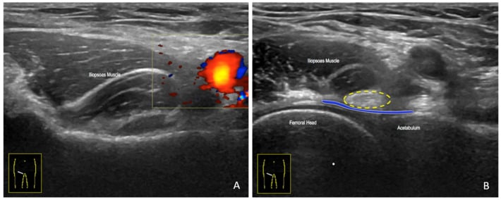
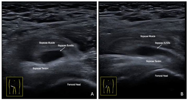
Clinical Presentation and Assessment
History. Ask for the precise location of the pain (adductor, inguinal, pubic, deep hip), its onset (acute or insidious, usually insidious), what aggravates it (kicking, sprinting, change of direction) and what relieves it (rest, but it returns with activity). Night pain, if present, suggests hip pathology.
Function and load. Can they kick, sprint, cut? Has training volume or intensity recently increased? Is there a previous adductor or groin problem? The contralateral side is often affected eventually, and the duration is weeks to months by presentation.
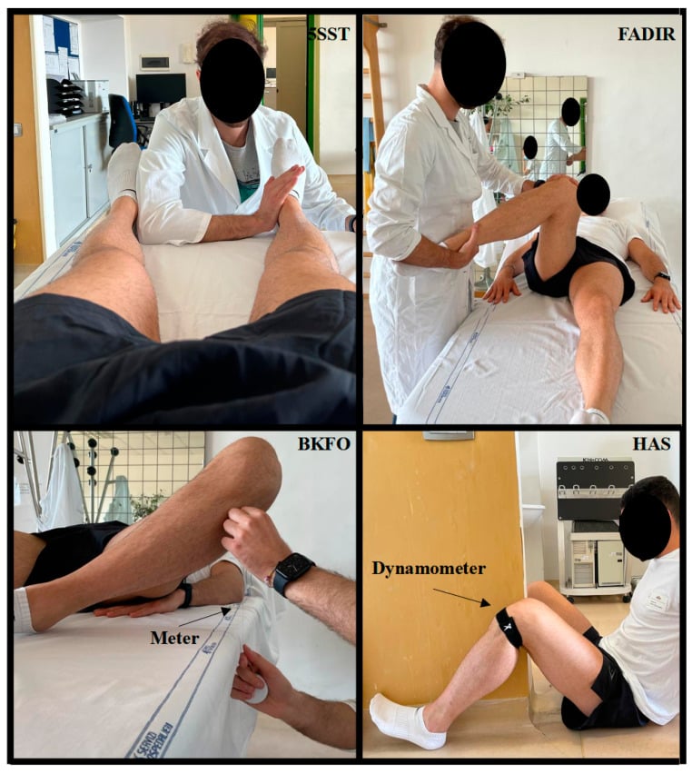
Examination sequence.
Systematic Groin Examination
- Gait (antalgic, waddling)
- Posture and pelvic alignment
- Muscle atrophy (adductors, core)
- Scars from previous surgery
- Adductor longus at the pubic insertion
- Pubic symphysis midline tenderness
- Inguinal canal along its course
- Anterior hip joint line
- Hip flexion and extension (compare sides)
- Hip internal rotation (limited in FAI)
- Hip external rotation (painful in labral tear)
- Straight leg raise (adductor stretch)
- Adductor squeeze test (0, 45, 90 degrees)
- FADIR test (hip impingement)
- FABER test (hip/SI joint)
- Resisted sit-up (rectus/core)
- Single-leg stance (pubic stress)
- Adductor strength (manual or dynamometer)
- Abductor strength (Trendelenburg)
- Core strength (plank hold time)
- Adductor:abductor ratio (target greater than 80%)
- Technique
- Supine, 45deg hip flexion, squeeze fist between knees
- Positive Finding
- Reproduces groin pain
- Significance
- Adductor-related groin pain
- Technique
- Supine, flex hip 90deg, adduct and internally rotate
- Positive Finding
- Anterior groin pain
- Significance
- Hip impingement (FAI)
- Technique
- Supine, flex-abduct-externally rotate hip (figure-4)
- Positive Finding
- Anterior or lateral hip pain
- Significance
- Labral tear or SI joint
- Technique
- Supine crunch with legs extended
- Positive Finding
- Inguinal canal or rectus pain
- Significance
- Inguinal-related (core deficiency)
- Technique
- Stand on affected leg for 30 seconds
- Positive Finding
- Pubic symphysis pain
- Significance
- Pubic-related (osteitis pubis)
Exclude serious pathology before attributing the pain to a Doha entity:
- Stress fracture of the femoral neck or pubic ramus (night pain, unable to weight-bear)
- Avascular necrosis of the hip (risk factors, limited range of motion)
- Infection: septic arthritis, osteomyelitis (fever, elevated inflammatory markers)
- Malignancy: bone tumour (rare, but consider it in an atypical presentation)
- Referred pain: lumbar radiculopathy, intra-abdominal pathology
Image urgently for inability to weight-bear, night pain that wakes the patient from sleep, constitutional symptoms (fever, weight loss) or progressive neurological symptoms.
The wider differential. The Doha categories sit within a wider differential. Beyond the Doha entities, the surgeon must actively exclude non-musculoskeletal and serious causes: many a "groin strain" has turned out to be a femoral neck stress fracture, a hernia or intra-abdominal pathology.
- Typical features
- Pain on kicking/cutting, medial groin
- Discriminating finding / test
- Positive adductor squeeze, tender adductor longus enthesis
- Confirmation
- Clinical; MRI adductor tendinopathy/tear
- Typical features
- Anterior groin pain, snapping hip
- Discriminating finding / test
- Pain on resisted hip flexion and on stretch
- Confirmation
- Clinical; MRI/US iliopsoas, dynamic US
- Typical features
- Deep groin ache on exertion, no bulge
- Discriminating finding / test
- Inguinal canal tenderness, pain on resisted sit-up
- Confirmation
- MRI posterior wall changes; exclude true hernia
- Typical features
- Midline symphyseal pain, often bilateral
- Discriminating finding / test
- Symphyseal point tenderness, single-leg stance pain
- Confirmation
- MRI pubic bone marrow oedema, secondary cleft sign
- Typical features
- Deep anterior groin, C-sign, clicking
- Discriminating finding / test
- Positive FADIR, limited internal rotation
- Confirmation
- X-ray (alpha angle, LCEA); MR arthrogram for labrum
- Typical features
- Load-related pain, night pain, unable to weight-bear
- Discriminating finding / test
- Pain on hop test, focal bony tenderness
- Confirmation
- MRI (marrow oedema/fracture line) — urgent
- Typical features
- Bulge, pain on Valsalva
- Discriminating finding / test
- Palpable reducible mass, cough impulse
- Confirmation
- Examination; dynamic US/CT
- Typical features
- Radiating pain, dysaesthesia (ilioinguinal/genitofemoral/obturator)
- Discriminating finding / test
- Neurological signs, positive nerve tests, no local pathology
- Confirmation
- MRI lumbar spine; diagnostic nerve block
- Typical features
- Pain unrelated to load, systemic or visceral symptoms
- Discriminating finding / test
- Abnormal abdominal/pelvic exam, urinalysis
- Confirmation
- Targeted imaging, specialist referral
Investigations
Radiographs first. A standing AP pelvis assesses the pubic symphysis and excludes fracture: look for pubic sclerosis and irregular pubic margins (osteitis pubis, a late finding), measure the symphysis width (normal under 10mm) and assess the hip joint space for arthritis. On the same film read the alpha angle (cam lesion), the lateral centre-edge angle (pincer) and the crossover sign. A frog-leg lateral of the hip is the best view of the anterior femoral head-neck junction and the best view for measuring the alpha angle: normal is under 55 degrees, and over 55 degrees is cam morphology; radial MRI cuts are the alternative. A bone scan shows increased uptake in pubic-related pain but is non-specific.
MRI pelvis and hips is the gold standard. Order "MRI pelvis and hips" rather than separate studies: the pelvis protocol captures the pubic symphysis and adductors, and dedicated hip sequences assess the labrum and cartilage. The protocol is coronal and axial T1, T2 and STIR sequences; intra-articular gadolinium (MR arthrogram) improves labral tear detection when hip pathology is suspected. What it shows, by entity:
- Adductor-related: adductor longus tendinopathy (increased signal), partial-thickness tear, bone marrow oedema at the pubic insertion
- Pubic-related: pubic bone marrow oedema (often bilateral), the secondary cleft sign, symphyseal fluid or irregularity, and adductor insertional changes
- Inguinal-related: posterior inguinal wall signal changes, conjoint tendon injury, rectus abdominis strain
- Hip-related: labral tear (needs intra-articular contrast for best sensitivity), cam or pincer morphology, cartilage damage
The secondary cleft sign. A fluid-filled cyst adjacent to the pubic symphysis, the parasymphyseal cyst. It is highly specific for chronic pubic stress, described as pathognomonic, and represents extension of symphyseal pathology into the adjacent bone.
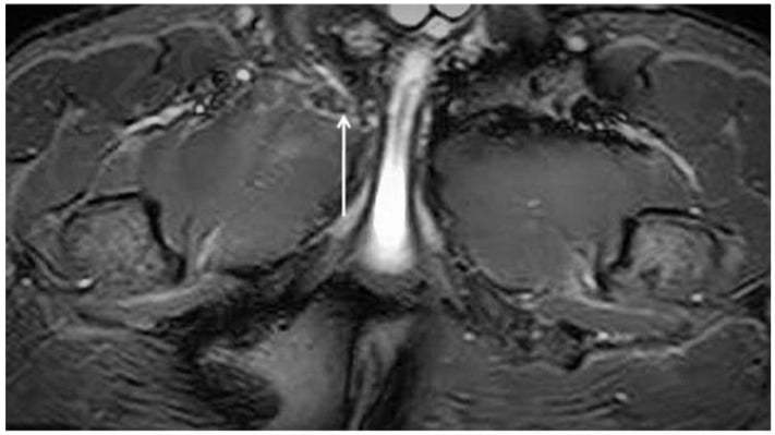
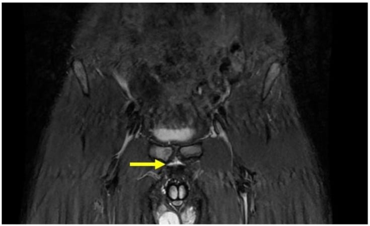
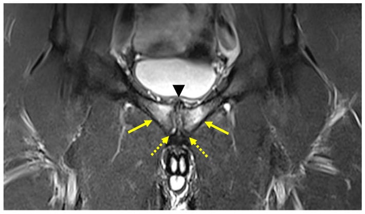
Ultrasound. Dynamic assessment of the adductor tendons and inguinal canal: operator-dependent, but a useful adjunct. It can assess the adductor during contraction and the inguinal canal during Valsalva, where it may show bulging, and it is less sensitive than MRI for bone oedema.
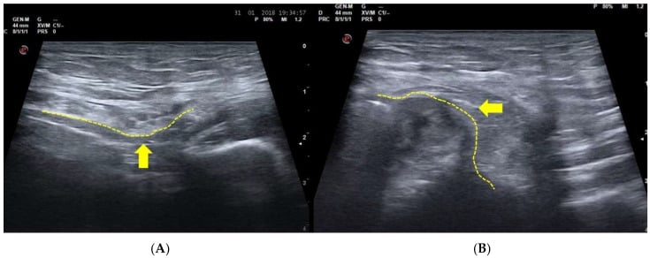
Diagnostic injection, if indicated. Image-guided local anaesthetic at the suspected source (the adductor enthesis, the hip joint) supports the diagnosis when the pain is relieved during the injection and can guide surgical decision-making. The targets and their interpretation follow.
Image-Guided Diagnostic and Therapeutic Injections
A diagnostic block resolves the central difficulty of groin pain in athletes: the Doha entities coexist, and imaging is frequently abnormal in athletes who have no pain. In the landmark MRI series, 98% of symptomatic patients had a finding capable of causing groin pain, but asymptomatic control athletes also had findings (Zoga 2008, PMID 18487535). MRI therefore localises a structure; it does not prove which structure is generating the symptoms. An image-guided (ultrasound or fluoroscopic) local-anaesthetic block adds the missing functional information: temporary abolition of the athlete's typical pain after anaesthetising a specific target confirms that target as the dominant pain generator and, for surgical candidates, predicts the response to addressing it.
- A positive block confirms
- Hip-related (intra-articular) pain — labrum/chondral/FAI
- Practical role
- Relief supports an intra-articular source and predicts a better response to hip arthroscopy; central to the 'hip-first' rule
- A positive block confirms
- Iliopsoas-related pain
- Practical role
- Confirms the often-missed anterior entity; can be therapeutic; distinguishes a painful iliopsoas from an incidental snap
- A positive block confirms
- Pubic-related pain (osteitis pubis)
- Practical role
- Diagnostic; corticosteroid can give short-term relief but is adjunctive to load management, not a substitute
- A positive block confirms
- Adductor-related pain
- Practical role
- Confirms the symptomatic adductor source and helps decide whether a tenotomy would target the right structure
- A positive block confirms
- Inguinal-related pain (athletic pubalgia / 'sports hernia')
- Practical role
- Relief supports the diagnosis and, in failed-conservative cases, predicts success of surgical repair
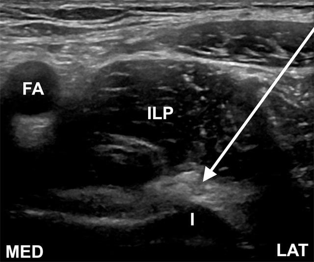
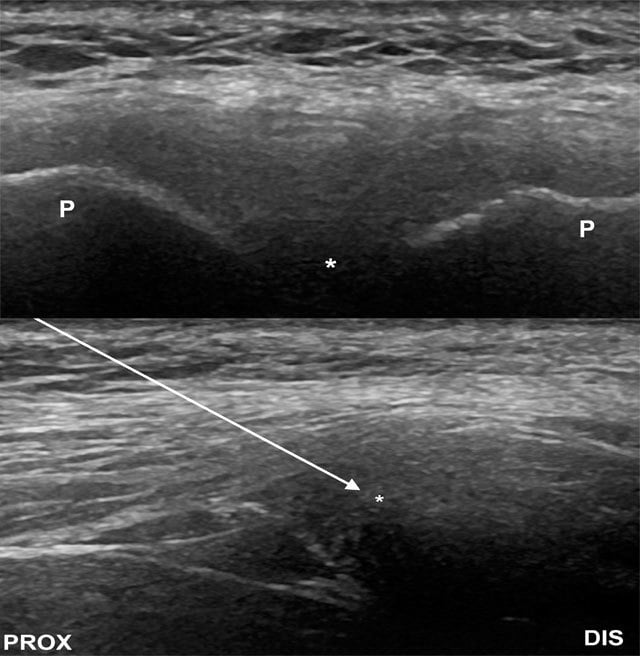
Image guidance is essential. Blind injections around the hip, symphysis and iliopsoas are inaccurate; ultrasound or fluoroscopy confirms placement and lets you document the volume and the immediate pain response.
The diagnostic value is in the response, not the drug. Record the athlete's pain with a provocative task (squeeze, resisted flexion, sport-specific movement) before and immediately after the anaesthetic; a clear, time-limited reduction is the positive result. Use a block above all when entities coexist or when imaging and examination disagree: it is the practical tool for picking the dominant generator before committing an athlete to surgery.
Confirmatory, not curative. A block identifies the generator but does not correct the underlying load or mechanics, so it never replaces the exercise-based rehabilitation that remains first line for every entity. Therapeutic corticosteroid is adjunctive and usually short-lived (for recalcitrant osteitis pubis, for example); repeated peritendinous steroid risks tendon weakening, and the evidence for biologic injections such as PRP in this setting remains limited and inconsistent.
Management Algorithm
Every athlete has a trial of conservative management before surgery is considered: 8-12 weeks of exercise-based rehabilitation, with surgery reserved for failed conservative treatment with clear structural pathology and multidisciplinary input. With proper rehabilitation, adductor-related pain settles in 70-80%; the figures for the other entities are tabulated under Outcomes and Prognosis.
Goal. Restore adductor strength and endurance and normalise the adductor:abductor ratio.
Conservative Protocol
- Relative rest (avoid aggravating activities)
- Ice after activity
- NSAIDs for symptom control
- Load management (reduce training volume by 50%)
- Maintain cardiovascular fitness (cycling, swimming)
- Copenhagen adductor protocol
- Progressive eccentric adductor exercises
- Core strengthening (planks, obliques)
- Adductor plank variations
- Sport-specific drills (gradual introduction)
- Restore the adductor:abductor ratio to target
- Graded return to training
- Monitor training load (acute:chronic workload ratio)
- Continue maintenance adductor strengthening
- Biomechanical assessment (kicking technique)
- Prevention programme ongoing
The Copenhagen adductor protocol. Developed in Denmark, it is a progressive eccentric adductor strengthening programme performed 3 times per week, and its core exercise is the Copenhagen adductor plank, a side plank with leg support, the Nordic hamstring exercise adapted for the adductors. In a cluster-randomised trial of semiprofessional football teams the programme lowered the risk of reporting groin problems by 41% (Harøy 2019, PMID 29891614); the outcome was self-reported prevalence rather than time-loss injury. The exercises, in progression:
- Isometric adductor squeeze (various angles)
- Copenhagen adductor plank (side plank with leg support)
- Eccentric adductor slide (standing slide-board)
- Single-leg adductor stability
- Sport-specific movements (kicking progressions)
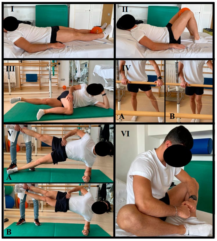
If conservative management fails. After 12 weeks without success, adductor tenotomy (release of adductor longus) can be considered, reserved for chronic recalcitrant cases; the technique and its variable results are under Surgical Techniques.
Surgical Techniques (When Conservative Fails)
Minimal repair technique (Muschaweck). Indicated for inguinal-related groin pain after 12 or more weeks of failed conservative management, with posterior wall deficiency on imaging and no true hernia present.
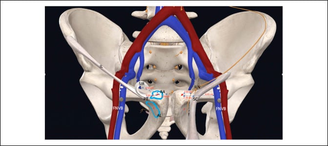
Surgical Steps
- Supine position
- Oblique inguinal incision (as for hernia repair)
- Incise the external oblique aponeurosis
- Protect the ilioinguinal nerve
- Assess the posterior inguinal wall
- Identify the transversalis fascia deficiency
- Evaluate the conjoint tendon
- Look for a true hernia (often absent)
- Reinforce the transversalis fascia with sutures
- Conjoint tendon to inguinal ligament (modified Bassini)
- Avoid mesh if possible (stiffness, decreased range of motion); some surgeons use it in selected cases
- Close the external oblique aponeurosis
- Subcutaneous and skin closure
- No drain typically required
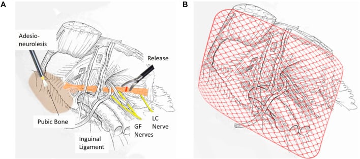
Outcomes. Return to sport is typically 8-12 weeks, with 85-95% success with proper patient selection. Complications include infection, haematoma and chronic pain (5-10%).
Complications
- Incidence
- 20-30% if undertreated
- Prevention/Management
- Proper diagnosis, adequate conservative trial, multidisciplinary approach
- Incidence
- Common in professional athletes
- Prevention/Management
- Structured return to sport protocol, objective criteria (strength, pain-free)
- Incidence
- Less than 5% post-surgery
- Prevention/Management
- Sterile technique, prophylactic antibiotics, wound care
- Incidence
- 5-10% after sports hernia repair
- Prevention/Management
- Proper patient selection, nerve protection during surgery
- Incidence
- Variable (10-40%)
- Prevention/Management
- Partial vs complete release, progressive rehabilitation
- Incidence
- If FAI untreated
- Prevention/Management
- Early recognition and treatment of hip pathology
Recurrent groin pain is the most common "complication": recurrent or persistent groin pain. Its causes include:
- Inadequate rehabilitation (the Copenhagen protocol was not completed)
- Premature return to sport (return before strength normalised)
- Missed hip pathology (FAI driving adductor overload)
- Wrong diagnosis (another cause of groin pain)
- Chronic pubic pathology (osteitis pubis takes months to resolve)
Preventing it.
- Structured return-to-sport criteria
- Ongoing maintenance strengthening
- Training-load monitoring (the acute:chronic workload ratio, under Prevention and Return to Sport)
- Biomechanical assessment and correction
- Early recognition and treatment of recurrent symptoms
Postoperative Care and Rehabilitation
Postoperative management varies by the procedure performed.
Rehabilitation Timeline
- Protected mobilisation, gentle walking
- No straining or Valsalva manoeuvres
- Ice and elevation for swelling
- Pain management with NSAIDs
- Avoid resisted hip flexion
- Progressive core activation (gentle transversus abdominis)
- Hip range-of-motion exercises (pain-free)
- Light cardiovascular work (stationary bike)
- No kicking or cutting movements
- Monitor wound healing
- Progressive core strengthening programme
- Begin straight-line jogging (week 6)
- Sport-specific drills (non-contact)
- Gradual increase in training volume
- Maintain core strengthening
- Full training participation (week 8-10)
- Contact drills as tolerated
- Match simulation
- Gradual return to competition
- Maintenance programme ongoing
Clearance. Pain-free with all movements, core strength normalised, functional testing passed and medical clearance obtained before full return to competition.
Across all procedures. Training load rises progressively, without rapid spikes in volume or intensity, and the acute:chronic workload ratio is monitored. Return is decided on objective criteria (strength tests, hop tests, sport-specific movements), not on time alone. Maintenance programmes continue, the Copenhagen protocol for the adductor and core strengthening for all, and the return-to-sport decision is made by the team: surgeon, physiotherapist, exercise physiologist and sports medicine physician.
Outcomes and Prognosis
Conservative management outcomes, by entity:
- Conservative Success
- 70-80% return to sport
- Timeframe
- 8-12 weeks
- Conservative Success
- 50-60% (many need surgery)
- Timeframe
- 12 weeks trial
- Conservative Success
- 80-90% (patience required)
- Timeframe
- 3-6 months
- Conservative Success
- 50-70% (many need arthroscopy)
- Timeframe
- 12 weeks trial
Surgical outcomes by procedure:
- Return to Sport
- 85-95%
- Timeframe
- 8-12 weeks
- Success Rate
- High with proper selection
- Return to Sport
- 60-85%
- Timeframe
- 12-16 weeks
- Success Rate
- Variable, controversial
- Return to Sport
- 85-90%
- Timeframe
- 4-6 months
- Success Rate
- Excellent in athletes
A good outcome goes with:
- Early recognition and treatment
- Proper diagnosis (Doha classification)
- An adequate conservative trial (8-12 weeks minimum)
- Good rehabilitation compliance
- Appropriate surgical selection (if indicated)
- A multidisciplinary team approach
A poor outcome goes with:
- Chronic duration (greater than 6 months) before presentation
- Premature return to sport
- Inadequate rehabilitation
- Missed hip pathology
- Multiple previous failed treatments
- Bilateral involvement
Prevention and Return to Sport
Preseason screening. Adductor strength, the adductor:abductor ratio, hip range of motion (screening for FAI) and core stability identify the at-risk athlete.
Prevention programmes. The Copenhagen adductor protocol, a core strengthening programme, hip mobility and strengthening, proper warm-up and cool-down, and progressive management of training load.
Load monitoring. Keep the acute:chronic workload ratio under 1.5 and avoid rapid spikes in training volume; monitor GPS data in field sports, periodise training and allow adequate recovery between sessions.
Early intervention. Address symptoms early rather than ignoring groin tightness, modify training load at the first sign, and involve a sports medicine physician, so that an acute problem does not become chronic.
Return to sport criteria. Objective criteria before full return:
- Pain-free with all sport-specific movements
- Adductor strength greater than 90% of the contralateral side
- Adductor:abductor ratio greater than 80%
- Functional tests passed (sprint, cut, kick)
- Graded training progression completed without symptoms
Return to Sport Protocol
- Straight-line jogging
- Progress to running
- No cutting or kicking
- Pain-free requirement
- Add lateral movements
- Progressive cutting drills
- Figure-8 running
- Continue to be pain-free
- Kicking progressions (if soccer/football)
- Sport-specific drills
- Non-contact training
- Increase intensity progressively
- Full team training
- Contact drills
- Match simulation
- Medical clearance required
- Gradual return to competition
- May start as a substitute
- Progress to full match play
- Continue maintenance strengthening
Guidelines, Registries & Global Practice
Athletic groin pain is a global problem of multidirectional field and court sports. The strongest epidemiological data come from prospective football (soccer) surveillance, but the principles apply to ice hockey, rugby codes, Australian Rules Football, Gaelic games and other change-of-direction sports.
Global epidemiology (PMID-backed):
- Key figure
- Hip/groin = 14% of all time-loss injuries; rate ~1.0/1000 h
- Most common entity
- Adductor-related = 63% of hip/groin injuries
- Reference
- Werner 2018, Br J Sports Med (PMID 29691289)
- Key figure
- Hip/groin = 12-16% of all injuries; mean 15 days absence; 15% re-injury
- Most common entity
- Adductor- then iliopsoas-related most common
- Reference
- Werner 2009, Br J Sports Med (PMID 19945984)
- Key figure
- Season prevalence of groin problems 21.3% untreated vs 13.5% with adductor programme
- Most common entity
- Adductor-related
- Reference
- Harøy 2019, Br J Sports Med (PMID 29891614)
Across these datasets the message is consistent: hip and groin complaints account for roughly one in eight time-loss injuries in men's professional football, adductor-related pain is by far the most common single diagnosis, and the burden has not fallen despite a modest decline in incidence — making prevention and accurate sub-classification a priority worldwide.
Where guidance converges and diverges:
- Position
- Use the unified clinical taxonomy (defined entities + hip-related + other); diagnose by history and examination
- Evidence basis
- Expert consensus + systematic reviews (Level 5)
- Position
- Exercise-based active rehabilitation is first line; image to confirm/exclude specific entities, not to screen
- Evidence basis
- Guideline synthesising RCT and cohort evidence
- Position
- Mandatory adequate conservative trial before surgery; reserve operative repair for failed rehabilitation with concordant findings
- Evidence basis
- Consensus + Level 4 surgical series
- Position
- Treat FAI syndrome with care/activity modification, physiotherapy-led rehabilitation, or arthroscopic surgery — shared decision-making
- Evidence basis
- International multidisciplinary consensus
There is broad international agreement on the core pathway: unified terminology (Doha), active exercise rehabilitation first, imaging to characterise rather than screen, and surgery only after a failed adequate conservative trial. Genuine practice variation centres on: (1) the threshold and technique for surgical repair of inguinal-related (so-called sports hernia) pain — open minimal repair, modified open repair, or laparoscopic/endoscopic repair, with no high-level head-to-head RCT establishing superiority; and (2) the role of adductor tenotomy, which remains controversial with variable reported outcomes.
Registry note: unlike arthroplasty, athletic groin pain has no implant joint registry. The closest registry-grade evidence is prospective injury surveillance (UEFA Elite Club Injury Study and similar national datasets), which functions as the field's outcome registry and is the source of the epidemiology above.
Global practice variation and resource setting:
Embedded multidisciplinary teams, early MRI, individualised load monitoring (GPS, acute:chronic workload), and rapid access to surgery. Pressure for early return is a major driver of chronicity.
Diagnosis rests on clinical examination and the Doha framework; MRI may be delayed or unavailable. Structured exercise rehabilitation (Copenhagen-based, Hölmich active-training principles) is low-cost, evidence-based and the mainstay everywhere.
- Comprehensive history and examination (including hip assessment)
- Imaging reports (MRI findings documented)
- Conservative management trial (duration, compliance, response)
- Multidisciplinary discussion notes (if surgery considered)
- Return to sport criteria and clearance
- Realistic success rates and return-to-sport timeframe for the specific procedure
- Risks: chronic/persistent pain, infection, recurrence, incomplete relief
- Alternative: continue or optimise conservative management
- Career and financial implications for professional athletes
Frame answers around the world standard of care, not one country's system: Doha terminology to classify, active exercise rehabilitation first line, imaging to characterise the entity, and surgery reserved for failed adequate conservative treatment with concordant findings. Be ready to state that adductor-related pain is the commonest entity (~63% of hip/groin injuries in elite football) and that hip pathology must always be excluded as a driver.
MCQ Practice Points
Q: What are the four clinical entities in the Doha classification of groin pain in athletes? A: Defined clinical entities = adductor-related (commonest, ~63% of hip/groin injuries in elite football), iliopsoas-related, inguinal-related (sports hernia) and pubic-related (osteitis pubis); plus hip-related (FAI/labral) and other causes. This classification replaced confusing historical terms and provides clinical examination standards for each entity.
Q: How is the adductor squeeze test performed and what does it assess? A: Patient supine, hip and knee flexed to 45 degrees, examiner places fist between knees, patient squeezes knees together against resistance. Positive test = reproduces groin pain, indicates adductor-related groin pain. Most sensitive at 45 degrees hip flexion for adductor longus pathology. Should also test at 0 and 90 degrees.
Q: What is the Copenhagen adductor strengthening protocol and what is its evidence base? A: Evidence-based progressive eccentric adductor strengthening program performed 3 times weekly for 8-12 weeks. Core exercise is the Copenhagen plank (side plank with adduction component). RCT showed 41% reduction in groin injury rates in soccer players. First-line treatment for adductor-related groin pain with 70-80% success rate.
Q: What is the secondary cleft sign and what does it indicate? A: Parasymphyseal cyst (fluid-filled cavity) adjacent to pubic symphysis seen on MRI. Pathognomonic for chronic pubic-related groin pain (osteitis pubis). Represents extension of symphyseal pathology into adjacent pubic bone. Indicates established chronic pathology requiring prolonged conservative management (3-6 months).
Q: Why is it critical to assess hip pathology in athletes with groin pain? A: Up to 50% of athletes with "groin pain" have underlying hip pathology (FAI, labral tear) that can cause secondary adductor overload. Hip pathology presents as groin pain. Limited hip internal rotation and positive FADIR test indicate hip impingement. Treating the adductor without addressing the hip leads to recurrence. Always perform FADIR test and assess hip ROM.
Q: What is a 'sports hernia' and how does it differ from a true inguinal hernia? A: Sports hernia (inguinal-related groin pain) is weakness or disruption of posterior inguinal wall (transversalis fascia, conjoint tendon) WITHOUT a peritoneal hernia sac. No palpable bulge on examination (unlike true hernia). MRI shows posterior wall signal changes. Treated with core strengthening; surgical repair if conservative fails (85-95% success). Term "hernia" is a misnomer.
Exam Viva Scenarios
Practise clinical reasoning and management decisions out loud
“A 24-year-old professional soccer player presents with 3 months of left groin pain. Pain is worst with kicking and change of direction. He has continued playing but performance is declining. Examination shows tenderness over the adductor longus insertion and positive adductor squeeze test at 45 degrees. What is your assessment and management?”
“A 28-year-old rugby player has inguinal-related groin pain for 6 months. He has completed 16 weeks of physiotherapy including core strengthening with only 30% improvement. MRI shows posterior inguinal wall signal changes consistent with sports hernia but no frank hernia. He wants to know about surgery. Walk me through your assessment and surgical decision-making.”
“An elite Australian Rules footballer has bilateral pubic-related groin pain for 4 months. MRI shows extensive pubic bone marrow oedema bilaterally and a secondary cleft sign. He has tried 4 weeks of rest and NSAIDs with minimal improvement. He is frustrated and wants to return to playing. How do you manage this situation?”
“A 26-year-old soccer player was treated for 'adductor-related groin pain' with 12 weeks of Copenhagen protocol. He improved initially but symptoms recurred when he returned to full training. On re-examination, you notice his hip internal rotation is 15 degrees (normal 45 degrees) and FADIR test reproduces his groin pain. What now?”
Doha Classification (4 Defined Entities + Hip-Related + Other)
- Adductor-related = Most common (~63% of hip/groin injuries), squeeze test positive, adductor tenderness
- Iliopsoas-related = Often forgotten defined entity, pain on resisted hip flexion, modified Thomas test
- Inguinal-related = Sports hernia, posterior wall deficiency, NO true hernia sac
- Pubic-related = Osteitis pubis, MRI shows bone oedema + secondary cleft sign
- Hip-related = FAI/labral, positive FADIR, limited hip internal rotation
Physical Examination Must-Do Tests
- Adductor squeeze test = 0, 45, 90 degrees hip flexion (most sensitive 45deg)
- FADIR test = Hip flexion-adduction-internal rotation (impingement)
- Hip internal rotation = Normal 45deg, limited if FAI (assess always)
- Resisted sit-up = Rectus/inguinal canal pain (inguinal-related)
- Pubic palpation = Point tenderness (pubic-related)
Imaging Protocol
- First-line = AP pelvis + frog-leg lateral (screen FAI, pubic pathology)
- Gold standard = MRI pelvis and hips (assess all entities)
- Secondary cleft sign = Parasymphyseal cyst, pathognomonic for chronic pubic stress
- Alpha angle = Frog-leg lateral, greater than 55deg = cam FAI
- MR arthrogram = If hip suspected, best for labral tears
Treatment Algorithm (Conservative First)
- Adductor-related = Copenhagen protocol 8-12wk, 70-80% success
- Inguinal-related = Core strengthening 12wk, surgery if fails (85-95% RTS)
- Pubic-related = Load management 3-6mo, patience critical, surgery rarely indicated
- Hip-related = Hip physio 12wk, arthroscopy if fails (85-90% RTS)
- All entities = Multidisciplinary approach, 12-week trial before surgery
Copenhagen Protocol Details
- Evidence-based = RCT showed 41% reduction in groin injuries
- Frequency = 3 times per week for 8-12 weeks
- Focus = Progressive eccentric adductor loading
- Core exercise = Copenhagen plank (side plank with adduction)
- Outcome = 70-80% return to sport with proper compliance
Key Exam Pearls
- Hip first always = 50% of 'groin pain' has hip pathology driving it
- Secondary cleft sign = Chronic osteitis pubis, requires months to heal
- Sports hernia misnomer = NO true hernia sac, posterior wall weakness
- Surgery last resort = 12-week conservative trial mandatory
- Premature RTS = Main cause of chronicity, objective criteria required
Evidence Base and Key Studies
Doha Agreement on Groin Pain Terminology
- One-day agreement meeting (4 Nov 2014), 24 international experts from 14 countries, informed by systematic reviews and a Delphi questionnaire
- Three major categories: (1) defined clinical entities (adductor-related, iliopsoas-related, inguinal-related, pubic-related); (2) hip-related groin pain; (3) other causes
- Classification is history- and examination-based, designed for both clinical practice and research
- Replaced confusing historical terms (Gilmore groin, sportsman hernia, etc.)
Adductor Strengthening Programme (Copenhagen Adduction Exercise) — Prevention RCT
- Cluster-randomised controlled trial: 35 semiprofessional Norwegian football teams, 652 players (intervention 339, control 313)
- Single-exercise programme based on the Copenhagen Adduction exercise, three progression levels (preseason 3x/week, in-season 1x/week)
- Average in-season prevalence of groin problems 13.5% (intervention) vs 21.3% (control)
- Risk of reporting groin problems 41% lower in the intervention group (OR 0.59, 95% CI 0.40 to 0.86, p=0.008)
Athletic Pubalgia and the 'Sports Hernia': MR Imaging Findings
- Retrospective MRI study of 141 patients (134 male) with clinical athletic pubalgia, compared with surgery (102), examination (141) and 25 asymptomatic controls
- 138 of 141 patients (98%) had MRI findings capable of causing groin pain
- MRI sensitivity/specificity vs surgery: 68%/100% for rectus abdominis tendon injury and 86%/89% for adductor tendon injury
- Only 2 patients had a true hernia at surgery; rectus abdominis and adductor injury were significantly more common than in controls (p less than 0.001)
- Findings grouped into osteitis pubis, adductor compartment injury, rectus abdominis injury, and disease remote from the symphysis
Experience with 'Sports Hernia' Spanning Two Decades
- Senior-author review of 8,490 patients and 5,460 operations over two decades
- Reframes 'sports hernia' as athletic pubalgia — a spectrum of abdominal/pelvic musculature injuries on both sides of the pubic symphysis, outside the hip joint
- Prospective data cited show greater than 95% success when timely, appropriate repair of selected injuries is performed
- Over the study period the proportion of female patients, age range, number of sports and number of identified syndromes all increased
- MRI substantially improved diagnosis of both hip and non-hip pelvic pathology
Return to Play After Hip Arthroscopy for FAI in Professional Soccer Players
- Single-surgeon case series of 24 professional soccer players (26 hips) undergoing hip arthroscopy for FAI, 2005-2015
- 96% returned to play at the professional level
- Mean time from surgery to first professional game 9.2 months (range 1.9-24.0)
- National-team players returned significantly earlier (median 5.7 vs 11.6 months, p=0.018)
- Severe chondral damage and microfracture did not preclude return to play
Active Physical Training for Long-Standing Adductor-Related Groin Pain (Landmark RCT)
- Single-blind randomised trial of 68 athletes with long-standing (median 40 weeks) adductor-related groin pain
- Active training (strength/coordination of hip and adductor muscles) vs physiotherapy without active training, over 8-12 weeks
- 23/29 in the active-training group vs 4/30 in the comparison group returned to sport without groin pain at 4 months
- Odds ratio 12.7 (95% CI 3.4 to 47.2) favouring active training