Junctional Fracture | Neck-Intertrochanteric Junction | ORIF vs Arthroplasty
- Basicervical fracture = extracapsular fracture at the base of the femoral neck (AO/OTA 31-B2.1); rotationally unstable
- Treat like an intertrochanteric fracture - cephalomedullary nail or sliding hip screw, NOT isolated cannulated screws (23% revision)
- Control rotation: add a derotation screw to a sliding hip screw; an IM nail with helical blade resists collapse best
- SHS and CMN are equivalent for cut-out and reoperation (~7-8% revision); choose by surgeon/fracture factors
- Collapse ~25%, conversion to arthroplasty ~8-9% - counsel about reoperation; arthroplasty for elderly/poor bone or salvage
- “Basicervical = extracapsular base-of-neck fracture, AO/OTA 31-B2.1, rotationally unstable
- “Manage as an intertrochanteric injury: CMN or SHS, never isolated cannulated screws (23% revision)
- “Add a derotation element / favour IM nail with helical blade to resist rotation and collapse
- “SHS vs CMN equivalent for cut-out and reoperation; arthroplasty for elderly poor bone or salvage
Basicervical fracture = fracture at the base of the femoral neck (AO/OTA 31-B2.1), just proximal to the intertrochanteric line. It is largely extracapsular and behaves biomechanically like an intertrochanteric fracture, not a true intracapsular neck fracture.
High collapse and rotational instability is the defining problem. Collapse occurs in roughly a quarter of cases and ~8-9% need conversion to arthroplasty. The single lag implant can allow the head-neck fragment to spin - rotational control is the key technical objective.
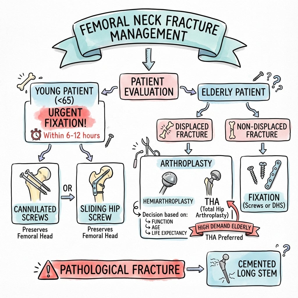
Treat as an intertrochanteric injury: cephalomedullary nail (CMN) or sliding hip screw (SHS) - these give equivalent cut-out and reoperation rates (~7-8%). Isolated cannulated screws are inadequate (23% revision) and should not be used.
Add a derotation screw to a sliding hip screw, or use an IM nail with a helical blade - the construct least associated with collapse in cohort data. Reserve arthroplasty for the elderly with poor bone, comminution, or failed fixation.
- Preferred Implant
- Sliding hip screw OR cephalomedullary nail
- Key Technical Point
- Add derotation screw to SHS
- Revision (pooled)
- ~7-8%
- Preferred Implant
- IM nail + helical blade
- Key Technical Point
- Lowest collapse risk in cohort data
- Revision (pooled)
- ~8%
- Preferred Implant
- Avoid
- Key Technical Point
- Inadequate rotational control
- Revision (pooled)
- 23% (unacceptable)
- Preferred Implant
- Arthroplasty (hemi / THA)
- Key Technical Point
- Salvage or primary in low-reserve patient
- Revision (pooled)
- ~0-8%
NAILConstruct Goals
Hook:NAIL: Neck base pattern, Anti-rotation essential, Intramedullary nail favoured, Lag screw central with low TAD!
Overview and Epidemiology
Basicervical fractures occur at the base of the femoral neck, just proximal to the intertrochanteric line (AO/OTA 31-B2.1). The fracture line lies largely outside the capsular reflection, so the injury is effectively extracapsular and behaves biomechanically far more like an intertrochanteric fracture than a true intracapsular neck fracture. The hallmark problem is rotational and collapse instability: a single lag implant can allow the proximal fragment to rotate, producing collapse, cut-out and a need for reoperation.
There is no universally agreed radiographic definition, which is the main reason reported failure rates vary so widely in the literature.
Mechanism of Injury
Similar to femoral neck fractures:
- Low-energy fall: Elderly patients (osteoporosis)
- High-energy trauma: Young patients (motor vehicle accident, fall from height)
- Torsion: Rotational force
- Direct trauma: Less common
The basicervical region is a transition zone between the femoral neck and intertrochanteric region. Because the line sits at the base of the neck, the fragment is short and the rotational lever arm is small, which is exactly why a single lag screw or blade is prone to allowing rotation and collapse.
Basicervical fracture = base-of-neck, AO/OTA 31-B2.1, functionally extracapsular. The exam point is that it should be managed like an intertrochanteric fracture (cephalomedullary nail or sliding hip screw with a derotation screw) and NOT with isolated cannulated screws, which carry a 23% revision rate. Because it is largely extracapsular, AVN risk is much lower than for a displaced intracapsular neck fracture.
Epidemiology
- Incidence: uncommon - 1.8% of all proximal femoral fractures in pooled review data; up to ~4% in single-centre series (the older "5-10%" figure is not supported)
- Age: predominantly elderly with osteoporotic bone; a minority result from high-energy trauma in younger patients (e.g. concurrent ipsilateral hip-and-shaft injuries, where basicervical was the commonest hip-fracture subtype)
- Gender: female predominance (osteoporosis)
- Laterality: usually unilateral
- Associated injuries: consider ipsilateral femoral shaft fracture in high-energy cases
Anatomy and Pathophysiology
Basicervical Region Anatomy
The basicervical region:
- Location: base of the femoral neck, immediately proximal to the intertrochanteric line
- Boundaries: distal to the true femoral neck, proximal to the lesser trochanter
- Capsule: the fracture line lies at or distal to the capsular reflection - functionally extracapsular
- Blood supply: the medial femoral circumflex artery and its retinacular branches enter more distally than the subcapital region, so the head fragment usually retains its blood supply
- Biomechanics: short proximal fragment with a small rotational lever arm
Pathophysiology
- Rotational instability - the short head-neck fragment can spin around a single lag screw or blade
- Collapse / shortening - axial and varus collapse at the fracture line
- Cut-out - lag screw migration through the head, worsened by a high tip-apex distance
- A purely extramedullary plate-and-screw construct showed substantially higher collapse and failure than an intramedullary helical-blade construct in cohort data
- Because the fracture is largely extracapsular and distal to the main retinacular entry, AVN is uncommon - far lower than for a displaced intracapsular neck fracture. Failure is a mechanical (collapse/cut-out) problem, not primarily a vascular one.
The head-neck fragment can rotate around a single lag implant. Build in rotational control: add a derotation screw to a sliding hip screw, or use a cephalomedullary nail with a helical blade. Avoid isolated cannulated screws (23% revision). Reserve arthroplasty for the elderly with poor bone, comminution, or failed fixation.
Classification Systems
AO/OTA Position
The basicervical fracture is coded 31-B2.1 within the femoral neck (31-B) group - a basicervical/transcervical base-of-neck fracture. It sits at the boundary between the neck (31-B) and trochanteric (31-A) groups, which is the source of much of the classification debate.
Key point: although coded under the neck group, its mechanical behaviour and treatment are those of an intertrochanteric (31-A) fracture, not a true intracapsular neck fracture.
Clinical Assessment
History
Mechanism: Similar to femoral neck fractures
- Low-energy fall: Elderly patients (osteoporosis)
- High-energy trauma: Young patients (motor vehicle accident, fall from height)
- Torsion: Rotational force
Symptoms:
- Immediate pain in hip/groin
- Inability to bear weight
- Leg shortening and external rotation (if displaced)
- Pain with movement
Physical Examination
Inspection:
- Leg shortening (if displaced)
- External rotation (if displaced)
- Swelling (minimal - deep location)
Palpation:
- Tenderness over hip/groin
- Crepitus (rare)
- Greater trochanter tenderness
Range of Motion:
- Limited hip ROM (pain)
- Pain with passive motion
- Inability to perform straight leg raise (if displaced)
Neurovascular Status:
- Usually intact
- Assess distal pulses and sensation
Leg shortening and external rotation suggest displacement. Basicervical fractures may present similarly to femoral neck or intertrochanteric fractures. Imaging is essential for diagnosis and classification.
Associated Injuries
- Other fractures: 10-15% (wrist, spine, other hip)
- Head injury: 5-10% (high-energy trauma)
- Soft tissue injuries: Less common
Investigations
Standard X-ray Protocol
Views: AP pelvis and lateral hip.
Key findings:
- Fracture location: Base of femoral neck, junction with intertrochanteric region
- Displacement: Assess displacement (under vs over 2mm)
- Comminution: Assess for comminution
- Bone quality: Assess for osteoporosis
AP and lateral views essential - shows fracture location and displacement.
Differential Diagnosis (Where Is The Fracture Line?)
The whole management decision turns on correctly distinguishing a basicervical fracture from its neighbours on the AP and lateral films.

- Location of line
- Just below head
- Capsule
- Intracapsular
- AVN risk
- High (displaced)
- Preferred fixation
- Arthroplasty (elderly) / urgent ORIF (young)
- Location of line
- Mid neck
- Capsule
- Intracapsular
- AVN risk
- Moderate-high
- Preferred fixation
- Cannulated screws / FNS / arthroplasty
- Location of line
- Base of neck, at/below capsular reflection
- Capsule
- Functionally extracapsular
- AVN risk
- Low
- Preferred fixation
- SHS + derotation OR cephalomedullary nail
- Location of line
- Between trochanters
- Capsule
- Extracapsular
- AVN risk
- Very low
- Preferred fixation
- Sliding hip screw or cephalomedullary nail
- Location of line
- Below/through lesser troch
- Capsule
- Extracapsular
- AVN risk
- Very low
- Preferred fixation
- Cephalomedullary nail (SHS contraindicated)
Management Algorithm
Management Pathway
Basicervical Fracture Management
Confirm a base-of-neck fracture on AP and lateral films and distinguish it from transcervical and intertrochanteric patterns. Assess comminution, bone quality and (in high-energy cases) an ipsilateral shaft fracture.
Most patients - including the elderly with reasonable bone - are fixed. Use a sliding hip screw with a derotation screw, or a cephalomedullary nail with a helical blade. Aim for a central lag position with tip-apex distance under 25mm.
For comminuted or rotationally unstable patterns, an intramedullary nail with a helical blade resists collapse best. Do not use isolated cannulated screws.
Reserve arthroplasty for the frail elderly with very poor bone, marked comminution, or as salvage after failed fixation (~8-9% of cases). Hemiarthroplasty for low demand; THA if active with acetabular wear.
Surgical Technique
Sliding Hip Screw (DHS) Technique
Indications:
- First-line extramedullary option for two-part basicervical fractures with good bone
Advantages:
- Familiar, low-cost, allows controlled compression
- Pooled revision ~7%, equivalent to cephalomedullary nailing
Critical addition: place an anti-rotation (derotation) screw above the lag screw. Without it the head-neck fragment can rotate during lag-screw insertion - the classic technical pitfall of this fracture.
Technique:
- Lateral approach
- Reduce fracture (anatomic reduction)
- Insert anti-rotation guide wire first
- Guide wire for lag screw (centre-centre position)
- Ream and insert lag screw, tip-apex distance under 25mm
- Attach side plate and fix
- Insert derotation screw; confirm reduction and rotational stability
Tip-Apex Distance: The Central Technical Target
Every construct in this topic is judged against one number - the tip-apex distance (TAD) - yet the metric deserves defining in its own right, because a low TAD is the single most reproducible way to reduce lag-screw cut-out in a rotationally unstable base-of-neck fracture.
Definition (Baumgaertner). TAD is the sum of two distances: the gap from the tip of the lag screw to the apex (subchondral surface) of the femoral head measured on the AP radiograph, plus the same distance measured on the lateral radiograph. Each distance is corrected for radiographic magnification, using the known diameter of the lag screw as the on-screen scale.
Why 25 mm. In the original 198-fracture series, none of the 120 screws with a TAD of 25 mm or less cut out; mean TAD was 24 mm in fractures that healed versus 38 mm in those that failed, with a strong dose-response - every additional millimetre raised the cut-out risk regardless of fracture pattern, reduction or implant.
How to hit it: aim the guide wire centre-centre or slightly inferior-central, deep to within 5-10 mm of subchondral bone, on both views before reaming. A calcar-referenced TAD (CalTAD), which measures the AP component from an inferiorly placed screw to the calcar rather than to the centre, has been proposed as a refinement, reflecting evidence that an inferior (rather than superior) AP screw position resists cut-out; the practical message is the same - deep, central-to-inferior, and never superior or short.
Tip-Apex Distance Predicts Cut-out - Original Series
- TAD = AP tip-apex distance + lateral tip-apex distance, corrected for magnification
- No cut-outs among the 120 screws with a TAD of 25 mm or less
- Rising TAD strongly predicts cut-out regardless of other fracture variables
TAD = (AP tip-to-apex) + (lateral tip-to-apex), corrected for magnification; target 25 mm or less. In the basicervical pattern a low TAD must be combined with explicit rotational control (a derotation screw or a helical blade) - a perfectly central screw still fails if the short head-neck fragment is free to spin.
Complications
- Incidence
- ~25%
- Risk Factors
- Comminution, rotational instability
- Prevention/Management
- Anti-rotation construct, central lag, low TAD
- Incidence
- ~8-9%
- Risk Factors
- Failed fixation, cut-out
- Prevention/Management
- IM nail + helical blade; salvage with arthroplasty
- Incidence
- Variable
- Risk Factors
- High TAD, eccentric screw, osteoporosis
- Prevention/Management
- TAD under 25mm, central screw position
- Incidence
- 23%
- Risk Factors
- Use of isolated screws
- Prevention/Management
- Avoid screws - use SHS or nail
- Incidence
- Uncommon
- Risk Factors
- Largely extracapsular pattern
- Prevention/Management
- Lower than intracapsular neck fractures
Fracture-Site Collapse and Fixation Failure
Collapse ~25%, conversion to arthroplasty ~8-9%:
- Cause: rotational instability and comminution of the short proximal fragment; an extramedullary plate (vs IM helical blade) independently raised failure (OR 12.2 in cohort data)
- Prevention: rotational control (derotation screw or helical blade), central lag screw, tip-apex distance under 25mm
- Management: revision fixation, or conversion to arthroplasty for established failure
AVN
Uncommon:
- Cause: the fracture is largely extracapsular and distal to the main retinacular entry, so the head usually keeps its blood supply - AVN is far less common than after a displaced intracapsular neck fracture
- Management: arthroplasty if symptomatic AVN does develop
Nonunion
Uncommon (lower than intracapsular neck fractures):
- Cause: inadequate fixation, poor reduction, poor bone quality
- Prevention: stable construct with good cortical apposition
- Management: revision fixation or conversion to arthroplasty
FAILComplications
Hook:FAIL: Failure/collapse ~25%, Arthroplasty conversion ~8-9%, Implant cut-out, Loss of length (shortening)!
Postoperative Care
Weight Bearing and Mobilisation
- Early mobilisation is the goal in this elderly population - prolonged restricted weight bearing is poorly tolerated and increases medical complications
- With a stable cephalomedullary nail or sliding hip screw, weight bear as tolerated under physiotherapy supervision; reserve protected weight bearing for very comminuted or tenuous constructs
- Arthroplasty: weight bear as tolerated
- Begin hip and knee range of motion and quadriceps work from day one
Recovery Trajectory
- Inpatient: orthogeriatric review, VTE prophylaxis, analgesia, delirium screening
- Weeks 0-6: supervised gait re-education, progressive loading, falls prevention
- Weeks 6-12: most patients consolidating; check radiographs for collapse and screw position
- Ongoing: bone-protection therapy, continued strength and balance work
Functional recovery to pre-injury mobility is the realistic endpoint; the priority is preventing collapse and a second fall, not return to sport.
Outcomes and Prognosis
Pooled Outcomes by Implant
Pooled systematic-review data (Dekhne 2021; Yoon 2022) give revision rates of approximately:
- Sliding hip screw: ~7%
- Cephalomedullary nail: ~8% (slightly faster union than SHS)
- Cannulated screws: 23% (unacceptable - avoid)
- Hemiarthroplasty: ~8%; total hip arthroplasty: ~0% (very small numbers)
Cohort data show fracture-site collapse in ~25% and conversion to arthroplasty in ~8-9% of fixed cases (Lee 2018).
Prognostic Factors
- Unfavourable
- Medial comminution / rotational instability
- Unfavourable
- Severe osteoporosis
- Unfavourable
- Isolated cannulated screws / no derotation screw
- Unfavourable
- High tip-apex distance, eccentric screw
Because the fracture is largely extracapsular, AVN and nonunion are uncommon - the dominant late problem is mechanical collapse, not vascular failure.
Prevention and Bone Health
This is predominantly a fragility fracture, so management does not end with the operation.
- Treat the osteoporosis: assess fracture risk and start bone-protective therapy (e.g. bisphosphonate or denosumab) with calcium/vitamin D as indicated
- Falls prevention: multifactorial assessment, gait and balance training, medication review, home-hazard modification
- Orthogeriatric co-management and early mobilisation reduce medical complications and mortality after hip fracture
- In the rare high-energy young patient, exclude an ipsilateral femoral shaft fracture and other injuries
The Concurrent Ipsilateral Femoral Neck-Shaft Fracture
The topic repeatedly flags the need to exclude an ipsilateral femoral shaft fracture in high-energy cases, and notes that the basicervical pattern was the commonest hip-fracture subtype among concurrent ipsilateral hip-and-shaft injuries - a combination that deserves its own plan, because it changes both the work-up and the implant strategy.
Recognise it. Ipsilateral neck-and-shaft fractures are a recognised association of high-energy femoral shaft fractures in younger patients. The neck component is notoriously easy to miss - it is often minimally displaced, vertical or basicervical, and overshadowed by the dramatic shaft injury, so a substantial minority are diagnosed late. Every high-energy femoral shaft fracture therefore warrants dedicated hip imaging: a true AP internal-rotation hip view, fine-cut CT of the femoral neck, and a deliberate fluoroscopic check of the neck before, during and after shaft fixation.
The neck fracture takes priority. Its risk of avascular necrosis and nonunion dominates the long-term prognosis, so it must be anatomically reduced and stabilised - do not let neck reduction be lost while passing an antegrade nail.
Two construct strategies:
- Single-implant reconstruction (cephalomedullary) nail - one device spans both fractures, but neck reduction and proximal-screw placement are constrained by the nail, and the neck can be left under-reduced.
- Dual-implant "separate" fixation - the neck is fixed independently (cannulated screws or a sliding hip screw) and the shaft with a retrograde nail or plate. This is increasingly favoured because it lets the higher-stakes neck fracture be reduced and fixed anatomically and independently of the shaft implant.
Whichever is chosen, the guiding principle is the same: treat the neck fracture as the priority injury and confirm its reduction is maintained after the shaft is stabilised.
In a young high-energy femur, actively hunt for an occult ipsilateral neck fracture - CT the neck and check it on fluoroscopy. If present, the neck fracture takes precedence: fix it anatomically, and prefer a strategy (often dual-implant with a retrograde shaft nail) that lets you reduce the neck independently rather than accepting a reduction compromised by a single spanning nail.
BASICBasicervical Fracture Features
Hook:BASIC: Base of neck, treat As intertrochanteric, Sliding hip screw/nail, Instability is rotational, Cannulated screws avoided!
Guidelines, Registries & Global Practice
Global Epidemiology
- Basicervical fractures are uncommon: ~1.8% of all proximal femoral fractures in pooled review data, up to ~4% in single-centre series
- Predominantly a fragility fracture of older adults with osteoporotic bone; female predominance
- In high-energy young patients, basicervical was the commonest hip-fracture subtype in concurrent ipsilateral hip-and-shaft injuries
Guidelines Side by Side
- Position relevant to basicervical fractures
- Codes the fracture as 31-B2.1; recommends managing the base-of-neck pattern with an angular-stable device (sliding hip screw or cephalomedullary nail) and controlling rotation
- Position relevant to basicervical fractures
- Hip-fracture guidance: surgery within 36 hours, orthogeriatric co-management, early mobilisation, extramedullary implant (e.g. SHS) for stable trochanteric-type fractures; intramedullary nail for subtrochanteric/reverse-oblique patterns
- Position relevant to basicervical fractures
- Hip-fracture CPG: prompt surgery, multidisciplinary care, VTE prophylaxis, and post-fracture bone-health/osteoporosis management
- Position relevant to basicervical fractures
- Supports treating the basicervical pattern as a trochanteric-type injury and avoiding isolated cannulated screws
Registry and Practice Variation
- Hip-fracture registries (e.g. UK National Hip Fracture Database, and national registries elsewhere) track time-to-surgery, orthogeriatric input and 30-day mortality; basicervical fractures are usually pooled within neck or trochanteric categories
- High-resource settings: routine cephalomedullary nails/helical blades, orthogeriatric pathways, systematic bone-protection
- Limited-resource settings: sliding hip screw remains a cost-effective, widely available workhorse with equivalent revision rates; implant choice is often driven by availability rather than marginal biomechanical advantage
Basicervical fractures are a classic viva trap. Know that it is a base-of-neck, AO/OTA 31-B2.1, functionally extracapsular fracture; that the problem is rotational instability and collapse (~25%) rather than AVN; that you treat it as an intertrochanteric injury with a sliding hip screw plus derotation screw or a cephalomedullary nail with helical blade; and that isolated cannulated screws are wrong (23% revision). Be ready to discuss rotational control, tip-apex distance and when to choose arthroplasty.
Controversies and Areas of Uncertainty
There is no consensus radiographic definition of a basicervical fracture. Series disagree on whether the line is judged against the intertrochanteric crest or the capsular reflection, so reported failure rates span 0% to over 50% and pooled estimates must be read cautiously (Yoo 2020; Dekhne 2021).
Meta-analysis found no significant difference in cut-out or reoperation between cephalomedullary nail and sliding hip screw, with slightly faster union for the nail (Yoon 2022). Yet a cohort identified the extramedullary plate as an independent risk factor for failure (Lee 2018). The pragmatic position: both work; favour an intramedullary helical-blade construct in osteoporotic/comminuted bone.
Once considered acceptable, isolated cannulated screws now have clear evidence against them - 23% revision in pooled data and lower load-to-failure biomechanically (Dekhne 2021; Panteli 2015). This is a genuine change in recommended practice.
Arthroplasty data are limited to small cohorts but suggest acceptable revision rates. Whether primary arthroplasty should be offered to selected frail elderly patients - rather than fixation followed by ~8-9% conversion - remains unresolved for want of comparative trials.
MCQ Practice Points
Q: Where is a basicervical fracture and how is it coded? A: At the base of the femoral neck, AO/OTA 31-B2.1. The line lies at or distal to the capsular reflection, so it is functionally extracapsular and behaves like an intertrochanteric fracture.
Q: Why are basicervical fractures prone to failure? A: Rotational and collapse instability of the short proximal fragment. Fracture-site collapse occurs in ~25% and ~8-9% require conversion to arthroplasty. The problem is mechanical, not vascular - AVN is uncommon.
Q: What implant should be used? A: Treat as an intertrochanteric fracture - a sliding hip screw with a derotation screw, or a cephalomedullary nail with a helical blade. SHS and CMN have equivalent cut-out and reoperation rates (~7-8%).
Q: Which fixation should be avoided for a basicervical fracture? A: Isolated cannulated screws - 23% revision rate in pooled data and lower load-to-failure biomechanically. The short fragment gives poor purchase and inadequate rotational control.
Q: When is arthroplasty indicated? A: The exception, not the default - frail elderly with very poor bone or marked comminution, or salvage after failed fixation. Most patients, including the elderly with reasonable bone, are fixed.
Q: What technical targets reduce failure? A: Rotational control (derotation screw or helical blade), a central lag screw, and a tip-apex distance under 25mm to minimise cut-out.
Exam Viva Scenarios
Practise clinical reasoning and management decisions out loud
“A 45-year-old man presents after a motor vehicle accident with a painful hip and inability to bear weight. AP and lateral radiographs show a base-of-neck femoral fracture with mild displacement. He is otherwise fit.”
“An 80-year-old woman presents after a fall. AP and lateral films show a base-of-neck fracture with medial comminution. She has osteoporosis but is independently mobile with a stick.”
“A 74-year-old man returns 3 months after sliding hip screw fixation of a basicervical fracture with increasing groin pain. Radiographs show varus collapse and the lag screw has cut out superiorly into the joint.”
Key Anatomy
- Base of femoral neck; line at/distal to capsular reflection - functionally extracapsular
- AO/OTA 31-B2.1; behaves like an intertrochanteric fracture
- Retinacular vessels enter distally, so AVN is uncommon
- Short proximal fragment, small lever arm - rotationally unstable
Classification / Definition
- AO/OTA 31-B2.1 (basicervical, within the 31-B neck group)
- No consensus radiographic definition - drives wide reported failure rates
- Stratify by comminution and bone quality, NOT age alone
- Differentiate from transcervical (intracapsular) and intertrochanteric lines
Treatment Algorithm
- Default: fixation, treated as an intertrochanteric injury
- Two-part good bone: SHS + derotation screw OR CMN (revision ~7-8%)
- Comminuted/osteoporotic: cephalomedullary nail + helical blade
- Arthroplasty: frail elderly poor bone, severe comminution, or salvage
Surgical Pearls
- Control rotation: derotation screw or helical blade
- Central lag screw, tip-apex distance under 25mm
- AVOID isolated cannulated screws (23% revision)
- SHS and CMN equivalent for cut-out and reoperation
Complications
- Fracture-site collapse: ~25%
- Conversion to arthroplasty: ~8-9%
- Lag screw cut-out: worse with high TAD
- AVN and nonunion: uncommon (extracapsular pattern)
Evidence Base
Definition, Treatment and Failure - Systematic Review
- Basicervical fractures = 1.8% of all proximal femoral fractures (uncommon)
- No consensus definition - heterogeneity drives variable failure rates
- Treat as an intertrochanteric-type injury, not with isolated cannulated screws
Treatment and Outcomes - Systematic Review (910 patients)
- Cannulated screws revision 23% - unacceptable for basicervical pattern
- SHS (7%) and CMN (8%) equivalent and acceptable
- Arthroplasty data limited but appears acceptable
Cephalomedullary Nail vs DHS - Meta-analysis
- No significant difference in cut-out or reoperation between CMN and DHS
- CMN achieved slightly faster union than DHS
- Implant selection can follow surgeon preference
Risk Factors for Fixation Failure (multicentre cohort)
- Collapse 24.6%, conversion to arthroplasty 8.6%
- Sliding hip screw plate had higher failure than IM nail + helical blade (OR 12.2)
- Inherent rotational and collapse instability drives failure
Biomechanical Comparison of Fixation Devices (cadaver)
- No clear biomechanical superiority of plate vs nail
- Add a derotation/anti-rotation element to resist rotational failure
- Select construct by anatomy and surgeon comfort
Biomechanical Rationale by Fracture Pattern
- Triangular cannulated screws are biomechanically weak in basicervical pattern
- Match implant to the specific fracture geometry
- Cephalomedullary devices favoured for high-shear patterns