The 'Stippled Epiphyses' Dysplasias
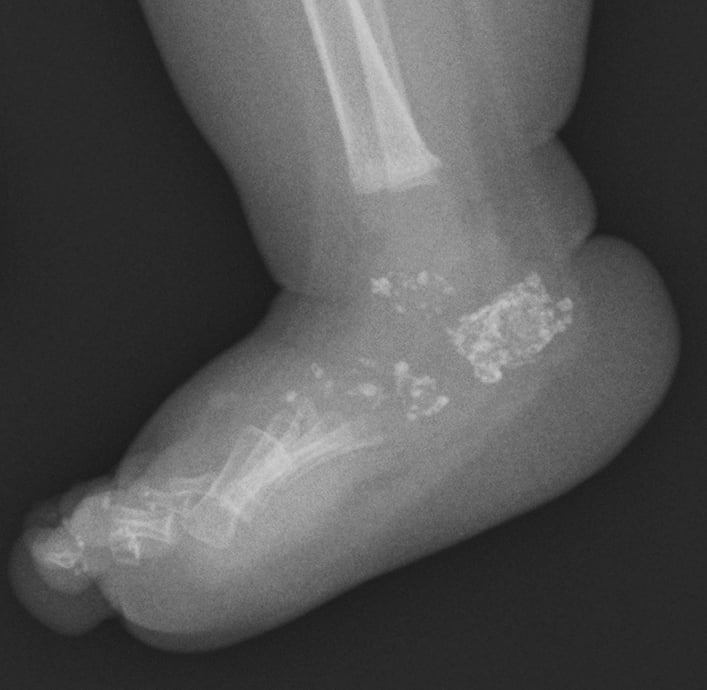
- CHONDRODYSPLASIA PUNCTATA (CDP) is a heterogeneous GROUP of skeletal dysplasias unified by a single radiographic HALLMARK in infancy: STIPPLED (punctate) epiphyseal and peri-articular CALCIFICATION (the calcific stippling characteristically RESOLVES as the child grows); chondrodysplasia punctata is recognised among the skeletal dysplasias diagnosed in the fetal/neonatal period.
- The RHIZOMELIC form is the most severe, but it is a SPECTRUM rather than one disease: an AUTOSOMAL-RECESSIVE peroxisomal disorder of PLASMALOGEN synthesis (five types - PEX7, GNPAT, AGPS, FAR1 and the long isoform of PEX5). CLASSIC disease, with negligible plasmalogens, gives symmetric rhizomelic shortening, profound developmental delay, congenital cataracts, contractures and often early death; NONCLASSIC patients with HIGHER residual plasmalogen levels have better growth and cognition and survive longer - so the plasmalogen level is prognostic, not merely diagnostic, and counselling should wait for it.
- CONRADI-HUNERMANN syndrome (CDPX2) is X-LINKED DOMINANT (EBP gene), affects mostly FEMALES (often lethal in males), and is milder/survivable: it causes ASYMMETRIC limb shortening (and hence limb-length DISCREPANCY), SCOLIOSIS, ICHTHYOSIS/skin changes following Blaschko's lines, and CATARACTS - the asymmetry is a key clue.
- There is also an X-LINKED RECESSIVE (brachytelephalangic) form (ARSL/arylsulfatase defects) with nasal and distal-phalangeal hypoplasia, and important SECONDARY/TERATOGENIC causes that phenocopy CDP - notably WARFARIN EMBRYOPATHY (maternal warfarin in pregnancy) and maternal AUTOIMMUNE disease (e.g. systemic lupus erythematosus) - so a maternal drug/autoimmune history matters.
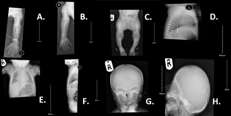
- The ORTHOPAEDIC sequelae depend on the form and include RHIZOMELIC/asymmetric limb SHORTENING and limb-length DISCREPANCY, SCOLIOSIS (especially Conradi-Hunermann), joint CONTRACTURES and (in milder survivors) deformity; ophthalmology (cataracts) and the systemic/metabolic aspects are co-managed.
- MANAGEMENT is largely SUPPORTIVE and form-dependent within a multidisciplinary team: genetic/metabolic diagnosis (distinguishing peroxisomal rhizomelic CDP from EBP/ARSL forms and from teratogenic causes), ORTHOPAEDIC management of limb-length discrepancy (epiphysiodesis/lengthening), SCOLIOSIS and contractures in survivable forms, cataract surgery, and family counselling - with the severe rhizomelic form being largely palliative.
- “Chondrodysplasia punctata = heterogeneous group; HALLMARK = STIPPLED (punctate) epiphyseal calcification in infancy (resolves with age).
- “RHIZOMELIC CDP = AUTOSOMAL-RECESSIVE PEROXISOMAL plasmalogen defect (RCDP1-5: PEX7, GNPAT, AGPS, FAR1, PEX5L) - a SPECTRUM graded by residual plasmalogen: classic (negligible) severe with early death, NONCLASSIC (higher) milder with better growth and cognition. CONRADI-HUNERMANN (CDPX2) = X-linked DOMINANT (EBP), females, ASYMMETRIC limb shortening + ichthyosis + cataracts.
- “Also X-linked recessive (brachytelephalangic) + TERATOGENIC phenocopies (WARFARIN embryopathy, maternal SLE/autoimmune). Orthopaedic: limb shortening/discrepancy, scoliosis, contractures - supportive + deformity correction.
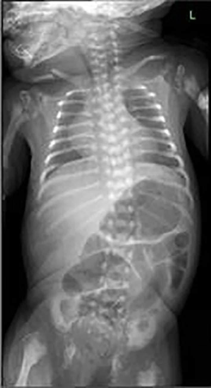
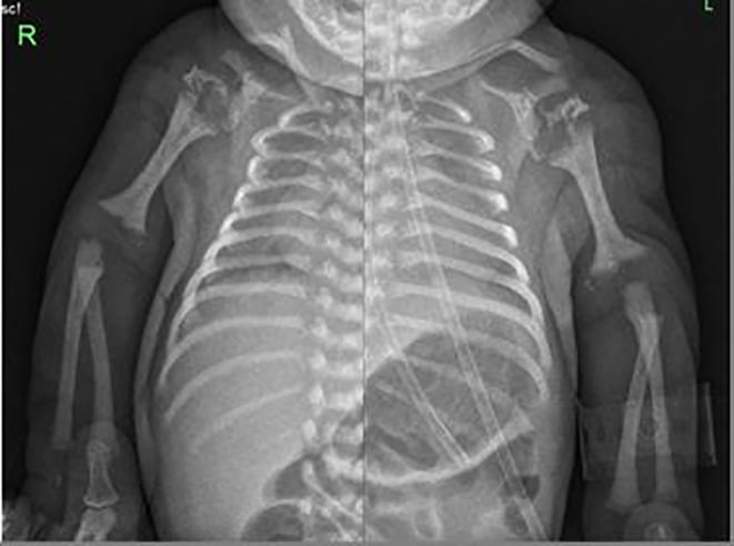
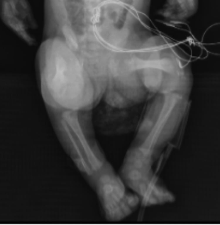
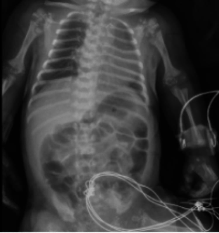
Infant radiograph: stippled (punctate) epiphyses. Rhizomelic (AR peroxisomal) = severe/symmetric; Conradi-Hunermann (X-linked dominant, EBP) = milder/asymmetric + ichthyosis + cataracts.
Teratogenic phenocopies: maternal warfarin (warfarin embryopathy) and maternal autoimmune disease (SLE). Take a maternal drug/autoimmune history.
Overview & Epidemiology
Chondrodysplasia punctata is not one disease but a radiographic sign shared by a group of disorders - stippled epiphyseal calcification in infancy - produced by defects in peroxisomal plasmalogen synthesis, cholesterol biosynthesis, arylsulfatase function, or by teratogenic insult.
- How rare: all forms are rare orphan diseases and no reliable population incidence was retrievable, so none is quoted here; the honest anchor is that CDP sits among the less common skeletal dysplasias encountered in fetal/neonatal practice - in the Jezova 10-year perinatal series, thanatophoric dysplasia and osteogenesis imperfecta made up about half of autopsy cases, with CDP among the less frequent diagnoses.
- The nosology context: more than 400 genetic skeletal disorders are recognised in the current classification, and an increasing proportion are confirmed by genetic testing.
- Who gets which form: the rhizomelic forms are autosomal recessive and affect either sex; Conradi- Hünermann (CDPX2) is X-linked dominant and seen almost entirely in females, because hemizygous males rarely survive; the brachytelephalangic form (CDPX1) is X-linked recessive; and the teratogenic phenocopies follow maternal exposures - warfarin, autoimmune disease - rather than any inheritance.
Pathophysiology: The Metabolic Basis of Each Form
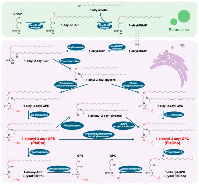
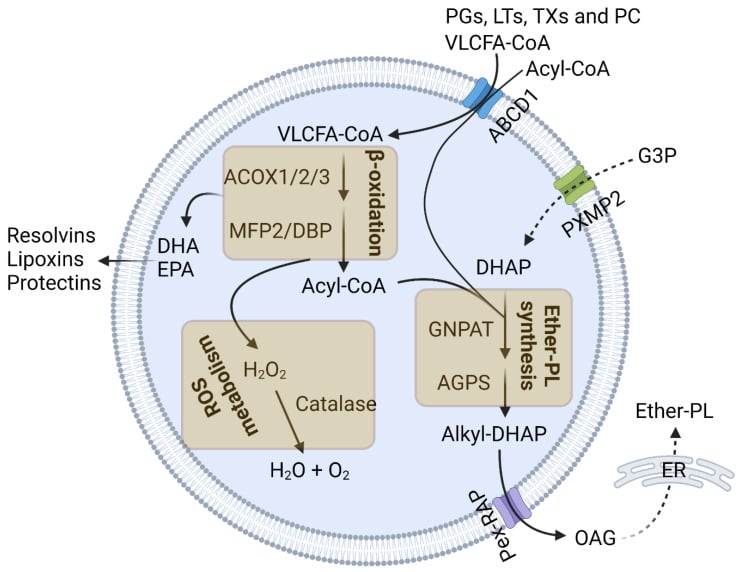
- Rhizomelic CDP - a plasmalogen defect. A peroxisomal disorder of plasmalogen (ether-phospholipid) biosynthesis: RCDP1 = PEX7 (the PTS2 receptor importing the plasmalogen-synthesis enzymes into the peroxisome), RCDP2-4 = GNPAT, AGPS and FAR1 (the enzymes themselves), and RCDP5 = loss of the long isoform of PEX5 (PEX5L), which co-receptors PTS2 import and so mimics PEX7 loss. The result is deficient plasmalogens (with raised phytanic acid in type 1) - low red-cell plasmalogens and raised phytanic acid confirm it biochemically.
- Conradi-Hunermann - a cholesterol-synthesis defect. EBP encodes emopamil-binding protein, a sterol Δ8-Δ7 isomerase in the post-squalene cholesterol biosynthesis pathway; its deficiency causes accumulation of sterol precursors (8-dehydrocholesterol, 8(9)-cholestenol - measurable in plasma). The male lethality reflects the essential cholesterol pathway, and the Blaschko-line skin reflects X-inactivation mosaicism in surviving females.
- Why the epiphyses are stippled. The punctate calcification is abnormal/ectopic calcific deposition within the un-ossified epiphyseal cartilage, which resolves as the cartilage ossifies normally with growth.
Q: What metabolic pathways underlie the rhizomelic and Conradi-Hünermann forms, and why are the epiphyses stippled?
A: Rhizomelic CDP = a peroxisomal plasmalogen-synthesis defect across five types (RCDP1 PEX7 [the PTS2 receptor], RCDP2-4 GNPAT/AGPS/FAR1, RCDP5 PEX5L) → low plasmalogens + raised phytanic acid (biochemical diagnosis), with the residual plasmalogen level grading severity. Conradi-Hünermann = EBP (emopamil-binding protein), a sterol Δ8-Δ7 isomerase in post-squalene cholesterol synthesis → accumulated sterol precursors (8-dehydrocholesterol); male-lethal, Blaschko-line skin from X-inactivation mosaicism. The stippling = ectopic calcification in un-ossified epiphyseal cartilage that resolves as it ossifies.
Clinical Presentation: The Forms & Their Features
Chondrodysplasia punctata is a heterogeneous group unified by stippled (punctate) epiphyseal calcification on infant radiographs (which resolves with growth). The rhizomelic form is a severe autosomal-recessive peroxisomal plasmalogen disorder spanning RCDP1 to RCDP5, in which classic disease gives symmetric rhizomelia, cataracts, contractures and often early death while nonclassic patients with higher residual plasmalogens do considerably better. Conradi-Hunermann (CDPX2) is X-linked dominant (EBP), mostly in females, milder, with asymmetric limb shortening (limb-length discrepancy), scoliosis, ichthyosis (along Blaschko's lines) and cataracts. An X-linked recessive (brachytelephalangic) form and teratogenic phenocopies - warfarin embryopathy and maternal autoimmune disease (SLE) - complete the group, so a maternal drug/autoimmune history is important.
- Genetics
- Autosomal recessive peroxisomal: PEX7, GNPAT, AGPS, FAR1, PEX5L
- Key features
- A spectrum graded by residual plasmalogen - classic: symmetric rhizomelia, delay, cataracts, early death; nonclassic: milder, better growth and cognition
- Genetics
- X-linked dominant (EBP); mostly females
- Key features
- Asymmetric limb shortening (discrepancy), scoliosis, ichthyosis, cataracts; milder
- Genetics
- ARSL/arylsulfatase
- Key features
- Brachytelephalangic - nasal + distal-phalangeal hypoplasia
- Genetics
- Warfarin embryopathy; maternal autoimmune (SLE)
- Key features
- Phenocopy of CDP - take maternal history
Investigations
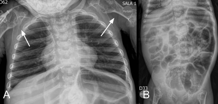
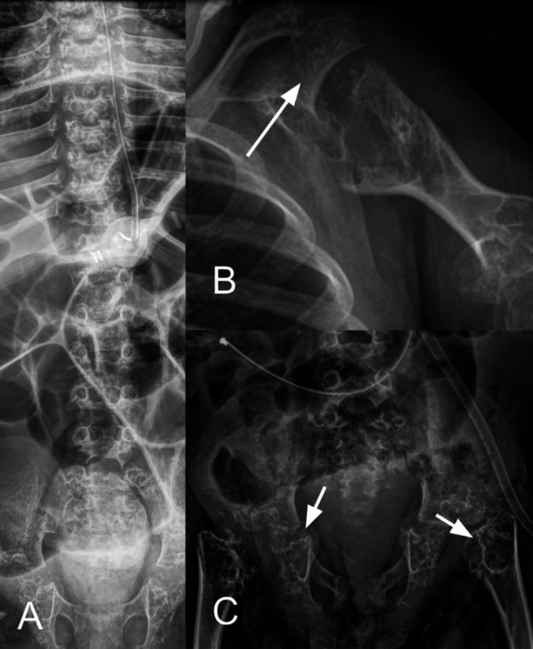
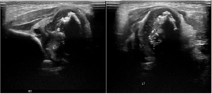
- Radiographs are the entry point: stippled (punctate) epiphyseal and peri-articular calcification in an infant, with the pattern of limb shortening suggesting the form - symmetric and rhizomelic in RCDP, asymmetric in Conradi-Hünermann. The stippling characteristically resolves as the epiphyses ossify, so later films show the sequelae (shortening, discrepancy, scoliosis) rather than the sign.
- Peroxisomal biochemistry for suspected rhizomelic disease: low red-cell plasmalogens with raised phytanic acid (type 1), confirmed in cultured fibroblasts; the residual plasmalogen level also grades severity.
- Plasma sterol profile for suspected Conradi-Hünermann: accumulation of the cholesterol precursors 8-dehydrocholesterol and 8(9)-cholestenol points to EBP.
- Genetic testing confirms the form: PEX7 and PEX5 exon 9 together when there is a selective PTS2-import defect (RCDP1 vs RCDP5), GNPAT/AGPS/FAR1 for RCDP2-4, EBP for CDPX2 and ARSL for CDPX1.
- Maternal history is an investigation in its own right: warfarin exposure in the first trimester, maternal autoimmune disease (SLE), vitamin-K deficiency and certain anticonvulsants.
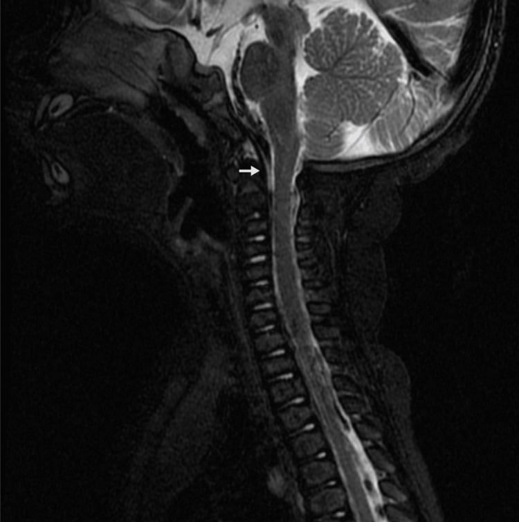
- Brain MRI when neurological signs are present: severe RCDP shows supratentorial white-matter abnormality with parieto-occipital predominance, and cerebellar atrophy in older patients.
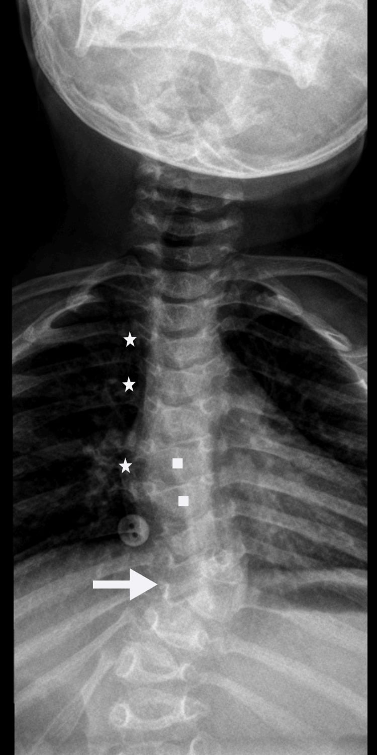
Warfarin Embryopathy and the Fuller Differential
- Warfarin embryopathy - the mechanism. Warfarin crosses the placenta and inhibits vitamin-K epoxide reductase, impairing γ-carboxylation of the vitamin-K-dependent bone protein matrix Gla protein (a normal inhibitor of cartilage calcification) - so cartilage calcifies abnormally, giving stippled epiphyses and nasal hypoplasia (fetal warfarin syndrome). The critical teratogenic window is the first trimester (about 6-9 weeks), which is why heparin/LMWH (which do not cross the placenta) are substituted in pregnancy.
- Other secondary causes. Maternal autoimmune disease (SLE), maternal vitamin-K deficiency and some anticonvulsants (phenytoin) can also produce a stippled-epiphyses phenocopy.
- The fuller differential of stippled epiphyses. Beyond CDP, consider Zellweger spectrum (another peroxisomal disorder), congenital hypothyroidism (epiphyseal dysgenesis), trisomy 18/21, GM1 gangliosidosis, congenital infection, and the cholesterol-pathway disorders CHILD syndrome (NSDHL) and Smith-Lemli-Opitz (DHCR7).
Q: What is the mechanism of warfarin embryopathy, and what is the fuller differential of stippled epiphyses?
A: Warfarin inhibits vitamin-K epoxide reductase → impaired γ-carboxylation of matrix Gla protein (a normal inhibitor of cartilage calcification) → abnormal cartilage calcification = stippled epiphyses + nasal hypoplasia (fetal warfarin syndrome); the critical window is the first trimester (~6-9 weeks) → substitute heparin/LMWH. Other causes: maternal SLE, vitamin-K deficiency, anticonvulsants. Fuller differential: Zellweger, congenital hypothyroidism (epiphyseal dysgenesis), trisomy 18/21, GM1 gangliosidosis, CHILD (NSDHL), Smith-Lemli-Opitz (DHCR7).
Rhizomelic CDP Is a Spectrum, Not a Single Lethal Disease
Calling the rhizomelic form uniformly severe and early-lethal is the traditional teaching and is wrong at the milder end. Severe (classic) RCDP has negligible plasmalogen levels with congenital cataracts, skeletal dysplasia, growth failure and neurodevelopmental deficit. Individuals with milder or nonclassic RCDP have higher residual plasmalogen levels, better growth and better cognitive outcomes, and survive well beyond infancy. The plasmalogen deficiency is itself described as the main pathogenic factor determining severity - so the red-cell plasmalogen level is not merely a diagnostic test but a prognostic one, and a family should not be counselled from the classic phenotype before the biochemistry is known.
Rhizomelic CDP is subdivided by the step that fails in plasmalogen synthesis: RCDP1 - PEX7 (the PTS2 receptor), RCDP2 - GNPAT, RCDP3 - AGPS, RCDP4 - FAR1, and RCDP5 - loss of the long isoform of PEX5 (PEX5L), reported in four patients from two families. RCDP5 matters conceptually: other PEX5 mutations cause Zellweger spectrum disease, but losing only PEX5L knocks out import of PTS2-tagged proteins alone - the same functional lesion as PEX7 - so it produces RCDP instead. Because RCDP1 and RCDP5 overlap biochemically, a selective PTS2-import defect warrants sequencing both PEX7 and PEX5 exon 9.
In the severe phenotype, MRI shows supratentorial white-matter abnormality with a parieto-occipital predominance, and cerebellar atrophy appears in older patients - relevant when a child with stippled epiphyses also has neurological signs, and a reminder that this is a peroxisomal leukoencephalopathy rather than a purely skeletal condition. Diagnosis of any peroxisomal disorder rests on a battery of blood and urine assays confirmed in cultured fibroblasts and by DNA analysis, and treatment remains largely symptomatic.
Do not answer "rhizomelic CDP is lethal in infancy" flatly. Say it is a spectrum graded by residual plasmalogen: classic disease with negligible plasmalogens is severe, while nonclassic RCDP has higher plasmalogens, better growth and better cognition. Five genetic types are recognised - PEX7, GNPAT, AGPS, FAR1 and PEX5L - and no reliable survival figure for the group was retrievable.
Management
- Diagnosis: identify the form - genetic/metabolic testing (peroxisomal studies for rhizomelic CDP; EBP/ ARSL for X-linked forms) and a maternal drug/autoimmune history (warfarin, SLE).
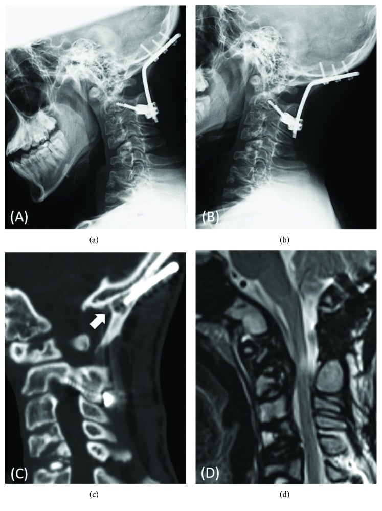
- Orthopaedic: manage limb-length discrepancy (epiphysiodesis/lengthening), scoliosis and contractures in survivable forms (especially Conradi-Hunermann).
- Multidisciplinary: ophthalmology (cataracts), dermatology (ichthyosis), metabolic/genetics and family counselling.
- Severe rhizomelic form: largely supportive/palliative given the poor prognosis.
Complications
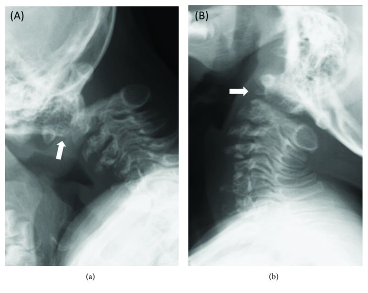
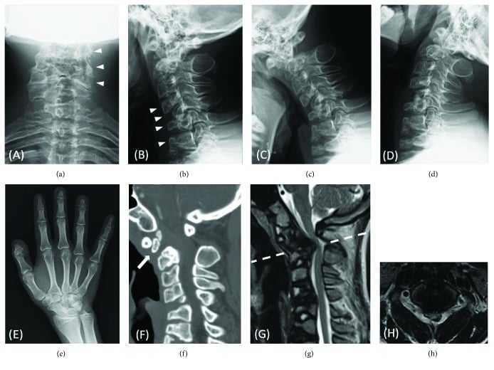
- Orthopaedic: limb-length discrepancy (asymmetric forms), progressive scoliosis, joint contractures, and hip instability; these generate the surgical workload in survivable forms such as Conradi-Hünermann.
- Neurological: profound developmental delay in classic RCDP; the peroxisomal leukoencephalopathy progresses with white-matter disease and cerebellar atrophy.
- Ophthalmic: congenital cataracts in both RCDP and CDPX2 threaten visual development and warrant early surgery.
- Cutaneous: ichthyosis along Blaschko's lines in CDPX2, usually improving with age.
- Systemic/prognostic: feeding difficulties, recurrent respiratory infections and early death in classic RCDP; nonclassic patients with higher residual plasmalogens survive longer with better growth and cognition - the reason counselling waits for the biochemistry.
Mnemonics & Memory Aids
STIPPLE
Hook:STIPPLE: Stippled epiphyses, Teratogenic phenocopies, Ichthyosis/cataracts, Peroxisomal rhizomelic (severe), Pattern (symmetric vs asymmetric), Limb sequelae, EBP gene.
PGAFP
Hook:PGAFP: the five rhizomelic types in order - PEX7, GNPAT, AGPS, FAR1, PEX5L. One receptor (PEX7), three enzymes, one co-receptor (PEX5L) - all fail plasmalogen synthesis.
GLA
Hook:GLA: warfarin blocks Gamma-carboxylation, Loses the calcification inhibitor (matrix Gla protein), and cartilage calcifies Abnormally - first-trimester window, so substitute heparin/LMWH in pregnancy.
Clinical Decision Scenarios
Practise clinical reasoning and management decisions out loud
“An infant radiograph shows stippled epiphyses. What is the diagnosis group, what forms do you consider, and what history is important?”
Hallmark
- Stippled (punctate) epiphyseal/peri-articular calcification in infancy
- Heterogeneous group; stippling resolves with growth
- Identify the form (prognosis/management differ greatly)
Genetic forms
- Rhizomelic CDP: AR peroxisomal plasmalogen defect, RCDP1-5 (PEX7, GNPAT, AGPS, FAR1, PEX5L)
- A SPECTRUM: classic (negligible plasmalogens) severe with early death; nonclassic (higher) milder - the level is prognostic
- Conradi-Hunermann (CDPX2): X-linked dominant (EBP), females, asymmetric shortening + ichthyosis + cataracts
- X-linked recessive (CDPX1, ARSL): brachytelephalangic (nasal/distal-phalangeal hypoplasia)
Secondary/teratogenic
- Warfarin embryopathy (maternal warfarin in pregnancy)
- Maternal autoimmune disease (e.g. SLE)
- Take a maternal drug/autoimmune history
Orthopaedic management
- Limb shortening/discrepancy (epiphysiodesis/lengthening)
- Scoliosis and contractures (survivable forms)
- Multidisciplinary: ophthalmology (cataracts), dermatology, genetics; severe form palliative
Evidence & Key Studies
Skeletal dysplasias of the fetus and infant (including chondrodysplasia punctata)
- More than 400 genetic skeletal disorders are included in the latest classification; the most severe/lethal phenotypes are identifiable in the prenatal period, and perinatal autopsy/post-mortem radiographs are crucial for definitive diagnosis.
- Chondrodysplasia punctata is among the recognised skeletal dysplasias encountered in fetal/neonatal practice (alongside thanatophoric dysplasia, osteogenesis imperfecta, campomelic dysplasia and achondroplasia).
- An increasing number of cases are confirmed by genetic testing, reflecting the heterogeneous genetic basis of these dysplasias.