Pain Out of Proportion | Pressure Monitoring | Urgent Fasciotomy
- Pain on passive stretch is earliest reliable sign - pain with passive finger extension
- 5 Ps are LATE findings - Pulselessness and pallor indicate damage already occurring
- Pressure threshold: ΔP under 30mmHg (diastolic minus compartment) SUSTAINED over 2 hours - an absolute reading over 30mmHg alone is NOT an indication
- Volar fasciotomy MUST include carpal tunnel release - oedema extends distally
- Volkmann's ischaemic contracture is the devastating end result of missed diagnosis
- “Clinical diagnosis sufficient - do NOT delay surgery for pressure measurement if high suspicion
- “Always release BOTH volar AND dorsal compartments if any doubt
- “Leave wounds completely open - never close primarily, DPC at 48-72 hours
- “High-risk fractures: supracondylar (children), both-bone forearm, floating elbow
Overview
Forearm compartment syndrome is a surgical emergency. Pressure rises inside the closed osteofascial compartments of the forearm, which have little compliance, until tissue perfusion is compromised and the muscles and nerves within suffer progressive ischaemic injury. Normal compartment pressure is 0-8 mmHg.
What the pressure does. Tissue perfusion depends on the pressure gradient between arterioles and venules. As compartment pressure rises, venous outflow is compromised first, venous pressure climbs and the arteriovenous gradient narrows, so capillary perfusion falls even though arterial inflow is still present. This is the arteriovenous gradient theory, and it is why the pulse is preserved until late.
What follows. Muscle ischaemia progresses to necrosis, and nerve ischaemia produces a sensory and then a motor deficit. Left untreated, the necrotic flexor muscle is replaced by fibrous tissue, and the hand is left with Volkmann's ischaemic contracture.
Aetiology and Risk Factors
Causes. Anything that adds volume to a compartment or compresses it from outside will do: a fracture bleeding into the muscle, a crush, a tight cast, or reperfusion after an arterial repair.
- Examples
- Both-bone forearm, supracondylar, distal radius
- Mechanism
- Bleeding, soft tissue injury
- Risk Level
- HIGH
- Examples
- Motor vehicle, industrial accidents
- Mechanism
- Direct muscle damage, oedema
- Risk Level
- HIGH
- Examples
- Tight casts, circumferential dressings
- Mechanism
- External compression
- Risk Level
- HIGH
- Examples
- Arterial injury with revascularisation
- Mechanism
- Reperfusion injury
- Risk Level
- HIGH
- Examples
- Anticoagulation, haemophilia
- Mechanism
- Compartment haemorrhage
- Risk Level
- MODERATE
- Examples
- Circumferential full-thickness
- Mechanism
- Eschar constriction + oedema
- Risk Level
- MODERATE
- Examples
- IV infiltration, drug injection
- Mechanism
- Fluid accumulation
- Risk Level
- MODERATE
- Examples
- Necrotising fasciitis
- Mechanism
- Oedema, tissue destruction
- Risk Level
- MODERATE
High-risk fractures. These are the injuries after which serial neurovascular examination is not optional.
- Supracondylar fractures in children - highest risk, especially displaced type III
- Both-bone forearm fractures - significant soft tissue injury
- Floating elbow - combined supracondylar and forearm fracture
- Monteggia and Galeazzi fractures - high-energy injury pattern
- Distal radius fractures with severe swelling or displacement
Patient factors. The typical patient is a young man after high-energy trauma.
- Factors
- Male gender (2:1), age under 35 years, anticoagulation, coagulopathy, high-energy trauma
- Factors
- Diabetes mellitus, peripheral vascular disease, drug or alcohol intoxication, unable to communicate
- Factors
- Circumferential casts, tight bandages, positioning in surgery, infiltrated IV lines, prolonged surgery
Anatomy and Pathophysiology
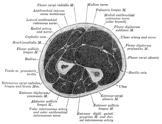
The compartments. The forearm has three to four compartments depending on the classification: two volar, one dorsal, and the mobile wad, which some consider part of the dorsal compartment and which is released through the dorsal approach. A fasciotomy is only complete if the surgeon knows what lies in each of them.
- Muscles
- PT, FCR, PL, FDS, FCU
- Nerve
- Median and ulnar nerves
- Note
- Most commonly affected
- Muscles
- FDP, FPL, PQ
- Nerve
- AIN (motor branch of the median)
- Note
- Weakness of FPL and index FDP is an early sign
- Muscles
- EDC, EDM, EIP, ECU, APL, EPB, EPL, supinator
- Nerve
- PIN
- Note
- Less commonly affected, but its pressure must still be checked
- Muscles
- BR, ECRL, ECRB
- Nerve
- Note
- Released with the dorsal approach
The cascade. Bleeding, oedema or external compression raises the pressure; venous outflow is obstructed first; the arteriovenous gradient falls and capillary perfusion with it. Muscle is more sensitive than nerve initially. Ischaemic cells swell, which raises the pressure further, a positive feedback loop that only decompression breaks.
- Tissue Effects
- Muscle injury begins
- Tissue Effects
- Reversible muscle injury
- Tissue Effects
- Nerve dysfunction (initially reversible)
- Tissue Effects
- Irreversible muscle necrosis begins
- Tissue Effects
- Permanent nerve damage, Volkmann's contracture
Classification Systems
Matsen classification (by clinical stage). The stage links what is found on examination to how urgently the compartment must be opened.
- Clinical Findings
- Pain on passive stretch, no neurological deficit
- Compartment Pressure
- Elevated (20-30 mmHg)
- Tissue Status
- Reversible ischaemia
- Urgency
- High suspicion, serial monitoring
- Clinical Findings
- Pain, paraesthesia, tense compartment
- Compartment Pressure
- ΔP under 30 mmHg sustained over 2 hours
- Tissue Status
- Progressive muscle ischaemia
- Urgency
- URGENT fasciotomy indicated
- Clinical Findings
- Pallor, paralysis, pulselessness
- Compartment Pressure
- Critically elevated (greater than 40 mmHg)
- Tissue Status
- Irreversible damage occurring
- Urgency
- EMERGENCY fasciotomy
- Clinical Findings
- Fixed contracture, tissue necrosis
- Compartment Pressure
- Variable (may normalise)
- Tissue Status
- Irreversible damage done
- Urgency
- Delayed reconstruction
Clinical Presentation
Pain on passive stretch is the most reliable early sign. Volar involvement hurts on passive finger extension; dorsal involvement hurts on passive finger flexion. The pain is disproportionate to the injury itself, and the analgesic requirement keeps climbing despite adequate initial dosing.
The five Ps are the classic list, and the last two are late:
- Pain - out of proportion to the injury, the earliest sign
- Pressure - a tense, wood-like compartment on palpation
- Paraesthesia - numbness as nerve ischaemia begins
- Pallor - late, indicating advanced ischaemia
- Pulselessness - very late, and irreversible damage is likely
A palpable pulse does NOT rule out compartment syndrome. Compartment syndrome affects the microcirculation while arterial inflow may be preserved until late. Waiting for pulse loss means irreversible damage has already occurred.
Nerve findings. Numbness in the first web space (AIN, median nerve) is the sensory sign; ulnar-distribution numbness is less common, and the PIN is motor only, with no sensory territory. Motor weakness follows the same nerves: an AIN palsy with a weak FPL (thumb IP) and weak FDP to the index, then weak finger flexion (FDS, FDP) and weak wrist flexion.
- Time Frame
- 0-2 hours
- Clinical Findings
- Pain out of proportion, pain on passive stretch
- Action Required
- HIGH SUSPICION - consider fasciotomy
- Time Frame
- 2-4 hours
- Clinical Findings
- Tense compartment, increasing analgesia needs, paraesthesia
- Action Required
- URGENT fasciotomy
- Time Frame
- 4-8 hours
- Clinical Findings
- Pallor, paralysis, weak/absent pulses
- Action Required
- EMERGENCY fasciotomy - damage likely
- Time Frame
- More than 8 hours
- Clinical Findings
- Pulselessness, complete sensorimotor loss, cold limb
- Action Required
- Fasciotomy +/- amputation consideration
- Analgesia - an increasing analgesic requirement (the child needs more and more opioid for a fracture that should be settling) is the most important and earliest objective red flag.
- Anxiety - rising distress and agitation in a child who was previously settling.
- Agitation - inconsolability and restlessness disproportionate to the injury.
In children you watch the trend in analgesia demand and behaviour, not a verbal pain-out-of-proportion report. An escalating opioid requirement after a supracondylar or both-bone forearm fracture is a compartment syndrome until proven otherwise, and the same low threshold for fasciotomy applies - children tolerate ischaemia no better than adults.
Investigations
When to measure. The diagnosis is clinical, and high clinical suspicion alone is sufficient: do not delay fasciotomy for pressure measurement, imaging or laboratory results, because time to fasciotomy is the critical factor. Pressure measurement is most valuable exactly where the examination fails you - the obtunded, intubated, blocked or very young patient who cannot report pain:
- Equivocal clinical examination
- Obtunded or uncooperative patient
- Unreliable examination (intoxication, head injury)
- Serial monitoring when clinical suspicion is moderate
Technique. The method matters:
- Use a calibrated device (Stryker STIC, arterial line transducer)
- Measure in the zone of maximum injury or swelling
- Measure all compartments if there is any suspicion
- Position the limb at the level of the heart
- Infiltrate 0.3ml of saline to confirm placement
The threshold. Delta P is the diastolic blood pressure minus the compartment pressure. It represents the perfusion pressure gradient and so accounts for the patient's haemodynamic status: a patient with a diastolic pressure of 70 mmHg and a compartment pressure of 45 mmHg has a ΔP of 25 mmHg, and fasciotomy is indicated. The threshold to quote is ΔP under 30 mmHg sustained over 2 hours; the older absolute reading of over 30 mmHg is superseded, because it is common and transient after injury, and on its own it is a prompt to reassess urgently rather than an indication to operate.
Why the differential is the one to quote. Perfusion of a compartment depends on the gradient between the patient's diastolic pressure and the pressure inside the compartment, so a hypotensive trauma patient can infarct muscle at an absolute pressure that would be harmless in someone normotensive. That is the whole argument, and it is why the threshold is expressed as diastolic minus compartment pressure rather than as a fixed number. It is also the threshold with validated diagnostic performance.
The performance figures belong to continuous monitoring, not to a one-off stab. In 850 patients with tibial diaphyseal fractures monitored continuously and analysed against the operative findings at fasciotomy and against the absence of contracture or neurological deficit at a mean of 59 weeks, sensitivity was 94%, specificity 98% and positive predictive value 93% (DOI). Of 152 fasciotomies, 141 were true positives and only 6 were false positives. Those numbers describe a monitored trend, and they do not transfer to a single needle reading taken once, which is far more vulnerable to catheter position, depth and technique.
Both errors are real, and missing it is the graver one. A missed forearm compartment syndrome produces a Volkmann's contracture and a permanently useless hand, so if the clinical picture says compartment syndrome you decompress regardless of what the needle says; a normal pressure never overrides convincing clinical signs. But the opposite error is not harmless: an unnecessary forearm fasciotomy means two long wounds, a likely skin graft, infection risk and lasting cosmetic and functional cost. That is why an isolated absolute reading over 30 in an awake, comfortable patient with a soft compartment and no pain on passive stretch is a reason to measure again and keep watching, not to operate on the spot. Practice genuinely varies between units, so know your own institution's written protocol.
Continuous monitoring has its own indications:
- Multiple trauma patient requiring sedation or ventilation
- Post-operative monitoring after high-risk surgery
- Burns with circumferential involvement
- Serial measurements impractical
Other tests. CK rises with muscle necrosis, myoglobin is the marker of rhabdomyolysis, lactate marks tissue ischaemia, renal function is monitored for myoglobinuric AKI, and coagulation studies are sent if a bleeding diathesis is suspected. Radiographs identify the underlying fracture and guide its management; CT and MRI are not routine and may delay treatment; Doppler ultrasound assesses arterial flow, but the presence of a pulse is not the same as adequate perfusion.
Compartment syndrome masked by a nerve block is a live controversy worth holding clearly, because the whole diagnosis rests on pain:
- The concern: a dense regional/peripheral nerve block, epidural, or opioid PCA abolishes the cardinal sign (pain out of proportion / pain on passive stretch) and can delay recognition of an evolving compartment syndrome in a high-risk limb.
- The modern, nuanced view: there is no high-level evidence that a block causes compartment syndrome or that it must always be withheld, and good analgesia is humane - but breakthrough pain "through" a working block, or escalating analgesic demand, is a major RED FLAG for compartment syndrome and must trigger urgent assessment, not a top-up.
- Practical rules: in a high-risk limb (both-bone forearm, supracondylar, crush, vascular repair) prefer shorter-acting, lower-concentration, single-shot or motor-sparing analgesia over a dense long-acting/continuous block, ensure the team knows the limb is at risk, and monitor the other signs and the analgesic trend (and compartment pressures if obtunded). Never let "the block is working" reassure you out of a diagnosis.
Management Algorithm
Remove every constrictor. Bivalve all casts, release all circumferential dressings and cut down to skin. Keep the limb at heart level and do not elevate it, because elevation reduces arterial perfusion. Removal of every constrictive element comes before definitive fasciotomy.
Optimise perfusion. Maintain the blood pressure and avoid hypotension, correct hypovolaemia and coagulopathy, give supplemental oxygen and keep the patient normothermic.
Book theatre. Contact theatre immediately and consent for fasciotomy. Plan for both volar and dorsal release, warn the patient that the wounds will be left open, and plan the return to theatre at 48-72 hours.
The decision. Time to fasciotomy is the most critical determinant of outcome, and the pathway depends on how sure you are:
- High clinical suspicion (pain out of proportion, pain on passive stretch): proceed directly to fasciotomy
- Uncertain diagnosis (equivocal examination, obtunded patient): measure pressures in all compartments, apply the Delta P threshold, and monitor serially if the values are borderline
- Established diagnosis: urgent fasciotomy within 6 hours of symptom onset, releasing volar and dorsal if the mechanism was high-energy or there is any doubt, and always including carpal tunnel decompression
Fasciotomy within 6 hours of symptom onset has significantly better outcomes than delayed decompression; after 8 hours, irreversible damage is highly likely.
Surgical Technique
Set-up. Supine, arm on a radiolucent hand table. A tourniquet is applied but used judiciously, inflated only if bleeding obscures the view, and there is a case for avoiding it altogether so that tissue perfusion can be assessed.
Consent. The patient should hear these before the anaesthetic:
- The wounds will be left open
- Risk of incomplete decompression
- Nerve and vessel injury
- Return to theatre at 48-72 hours
- A skin graft may be required
- Ongoing muscle or nerve damage despite surgery
The incision. A curvilinear or zigzag incision from the medial epicondyle to the palm, crossing the antecubital fossa obliquely so that a linear scar does not contract across the flexion crease, curving ulnar at the wrist and extending into the palm for the carpal tunnel release. Total length is 15-20cm in the forearm plus a 3-4cm palmar extension.
The steps. Each compartment is opened in turn, and the muscle is inspected at the end:
- Skin incision - full-thickness, generous length
- Identify and protect the superficial veins - ligate if necessary
- Release the lacertus fibrosus (bicipital aponeurosis) at the antecubital fossa
- Identify the median nerve - it runs between FDS and FDP
- Release the superficial volar compartment - incise the fascia overlying PT, FCR, PL, FCU and FDS along its full length; the muscles should decompress and bulge through the fasciotomy
- Release the deep volar compartment - retract FDS, incise the deep fascia overlying FDP, FPL and PQ, and protect the AIN on the interosseous membrane
- Carpal tunnel release (mandatory) - extend the incision into the palm and divide the transverse carpal ligament under direct vision, protecting the median nerve and its palmar cutaneous branch
- Inspect all muscles - viable muscle is pink, contracts with stimulation and bleeds when cut; non-viable muscle is dark, does not contract and does not bleed; debride clearly necrotic tissue
Complications
Missed or delayed diagnosis. The first of the sequelae is permanent.
- Description
- Irreversible flexor muscle fibrosis and shortening
- Timing
- Weeks-months
- Treatment
- Tendon lengthening, muscle slide, free flap
- Description
- Median and/or ulnar nerve damage
- Timing
- Immediate
- Treatment
- Nerve exploration, possible grafting
- Description
- Dead muscle requiring debridement
- Timing
- Days
- Treatment
- Serial debridement, coverage
- Description
- Myoglobin release causing AKI
- Timing
- Hours-days
- Treatment
- Aggressive hydration, ?dialysis
- Description
- Neuropathic and ischaemic pain
- Timing
- Months
- Treatment
- Multidisciplinary management
- Description
- Loss of grip strength, dexterity
- Timing
- Permanent
- Treatment
- Reconstructive surgery, therapy
Volkmann's ischaemic contracture is the irreversible fibrotic contracture of the forearm flexors that follows untreated compartment syndrome. Severity is graded by Tsuge, in the classification section above, and the shortened flexors set the hand in a classic posture:
- Wrist flexion
- MCP hyperextension
- IP joint flexion
- Thumb adduction
The cascade sign is its signature: passive wrist extension makes the fingers flex further, because the muscle is shortened.
Complications of the fasciotomy itself. Early: wound infection, bleeding, iatrogenic nerve or vessel injury, skin edge necrosis and incomplete release. Late: unsightly scars, skin graft contracture, chronic wound healing, tethering of tendons, altered sensation and the need for secondary reconstruction.
Postoperative Care and Rehabilitation
The wounds. Leave them completely open and never close primarily. Apply a loose non-adherent dressing (Jelonet, Adaptic), and consider negative pressure wound therapy if significant oedema persists, the wound is large with exposed structures, or a dressing is difficult to maintain.
The splint holds the position of function to prevent contracture, and it must not itself compress the compartments, so avoid tight circumferential dressings:
- Wrist: 20-30° extension
- MCP joints: 70-90° flexion
- IP joints: full extension
- Thumb: abduction and opposition
Monitoring. Neurovascular observations every 2 hours for the first 24 hours, watching for persistent pain (inadequate decompression), a worsening motor or sensory deficit, signs of bleeding, and the systemic complication, rhabdomyolysis.
Rhabdomyolysis. Check CK daily initially, serum and urine myoglobin, creatinine and eGFR, and lactate as a marker of tissue ischaemia, and maintain a urine output over 1ml/kg/h. Treatment is aggressive IV hydration to that urine output target, urinary alkalinisation with IV sodium bicarbonate, potassium monitoring for hyperkalaemia, dialysis if AKI develops, and treatment of the underlying cause.
Return to theatre at 48-72 hours. The wound is reassessed and closed by whichever method the tissues allow.
Who. Delayed primary closure is ideal when the muscle remains viable (pink, contractile), the oedema has resolved, there is no sign of infection, and the skin edges can be approximated without tension.
How. Irrigate thoroughly, reassess muscle viability and debride any necrotic tissue, then approximate the skin edges with interrupted sutures or staples, avoiding tension. A significant gap may need staged closure.
Rehabilitation follows the wound, and the hand therapist is involved from the start.
- Timing
- 0-2 weeks
- Content
- Wound healing priority; gentle passive ROM when wounds closed or stable; oedema control with elevation and compression; hand therapy referral
- Timing
- 2-6 weeks
- Content
- Active ROM; scar massage and desensitisation; gentle strengthening; functional activities
- Timing
- 6+ weeks
- Content
- Progressive strengthening; return to activities of daily living; monitor for contracture; long-term follow-up
Watch for the late complication. A progressive finger flexion posture, a positive cascade sign and grip weakness are the signs of a developing Volkmann's contracture, and a contracture needs prompt referral to hand surgery.
Outcomes and Prognosis
Timing. Time to fasciotomy is the dominant determinant of outcome. In the series that defined the early-versus-late divide (Sheridan and Matsen, in the evidence section), fasciotomy performed early, under 12 hours after onset, gave normal function in 68% of extremities, and only 8% of late fasciotomies did, with complication rates of 4.5% early against 54% late. Outcome worsens continuously with delay, which is why contemporary practice aims for decompression as soon as the diagnosis is made, ideally within 6 hours.
- Normal Function
- 68%
- Complications
- 4.5%
- Normal Function
- 8%
- Complications
- 54%
Other prognostic factors are the severity of the initial injury, the associated fractures and soft tissue trauma, the patient's age (younger patients have better recovery potential), rehabilitation compliance, and complications such as infection and rhabdomyolysis.
Recovery runs over months to years.
- Milestones
- First sensation 2-4 weeks; protective sensation 6-12 weeks; discriminative touch 3-6 months
- Caveat
- May remain incomplete in severe cases
- Milestones
- Muscle contraction 4-8 weeks; functional strength 3-6 months; maximal recovery 12-18 months
- Caveat
- Depends on the extent of muscle necrosis
- Milestones
- ADL independence 3-6 months; return to work 6-12 months; full recovery 12-24 months
- Caveat
- May have persistent weakness
Volkmann's contracture. An established contracture is irreversible, and the only treatment is reconstruction, so prevention by early fasciotomy is the whole strategy. When reconstruction is needed, the result tracks the Tsuge grade.
- Surgery
- Muscle slide, tendon lengthening
- Expected Function
- Good - 70-80% normal
- Patient Satisfaction
- High
- Surgery
- Muscle slide + tendon transfers
- Expected Function
- Fair - 40-60% normal
- Patient Satisfaction
- Moderate
- Surgery
- Free functioning muscle transfer
- Expected Function
- Poor - 20-30% normal
- Patient Satisfaction
- Low to moderate
- Incidence
- 10-20%
- Impact
- Cosmetic, possible functional limitation
- Management
- Scar revision, Z-plasty
- Incidence
- 15-25%
- Impact
- Neuropathic pain, impaired function
- Management
- Multidisciplinary pain management
- Incidence
- 30-50%
- Impact
- Reduced grip strength, endurance
- Management
- Ongoing therapy, adaptive strategies
- Incidence
- 20-35%
- Impact
- Protective sensation usually preserved
- Management
- Desensitisation therapy
- Incidence
- 5-10% (early surgery), 20-40% (late surgery)
- Impact
- Severe functional impairment
- Management
- Reconstructive surgery
- Incidence
- 5-15%
- Impact
- Delayed healing, possible amputation
- Management
- Antibiotics, serial debridement
Medicolegal. Missed or delayed compartment syndrome is one of the most common and costly sources of orthopaedic litigation internationally, and claims are frequent and high-value in multiple jurisdictions (analyses of UK NHS Resolution and US closed-claims data), driven by the catastrophic and irreversible nature of the sequelae. The common allegations are delayed diagnosis, failure to monitor or document neurovascular status, and an inadequate, incomplete fasciotomy. Prevention is meticulous time-stamped documentation, a low threshold for fasciotomy, and early senior or specialist involvement; the record should contain:
- Time-stamped neurovascular examinations
- Compartment pressure measurements (if performed)
- Clinical decision-making rationale
- Patient and family discussions about risks
- Informed consent including the risk of Volkmann's contracture
- Time from symptom onset to surgical decompression
Guidelines, Registries & Global Practice
Differential Diagnosis
Compartment syndrome must be distinguished from other causes of a painful, swollen or neurologically compromised forearm. The two most dangerous mimics to exclude are arterial injury and an evolving compartment syndrome masked by a nerve block.
- Distinguishing Features
- Pain out of proportion, pain on passive stretch, tense compartment, escalating analgesia
- Key Test
- Clinical; compartment pressure / ΔP if equivocal
- Pitfall
- Pulse often preserved until late - do not wait for it
- Distinguishing Features
- Cold pale limb, absent pulse, hard signs of vascular injury, ABI reduced
- Key Test
- Doppler, CT angiography
- Pitfall
- Can coexist with compartment syndrome (e.g. supracondylar fracture)
- Distinguishing Features
- Focal sensorimotor deficit in single nerve territory, soft compartment, no disproportionate pain
- Key Test
- Clinical exam, nerve conduction (delayed)
- Pitfall
- Mislabelling early compartment ischaemia as primary nerve injury
- Distinguishing Features
- Erythema, warmth, fever, raised inflammatory markers, soft compartment
- Key Test
- Bloods (WCC, CRP), clinical
- Pitfall
- Necrotising fasciitis can mimic and coexist - look for crepitus, systemic toxicity
- Distinguishing Features
- Swelling, less severe pain, no pain on passive stretch
- Key Test
- Duplex ultrasound
- Pitfall
- Rare in upper limb; do not anticoagulate if compartment syndrome possible
- Distinguishing Features
- Pain proportionate to injury, settles with analgesia and splintage
- Key Test
- Clinical response to analgesia
- Pitfall
- Attributing escalating pain to 'just the fracture'
Global Epidemiology
The largest population series (McQueen et al, J Bone Joint Surg Br 2000, PMID 10755426) of 164 consecutive acute compartment syndromes found an associated fracture in 69%, with forearm cases linked to distal radius fractures and occurring predominantly in young men under 35 years. The Lancet Emergency Surgery Series review (von Keudell et al 2015, PMID 26460664) confirms acute compartment syndrome affects upper and lower limbs in both adults and children and that there remains no internationally agreed diagnostic gold standard. Forearm compartment syndrome in children is most often associated with displaced supracondylar humeral fractures (Mubarak & Carroll, PMID 479251).
Guideline & Registry Comparison
- Region
- UK
- Key Position
- Urgent senior decompression; clinical diagnosis paramount; document neurovascular status serially
- Evidence Level
- Consensus standard
- Region
- International
- Key Position
- Clinical suspicion drives early fasciotomy; continuous pressure monitoring for obtunded/unreliable patients
- Evidence Level
- Narrative review (Level V)
- Region
- USA
- Key Position
- ΔP under 30 mmHg sustained over 2 hours; fasciotomy if clinically suspected - the absolute reading alone is not an indication
- Evidence Level
- Expert consensus
- Region
- International
- Key Position
- Complete volar + dorsal release with mandatory carpal tunnel decompression; wounds left open
- Evidence Level
- Technique consensus
- Region
- Europe
- Key Position
- ΔP threshold (McQueen) favoured over absolute pressure; early decompression
- Evidence Level
- Based on Level II-III data
There is no orthopaedic registry that systematically captures forearm compartment syndrome in the way arthroplasty registries (AOANJRR, NJR, AJRR) capture joint replacement; incidence and outcome data derive from single-centre and national trauma series rather than registry datasets.
Practice Variation & Regional Considerations
The principal area of international practice variation is the diagnostic pathway: continuous intracompartmental pressure monitoring is favoured in some UK and European units for high-risk or obtunded patients, whereas a clinically-driven "decompress if suspected" approach predominates in many trauma centres internationally (von Keudell 2015, PMID 26460664). The ΔP less than 30 mmHg threshold (McQueen, PMID 8898137) is the most widely adopted pressure criterion worldwide.
In trauma centres internationally, standardised serial neurovascular observation is applied after high-risk fractures, and first-generation cephalosporin prophylaxis (e.g. cefazolin) is the standard at fasciotomy. In rural and remote settings where transfer time may exceed 4-6 hours, local surgeons should have a low threshold to perform fasciotomy before transfer rather than risk irreversible ischaemia in transit. VTE prophylaxis follows standard trauma protocols, with mechanical methods preferred initially given the bleeding risk into freshly decompressed compartments.
MCQ Practice Points
Q: What is the earliest and most reliable clinical sign of compartment syndrome?
A: Pain out of proportion to the injury, especially pain on passive stretch of the affected compartment. For volar forearm compartment, this is pain with passive finger extension.
Q: What are the compartment pressure thresholds indicating need for fasciotomy?
A: A Delta P (diastolic blood pressure minus compartment pressure) under 30 mmHg, SUSTAINED for more than 2 hours. Delta P accounts for individual patient perfusion status. An absolute compartment pressure over 30 mmHg is NOT on its own an indication - it is common and usually transient after injury, and acting on it alone would have sent 43% of continuously monitored fractures to an unnecessary fasciotomy in the study that defined the delta-P rule.
Q: What MUST be included with volar forearm fasciotomy?
A: Carpal tunnel decompression - swelling extends distally into the carpal tunnel and must be released to prevent median nerve compression at the wrist.
Q: Which of the 5 Ps are considered LATE signs of compartment syndrome?
A: Pallor and pulselessness are late signs. Waiting for these findings means irreversible muscle and nerve damage has likely already occurred. Pain, pressure, and paresthesia are earlier findings.
Q: What is the pathological basis of Volkmann's ischemic contracture?
A: Ischemic necrosis of forearm flexor muscles leading to fibrotic replacement. The shortened, fibrotic muscles cause the characteristic posture: wrist flexion, MCP hyperextension, IP joint flexion.
Q: What is the critical time window for fasciotomy in compartment syndrome?
A: 6-8 hours from onset of ischemia. Fasciotomy within 6 hours has significantly better outcomes. After 8 hours, irreversible muscle necrosis and nerve damage are highly likely.
Clinical Decision Scenarios
Practise clinical reasoning and management decisions out loud
“8-year-old boy, 6 hours post-supracondylar fracture ORIF. Nurses report severe forearm pain despite regular analgesia. Pain with passive finger extension. Cast has been bivalved.”
“25-year-old motorcyclist with closed both-bone forearm fracture. Moderate forearm pain, compartment feels somewhat tense but not rock-hard. Fingers move but patient reports altered sensation in thumb web space. You are uncertain about the diagnosis.”
“28-year-old presents 6 months after forearm fracture treated elsewhere. He has a fixed flexion deformity of fingers with wrist in flexed position. Extending wrist causes fingers to flex further. He has weak grip and altered sensation.”
Clinical Signs
- Pain OUT OF PROPORTION (earliest)
- Pain on PASSIVE STRETCH (most reliable)
- Tense, wooden compartment
- Paresthesia (nerve ischemia)
- Pallor and pulselessness = TOO LATE
Pressure Thresholds
- Delta P under 30 (DBP - compartment pressure) SUSTAINED over 2 hours
- Absolute over 30 alone is NOT an indication
- Clinical diagnosis SUFFICIENT
- Do NOT delay for pressures if high suspicion
Volar Fasciotomy
- Curvilinear incision elbow to palm
- Release lacertus fibrosus
- Release superficial + deep volar
- ALWAYS add carpal tunnel release
- Protect median nerve throughout
Post-operative
- Leave wounds OPEN
- Loose dressings or VAC
- Splint in position of function
- DPC at 48-72 hours
- STSG if cannot close
High-Risk Fractures
- Supracondylar (children)
- Both-bone forearm
- Floating elbow
- Monteggia/Galeazzi
- High-energy distal radius
Volkmann's Contracture
- Irreversible flexor fibrosis
- Wrist flexed, MCP extended, IP flexed
- Cascade sign positive
- PREVENTION is key - early fasciotomy
Evidence Base
Delta P Threshold for Fasciotomy (Landmark)
- Prospective study of 116 tibial diaphyseal fractures with continuous anterior compartment monitoring for 24 hours; 3 (2.6%) developed acute compartment syndrome.
- Using a differential pressure (diastolic BP minus compartment pressure) threshold of less than 30 mmHg led to NO missed cases; no patient had compartment-syndrome sequelae at minimum six-month review.
- An absolute threshold of greater than 30 mmHg would have led to fasciotomy in 43% of patients, many unnecessary.
Timing of Fasciotomy and Outcomes (Landmark)
- 66 cases of acute compartment syndrome treated by fasciotomy in 46 extremities of 44 patients.
- Fasciotomy performed early (less than 12 hours after onset) gave normal function in 68% of extremities; only 8% of late fasciotomies achieved normal function.
- Complication rates were 4.5% (early) versus 54% (late).
Volkmann's Contracture, Supracondylar Fractures and Prevention (Landmark)
- Review of 55 children (58 limbs) with Volkmann's contracture treated in Toronto 1955-1975.
- Supracondylar fractures of the elbow that progressed to Volkmann's contracture frequently had BOTH an arterial injury and a compartment syndrome.
- Most children had not received early appropriate treatment, and the frequency of contracture had not declined over 21 years despite increased awareness.
Acute Compartment Syndrome of the Upper Extremity (Review)
- Comprehensive review of acute compartment syndrome of the hand, forearm and upper arm.
- Pain out of proportion to injury is the most reliable early symptom; diagnosis is particularly difficult in obtunded patients and young children.
- Early recognition and expeditious fasciotomy are essential to obtain good outcomes and prevent permanent disability and ischaemic contracture.
Acute Compartment Syndrome: Who Is at Risk? (Epidemiology)
- Analysis of 164 consecutive patients with acute compartment syndrome over eight years.
- An associated fracture was present in 69%; forearm compartment syndrome was associated with distal radius fractures and, like the cohort overall, occurred most commonly in young men (typically under 35 years).
- Soft-tissue injury without fracture was the second most common cause; one-tenth had a bleeding disorder or were anticoagulated.
Diagnosis and Treatment of Acute Extremity Compartment Syndrome (Lancet Series)
- Lancet Emergency Surgery Series review summarising upper- and lower-limb acute compartment syndrome in adults and children.
- There is no universally agreed diagnostic standard; some advocate continuous intracompartmental pressure monitoring in high-risk injuries while others favour aggressive fasciotomy when the syndrome is even suspected.
- Ineffective treatment risks permanent dysaesthesia, ischaemic contracture, muscle dysfunction, limb loss and death; fasciotomy itself carries long-term morbidity.