Pain Out of Proportion | Pressure Monitoring | Urgent Fasciotomy
FOREARM COMPARTMENTS
Critical Must-Knows
- Pain on passive stretch is earliest reliable sign - pain with passive finger extension
- 5 Ps are LATE findings - Pulselessness and pallor indicate damage already occurring
- Pressure threshold: greater than 30mmHg absolute OR ΔP less than 30mmHg from diastolic
- Volar fasciotomy MUST include carpal tunnel release - edema extends distally
- Volkmann's ischemic contracture is the devastating end result of missed diagnosis
Clinical Pearls
- "Clinical diagnosis sufficient - do NOT delay surgery for pressure measurement if high suspicion
- "Always release BOTH volar AND dorsal compartments if any doubt
- "Leave wounds completely open - never close primarily, DPC at 48-72 hours
- "High-risk fractures: supracondylar (children), both-bone forearm, floating elbow
Clinical Imaging
Imaging Gallery

Critical Forearm Compartment Syndrome Exam Points
Pain on Passive Stretch
EARLIEST and MOST RELIABLE sign. Pain with passive finger extension (volar) or flexion (dorsal) indicates rising compartment pressures. Do NOT wait for other signs.
5 Ps are LATE
Pain, Pressure, Pallor, Pulselessness, Paresthesia. Pallor and pulselessness are LATE signs - fasciotomy is needed BEFORE these appear or damage is irreversible.
Pressure Thresholds
Absolute greater than 30mmHg OR ΔP less than 30mmHg (diastolic BP minus compartment pressure = perfusion pressure). Either indicates need for urgent fasciotomy.
Always Release CT
Volar fasciotomy MUST include carpal tunnel decompression - swelling extends distally. Add dorsal release if pressures elevated or any clinical doubt whatsoever.
At a Glance Table
Forearm Compartment Syndrome Quick Reference
| Category | Key Points | Critical Values | Action |
|---|---|---|---|
| Diagnosis | Clinical - pain on passive stretch | Absolute pressure greater than 30mmHg OR ΔP less than 30mmHg | Urgent fasciotomy |
| Timing | Muscle necrosis starts at 30 mins | Irreversible damage after 6-8 hours | Decompress within 6 hours |
| Compartments | Volar superficial, volar deep, dorsal, mobile wad | Release volar AND dorsal if any doubt | Always include carpal tunnel |
| High-Risk Fractures | Supracondylar, both-bone forearm, floating elbow | Maintain high index of suspicion | Serial neurovascular exams |
| Complications | Volkmann's contracture if untreated | Irreversible flexor muscle fibrosis | Prevention is key |
MnemonicCards
PRESSCompartment Syndrome Pathophysiology
| P | Pressure rises Bleeding/edema in closed space |
| R | Reduced venous outflow Venous compression first |
| E | Edema worsens Positive feedback loop |
| S | Starving tissues Ischemia of muscle/nerve |
| S | Scarring if untreated Volkmann's contracture |
| P | Pressure rises Bleeding/edema in closed space | S | Starving tissues Ischemia of muscle/nerve |
| R | Reduced venous outflow Venous compression first | S | Scarring if untreated Volkmann's contracture |
| E | Edema worsens Positive feedback loop |
Hook:PRESS-ure leads to tissue death - urgent decompression breaks the cycle
5 Ps5 Ps of Compartment Syndrome
| P | Pain OUT OF PROPORTION to injury - EARLIEST sign |
| P | Pressure Tense, wood-like compartment on palpation |
| P | Paresthesia Numbness - nerve ischemia beginning |
| P | Pallor LATE sign - indicates advanced ischemia |
| P | Pulselessness VERY LATE - irreversible damage likely |
| P | Pain OUT OF PROPORTION to injury - EARLIEST sign | P | Pallor LATE sign - indicates advanced ischemia |
| P | Pressure Tense, wood-like compartment on palpation | P | Pulselessness VERY LATE - irreversible damage likely |
| P | Paresthesia Numbness - nerve ischemia beginning |
Hook:Pain out of Proportion is the KEY - do NOT wait for the late Ps!
CURLSVolar Fasciotomy Steps
| C | Curvilinear incision Elbow to palm, cross flexion creases obliquely |
| U | Undo lacertus Release bicipital aponeurosis |
| R | Release compartments Superficial then deep volar fascia |
| L | Liberate carpal tunnel ALWAYS include CT release |
| S | Safe structures Protect median nerve, vessels throughout |
| C | Curvilinear incision Elbow to palm, cross flexion creases obliquely | L | Liberate carpal tunnel ALWAYS include CT release |
| U | Undo lacertus Release bicipital aponeurosis | S | Safe structures Protect median nerve, vessels throughout |
| R | Release compartments Superficial then deep volar fascia |
Hook:CURLS protects fingers - from contracture!
Overview
Forearm compartment syndrome is a surgical emergency characterized by elevated pressure within the closed fascial compartments of the forearm, leading to compromised tissue perfusion and progressive ischemic injury to muscles and nerves.
Key Concepts:
- Forearm contains 3-4 distinct osteofascial compartments with limited compliance
- Elevated compartment pressure reduces capillary perfusion (normal compartment pressure: 0-8 mmHg)
- Muscle necrosis begins at 30 minutes of complete ischemia; irreversible after 6-8 hours
- Volkmann's ischemic contracture is the devastating sequela of missed or delayed diagnosis
- High medicolegal risk - one of the most common causes of orthopaedic litigation
Definition: Compartment syndrome occurs when pressure within a closed osteofascial space rises sufficiently to compromise tissue perfusion, leading to:
- Muscle ischemia progressing to necrosis
- Nerve ischemia causing sensory then motor deficits
- If untreated: Volkmann's ischemic contracture
Pathophysiology Principle
Arteriovenous (AV) gradient theory: Tissue perfusion depends on the pressure gradient between arterioles and venules. When compartment pressure rises, venous outflow is first compromised, increasing venous pressure and reducing the AV gradient. This leads to decreased capillary perfusion even when arterial inflow remains present (pulse is preserved until late).
Anatomy and Pathophysiology
Forearm Compartments
The forearm contains three to four distinct compartments depending on classification. Understanding anatomy is critical for complete fasciotomy.
Volar Superficial Compartment
Muscles:
- Pronator teres (PT)
- Flexor carpi radialis (FCR)
- Palmaris longus (PL)
- Flexor digitorum superficialis (FDS)
- Flexor carpi ulnaris (FCU)
Nerves: Median nerve, Ulnar nerve
Key: Most commonly affected in compartment syndrome
Volar Deep Compartment
Muscles:
- Flexor digitorum profundus (FDP)
- Flexor pollicis longus (FPL)
- Pronator quadratus (PQ)
Nerves: Anterior interosseous nerve (AIN) - motor branch of median
Key: Contains AIN - FPL/FDP index weakness is early sign
Dorsal Compartment
Muscles:
- Extensor digitorum communis (EDC)
- Extensor digiti minimi (EDM)
- Extensor indicis proprius (EIP)
- Extensor carpi ulnaris (ECU)
- Abductor pollicis longus (APL)
- Extensor pollicis brevis (EPB)
- Extensor pollicis longus (EPL)
- Supinator
Nerves: Posterior interosseous nerve (PIN)
Key: Less commonly affected but must check pressures
Mobile Wad (Lateral)
Muscles:
- Brachioradialis (BR)
- Extensor carpi radialis longus (ECRL)
- Extensor carpi radialis brevis (ECRB)
Key: Some consider part of dorsal compartment; released with dorsal approach
Pathophysiology
Ischemia-Reperfusion Cascade:
- Increased compartment pressure (from bleeding, edema, external compression)
- Venous outflow obstruction - first to be affected
- Decreased arteriovenous gradient - reduced capillary perfusion
- Tissue ischemia - muscle more sensitive than nerve initially
- Cellular swelling - further increases pressure (positive feedback)
- Muscle necrosis - begins within 30 minutes of complete ischemia
- Nerve damage - reversible initially, irreversible after 6-8 hours
- Volkmann's contracture - fibrotic replacement of necrotic muscle
Critical Time Windows
| Duration of Ischemia | Tissue Effects |
|---|---|
| 30 minutes | Muscle injury begins |
| 2-4 hours | Reversible muscle injury |
| 4-6 hours | Nerve dysfunction (initially reversible) |
| 6-8 hours | Irreversible muscle necrosis begins |
| More than 8 hours | Permanent nerve damage, Volkmann's contracture |
6-Hour Window
Fasciotomy performed within 6 hours of symptom onset has significantly better outcomes than delayed decompression. After 8 hours, irreversible damage is highly likely.
Etiology and Risk Factors
Common Causes
Causes of Forearm Compartment Syndrome
| Category | Examples | Mechanism | Risk Level |
|---|---|---|---|
| Fractures | Both-bone forearm, supracondylar, distal radius | Bleeding, soft tissue injury | HIGH |
| Crush injury | Motor vehicle, industrial accidents | Direct muscle damage, edema | HIGH |
| Iatrogenic | Tight casts, circumferential dressings | External compression | HIGH |
| Vascular | Arterial injury with revascularization | Reperfusion injury | HIGH |
| Bleeding | Anticoagulation, hemophilia | Compartment hemorrhage | MODERATE |
| Burns | Circumferential full-thickness | Eschar constriction + edema | MODERATE |
| Injection/extravasation | IV infiltration, drug injection | Fluid accumulation | MODERATE |
| Infection | Necrotizing fasciitis | Edema, tissue destruction | MODERATE |
High-Risk Fracture Patterns
High-Risk Fractures - Maintain Vigilance
- Supracondylar fractures in children - highest risk, especially displaced type III
- Both-bone forearm fractures - significant soft tissue injury
- Floating elbow - combined supracondylar + forearm fracture
- Monteggia and Galeazzi fractures - high-energy injury pattern
- Distal radius fractures with severe swelling/displacement
Patient Risk Factors
High Risk
- Male gender (2:1)
- Age less than 35 years
- Anticoagulation
- Coagulopathy
- High-energy trauma
Moderate Risk
- Diabetes mellitus
- Peripheral vascular disease
- Drug/alcohol intoxication
- Unable to communicate
Iatrogenic Risk
- Circumferential casts
- Tight bandages
- Positioning in surgery
- Infiltrated IV lines
- Prolonged surgery
Classification Systems
Matsen Classification (By Clinical Stage):
| Stage | Clinical Findings | Compartment Pressure | Tissue Status | Urgency |
|---|---|---|---|---|
| Threatened | Pain on passive stretch, no neurologic deficit | Elevated (20-30 mmHg) | Reversible ischemia | High suspicion, serial monitoring |
| Moderate | Pain, paresthesia, tense compartment | Significantly elevated (greater than 30 mmHg or ΔP less than 30) | Progressive muscle ischemia | URGENT fasciotomy indicated |
| Severe | Pallor, paralysis, pulselessness | Critically elevated (greater than 40 mmHg) | Irreversible damage occurring | EMERGENCY fasciotomy |
| Established | Fixed contracture, tissue necrosis | Variable (may normalize) | Irreversible damage done | Delayed reconstruction |
The Matsen classification helps guide urgency of intervention based on clinical and pressure findings.
Clinical Presentation
Clinical Features by Stage
Progression of Clinical Signs
| Stage | Time Frame | Clinical Findings | Action Required |
|---|---|---|---|
| Early | 0-2 hours | Pain out of proportion, pain on passive stretch | HIGH SUSPICION - consider fasciotomy |
| Progressive | 2-4 hours | Tense compartment, increasing analgesia needs, paresthesia | URGENT fasciotomy |
| Late | 4-8 hours | Pallor, paralysis, weak/absent pulses | EMERGENCY fasciotomy - damage likely |
| Established | More than 8 hours | Pulselessness, complete sensorimotor loss, cold limb | Fasciotomy +/- amputation consideration |
Key Clinical Signs
Pain on Passive Stretch - THE most reliable early sign:
- Volar compartment: Pain with passive finger EXTENSION
- Dorsal compartment: Pain with passive finger FLEXION
- Pain is disproportionate to the injury itself
- Increasing analgesic requirements despite adequate initial dosing
Sensory Changes:
- First web space numbness (AIN - median nerve)
- Ulnar nerve distribution numbness (less common)
- PIN (posterior interosseous nerve) - motor only, no sensory
Motor Weakness:
- AIN palsy: Weak FPL (thumb IP), FDP to index finger
- Weak finger flexion (FDS, FDP)
- Weak wrist flexion
Critical Clinical Point
A palpable pulse does NOT rule out compartment syndrome. Compartment syndrome affects the microcirculation while arterial inflow may be preserved until late. Waiting for pulse loss means irreversible damage has already occurred.
Investigations
Compartment Pressure Measurement
Indications for Pressure Measurement:
- Equivocal clinical examination
- Obtunded/uncooperative patient
- Unreliable examination (intoxication, head injury)
- Serial monitoring when clinical suspicion moderate
Technique:
- Use calibrated device (Stryker STIC, arterial line transducer)
- Measure in zone of maximum injury/swelling
- Measure all compartments if any suspicion
- Position limb at level of heart
- Infiltrate 0.3ml saline to confirm placement
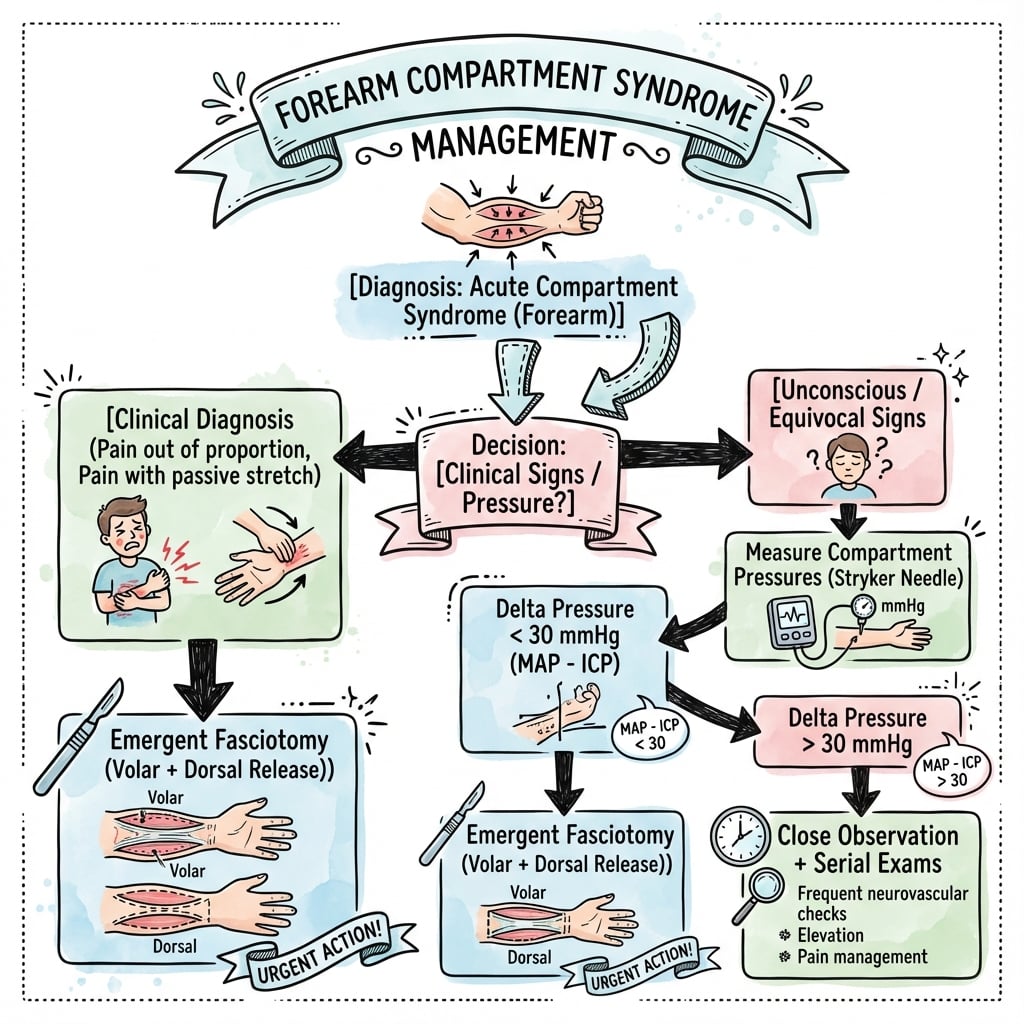
Pressure Thresholds for Fasciotomy
PRESSURE THRESHOLDS
Delta P Concept
ΔP (Delta P) = Diastolic BP - Compartment Pressure
This represents the perfusion pressure gradient. When ΔP is less than 30 mmHg, tissue perfusion is inadequate regardless of absolute compartment pressure.
Example: Patient with DBP 70 mmHg and compartment pressure 45 mmHg:
- ΔP = 70 - 45 = 25 mmHg (LESS THAN 30 = fasciotomy indicated)
Other Investigations
Laboratory:
- CK (creatine kinase) - elevated with muscle necrosis
- Myoglobin - rhabdomyolysis marker
- Serum lactate - tissue ischemia
- Renal function - monitor for myoglobinuric AKI
- Coagulation studies - if bleeding diathesis suspected
Imaging:
- X-rays - identify underlying fracture, guide fracture management
- CT/MRI - NOT routine, may delay treatment
- Doppler ultrasound - assess arterial flow (pulse presence ≠ adequate perfusion)
Do NOT Delay for Investigations
Clinical diagnosis is sufficient. Do not delay fasciotomy for pressure measurement, imaging, or laboratory results if clinical suspicion is high. Time to fasciotomy is the critical factor.
Continuous Monitoring
Indications for continuous compartment pressure monitoring:
- Multiple trauma patient requiring sedation/ventilation
- Post-operative monitoring after high-risk surgery
- Burns with circumferential involvement
- Serial measurements impractical
Management Algorithm
Initial Actions:
Remove Constrictors
- Bivalve ALL casts
- Release ALL circumferential dressings
- Cut down to skin
- Position limb at heart level
- Do NOT elevate (reduces arterial perfusion)
Optimize Perfusion
- Maintain blood pressure (avoid hypotension)
- Correct hypovolemia
- Supplemental oxygen
- Maintain normothermia
- Correct coagulopathy
Urgent Surgery
- Contact theatre immediately
- Consent for fasciotomy
- Plan for both volar AND dorsal release
- Warn patient wounds will be left open
- Plan for return to theatre 48-72h
Immediate removal of all constrictive elements is critical before definitive fasciotomy.
Surgical Technique
Preoperative Preparation
Patient Positioning:
- Supine position, arm on radiolucent hand table
- Tourniquet applied but use judiciously (inflate only if bleeding obscures view)
- Consider avoiding tourniquet to assess tissue perfusion
Consent Discussion:
- Explain wounds will be left open
- Risk of incomplete decompression
- Nerve and vessel injury risk
- Need for return to theatre at 48-72 hours
- Skin graft may be required
- Risk of ongoing muscle/nerve damage despite surgery
Volar Fasciotomy Technique
Incision Planning:
- Curvilinear/zigzag incision from medial epicondyle to palm
- Cross antecubital fossa obliquely (avoid linear scar contracture across flexion crease)
- Curve ulnar at wrist, then extend into palm for carpal tunnel release
- Total length: 15-20cm forearm + 3-4cm palm extension
Step-by-Step Technique:
- Skin Incision - Full-thickness skin incision, generous length
- Identify and protect superficial veins - ligate if necessary
- Release lacertus fibrosus (bicipital aponeurosis) at antecubital fossa
- Identify median nerve - runs between FDS and FDP
- Release superficial volar compartment:
- Incise fascia overlying PT, FCR, PL, FCU, FDS
- Full-length fascial release
- Palpate muscles - should decompress and bulge through fasciotomy
- Release deep volar compartment:
- Retract FDS muscles
- Incise deep fascia overlying FDP, FPL, PQ
- Protect AIN (runs on interosseous membrane)
- Carpal tunnel release (MANDATORY):
- Extend incision into palm
- Divide transverse carpal ligament under direct vision
- Protect median nerve and palmar cutaneous branch
- Inspect all muscles:
- Viable muscle: pink, contracts with stimulation, bleeds when cut
- Non-viable muscle: dark, does not contract, does not bleed
- Debride clearly necrotic tissue
A systematic approach to volar fasciotomy ensures complete decompression and protection of critical structures.
Mandatory Components
Volar Fasciotomy MUST Include:
- Superficial compartment release (complete)
- Deep compartment release (complete)
- Carpal tunnel decompression (ALWAYS)
- Protection of median nerve throughout
- Assessment of muscle viability
Complications
Complications of Delayed/Missed Diagnosis
Sequelae of Untreated Compartment Syndrome
| Complication | Description | Timing | Treatment |
|---|---|---|---|
| Volkmann's ischemic contracture | Irreversible flexor muscle fibrosis and shortening | Weeks-months | Tendon lengthening, muscle slide, free flap |
| Permanent nerve injury | Median and/or ulnar nerve damage | Immediate | Nerve exploration, possible grafting |
| Muscle necrosis | Dead muscle requiring debridement | Days | Serial debridement, coverage |
| Rhabdomyolysis | Myoglobin release causing AKI | Hours-days | Aggressive hydration, ?dialysis |
| Chronic pain | Neuropathic and ischemic pain | Months | Multidisciplinary management |
| Functional loss | Loss of grip strength, dexterity | Permanent | Reconstructive surgery, therapy |
Volkmann's Ischemic Contracture
Volkmann's Contracture - The Devastating Outcome
Irreversible fibrotic contracture of forearm flexor muscles following untreated compartment syndrome.
Classic Posture:
- Wrist flexion
- MCP hyperextension
- IP joint flexion
- Thumb adduction
Cascade Sign: Passive wrist extension causes fingers to flex further (muscle shortening)
Classification (Tsuge):
| Type | Muscle Involvement | Features | Treatment |
|---|---|---|---|
| Mild | FDP to 2-3 fingers, FPL | Limited contracture | Muscle slide, tendon lengthening |
| Moderate | All flexors involved | Significant deformity | Muscle slide + tendon transfer |
| Severe | Both flexors AND extensors | Severe dysfunction | Free functioning muscle transfer |
Complications of Fasciotomy
Early Complications
- Wound infection
- Bleeding
- Nerve injury (iatrogenic)
- Vessel injury
- Skin edge necrosis
- Incomplete release
Late Complications
- Unsightly scars
- Skin graft contracture
- Chronic wound healing
- Tethering of tendons
- Altered sensation
- Need for secondary reconstruction
Postoperative Care and Rehabilitation
Immediate Post-operative Management
Wound Care:
- Leave wounds completely OPEN - NEVER close primarily
- Apply loose non-adherent dressing (Jelonet, Adaptic)
- Consider negative pressure wound therapy (VAC) if:
- Significant edema persists
- Large wound with exposed structures
- Difficult to maintain dressing
Splinting:
- Position of function to prevent contracture:
- Wrist: 20-30° extension
- MCP joints: 70-90° flexion
- IP joints: Full extension
- Thumb: Abduction and opposition
- Avoid tight circumferential dressings
- Ensure splint does not compress compartments
Monitoring:
- Neurovascular observations every 2 hours for first 24 hours
- Monitor for:
- Persistent pain (inadequate decompression)
- Worsening motor/sensory deficit
- Signs of bleeding
- Systemic complications (rhabdomyolysis)
Laboratory Monitoring
Rhabdomyolysis Monitoring
- CK (creatine kinase) - daily initially
- Myoglobin - serum and urine
- Renal function - creatinine, eGFR
- Urine output - maintain greater than 1ml/kg/h
- Lactate - marker of tissue ischemia
Management of Rhabdomyolysis
- Aggressive IV hydration - aim UOP greater than 1ml/kg/h
- Alkalinize urine - IV sodium bicarbonate
- Monitor potassium - hyperkalemia risk
- Consider dialysis if AKI develops
- Treat underlying cause
Return to Theatre (48-72 Hours)
Assessment for Wound Closure:
Criteria for DPC:
- Muscle remains viable (pink, contractile)
- Edema has resolved
- No signs of infection
- Skin edges can be approximated without tension
Technique:
- Thorough wound irrigation
- Reassess muscle viability - debride any necrotic tissue
- Approximate skin edges with interrupted sutures or staples
- Avoid tension on closure
- May need staged closure if significant gap
Delayed primary closure is ideal when edema resolves and skin edges can be approximated without tension.
Rehabilitation Protocol
Phase 1 (0-2 weeks):
- Wound healing priority
- Gentle passive ROM when wounds closed/stable
- Edema control - elevation, compression
- Hand therapy referral
Phase 2 (2-6 weeks):
- Active ROM exercises
- Scar massage and desensitization
- Gentle strengthening
- Functional activities
Phase 3 (6+ weeks):
- Progressive strengthening
- Return to activities of daily living
- Monitor for contracture development
- Long-term follow-up
Watch for Late Complications
Monitor for signs of developing Volkmann's contracture:
- Progressive finger flexion posture
- Cascade sign (wrist extension worsens finger flexion)
- Grip weakness
- Prompt referral to hand surgery if contracture develops
Outcomes and Prognosis
Factors Affecting Outcome
Timing of Fasciotomy:
| Time to Fasciotomy | Expected Outcome | Functional Recovery |
|---|---|---|
| Less than 6 hours | Excellent - minimal muscle/nerve damage | 68% near-normal function |
| 6-12 hours | Good - some muscle fibrosis, nerve recovery | 40% near-normal function |
| 12-24 hours | Fair - significant muscle damage, incomplete nerve recovery | 20% near-normal function |
| Greater than 24 hours | Poor - established damage, Volkmann's likely | 8% near-normal function |
Other Prognostic Factors:
- Severity of initial injury
- Associated fractures and soft tissue trauma
- Patient age (younger = better recovery potential)
- Rehabilitation compliance
- Presence of complications (infection, rhabdomyolysis)
Expected Recovery Timeline
Sensory Recovery
Timeline:
- First sensation: 2-4 weeks
- Protective sensation: 6-12 weeks
- Discriminative touch: 3-6 months
- May remain incomplete in severe cases
Motor Recovery
Timeline:
- Muscle contraction: 4-8 weeks
- Functional strength: 3-6 months
- Maximal recovery: 12-18 months
- Depends on extent of muscle necrosis
Functional Outcomes
Timeline:
- ADL independence: 3-6 months
- Return to work: 6-12 months
- Full recovery: 12-24 months
- May have persistent weakness
Volkmann's Contracture Outcomes
Prevention is Key:
- Incidence with early fasciotomy (less than 6h): less than 5%
- Incidence with delayed fasciotomy (greater than 12h): 20-40%
- Established contracture is IRREVERSIBLE - reconstructive surgery only
Reconstructive Surgery Outcomes:
| Severity | Surgery | Expected Function | Patient Satisfaction |
|---|---|---|---|
| Mild | Muscle slide, tendon lengthening | Good - 70-80% normal | High |
| Moderate | Muscle slide + tendon transfers | Fair - 40-60% normal | Moderate |
| Severe | Free functioning muscle transfer | Poor - 20-30% normal | Low to moderate |
Long-term Complications Rates
Long-term Complication Rates After Forearm Fasciotomy
| Complication | Incidence | Impact | Management |
|---|---|---|---|
| Scar contracture | 10-20% | Cosmetic, possible functional limitation | Scar revision, Z-plasty |
| Chronic pain | 15-25% | Neuropathic pain, impaired function | Multidisciplinary pain management |
| Residual weakness | 30-50% | Reduced grip strength, endurance | Ongoing therapy, adaptive strategies |
| Numbness | 20-35% | Protective sensation usually preserved | Desensitization therapy |
| Volkmann's contracture | 5-10% (early surgery), 20-40% (late surgery) | Severe functional impairment | Reconstructive surgery |
| Infection | 5-15% | Delayed healing, possible amputation | Antibiotics, serial debridement |
Medicolegal Outcomes
High Medicolegal Risk
Missed or delayed compartment syndrome is one of the most common and costly sources of orthopaedic litigation internationally:
- Common allegations: delayed diagnosis, failure to monitor or document neurovascular status, and inadequate (incomplete) fasciotomy
- Claims are frequent and high-value in multiple jurisdictions (e.g. analyses of UK NHS Resolution and US closed-claims data), driven by the catastrophic, irreversible nature of the sequelae
- Prevention: meticulous time-stamped documentation, a low threshold for fasciotomy, and early senior/specialist involvement
Key Documentation for Medicolegal Protection:
- Time-stamped neurovascular examinations
- Compartment pressure measurements (if performed)
- Clinical decision-making rationale
- Patient/family discussions about risks
- Informed consent including Volkmann's contracture risk
- Time from symptom onset to surgical decompression
Evidence Base
Delta P Threshold for Fasciotomy (Landmark)
- Prospective study of 116 tibial diaphyseal fractures with continuous anterior compartment monitoring for 24 hours; 3 (2.6%) developed acute compartment syndrome.
- Using a differential pressure (diastolic BP minus compartment pressure) threshold of less than 30 mmHg led to NO missed cases; no patient had compartment-syndrome sequelae at minimum six-month review.
- An absolute threshold of greater than 30 mmHg would have led to fasciotomy in 43% of patients, many unnecessary.
Timing of Fasciotomy and Outcomes (Landmark)
- 66 cases of acute compartment syndrome treated by fasciotomy in 46 extremities of 44 patients.
- Fasciotomy performed early (less than 12 hours after onset) gave normal function in 68% of extremities; only 8% of late fasciotomies achieved normal function.
- Complication rates were 4.5% (early) versus 54% (late).
Volkmann's Contracture, Supracondylar Fractures and Prevention (Landmark)
- Review of 55 children (58 limbs) with Volkmann's contracture treated in Toronto 1955-1975.
- Supracondylar fractures of the elbow that progressed to Volkmann's contracture frequently had BOTH an arterial injury and a compartment syndrome.
- Most children had not received early appropriate treatment, and the frequency of contracture had not declined over 21 years despite increased awareness.
Acute Compartment Syndrome of the Upper Extremity (Review)
- Comprehensive review of acute compartment syndrome of the hand, forearm and upper arm.
- Pain out of proportion to injury is the most reliable early symptom; diagnosis is particularly difficult in obtunded patients and young children.
- Early recognition and expeditious fasciotomy are essential to obtain good outcomes and prevent permanent disability and ischaemic contracture.
Acute Compartment Syndrome: Who Is at Risk? (Epidemiology)
- Analysis of 164 consecutive patients with acute compartment syndrome over eight years.
- An associated fracture was present in 69%; forearm compartment syndrome was associated with distal radius fractures and, like the cohort overall, occurred most commonly in young men (typically under 35 years).
- Soft-tissue injury without fracture was the second most common cause; one-tenth had a bleeding disorder or were anticoagulated.
Diagnosis and Treatment of Acute Extremity Compartment Syndrome (Lancet Series)
- Lancet Emergency Surgery Series review summarising upper- and lower-limb acute compartment syndrome in adults and children.
- There is no universally agreed diagnostic standard; some advocate continuous intracompartmental pressure monitoring in high-risk injuries while others favour aggressive fasciotomy when the syndrome is even suspected.
- Ineffective treatment risks permanent dysaesthesia, ischaemic contracture, muscle dysfunction, limb loss and death; fasciotomy itself carries long-term morbidity.
Clinical Decision Scenarios
Use these scenarios to practise clinical reasoning and management decisions
Post-Fracture Compartment Syndrome
"8-year-old boy, 6 hours post-supracondylar fracture ORIF. Nurses report severe forearm pain despite regular analgesia. Pain with passive finger extension. Cast has been bivalved."
Equivocal Clinical Presentation
"25-year-old motorcyclist with closed both-bone forearm fracture. Moderate forearm pain, compartment feels somewhat tense but not rock-hard. Fingers move but patient reports altered sensation in thumb web space. You are uncertain about the diagnosis."
Established Volkmann's Contracture
"28-year-old presents 6 months after forearm fracture treated elsewhere. He has a fixed flexion deformity of fingers with wrist in flexed position. Extending wrist causes fingers to flex further. He has weak grip and altered sensation."
MCQ Practice Points
Question: Earliest Sign
Q: What is the earliest and most reliable clinical sign of compartment syndrome?
A: Pain out of proportion to the injury, especially pain on passive stretch of the affected compartment. For volar forearm compartment, this is pain with passive finger extension.
Question: Pressure Thresholds
Q: What are the compartment pressure thresholds indicating need for fasciotomy?
A: Either absolute pressure greater than 30mmHg OR Delta P less than 30mmHg (where Delta P = diastolic blood pressure minus compartment pressure). Delta P accounts for individual patient perfusion status.
Question: Fasciotomy Components
Q: What MUST be included with volar forearm fasciotomy?
A: Carpal tunnel decompression - swelling extends distally into the carpal tunnel and must be released to prevent median nerve compression at the wrist.
Question: Late Signs
Q: Which of the 5 Ps are considered LATE signs of compartment syndrome?
A: Pallor and pulselessness are late signs. Waiting for these findings means irreversible muscle and nerve damage has likely already occurred. Pain, pressure, and paresthesia are earlier findings.
Question: Volkmann's Pathology
Q: What is the pathological basis of Volkmann's ischemic contracture?
A: Ischemic necrosis of forearm flexor muscles leading to fibrotic replacement. The shortened, fibrotic muscles cause the characteristic posture: wrist flexion, MCP hyperextension, IP joint flexion.
Question: Time Window
Q: What is the critical time window for fasciotomy in compartment syndrome?
A: 6-8 hours from onset of ischemia. Fasciotomy within 6 hours has significantly better outcomes. After 8 hours, irreversible muscle necrosis and nerve damage are highly likely.
Guidelines, Registries & Global Practice
Differential Diagnosis
Compartment syndrome must be distinguished from other causes of a painful, swollen or neurologically compromised forearm. The two most dangerous mimics to exclude are arterial injury and an evolving compartment syndrome masked by a nerve block.
Differential Diagnosis of the Painful Swollen Forearm
| Condition | Distinguishing Features | Key Test | Pitfall |
|---|---|---|---|
| Acute compartment syndrome | Pain out of proportion, pain on passive stretch, tense compartment, escalating analgesia | Clinical; compartment pressure / ΔP if equivocal | Pulse often preserved until late - do not wait for it |
| Arterial injury / ischaemia | Cold pale limb, absent pulse, hard signs of vascular injury, ABI reduced | Doppler, CT angiography | Can coexist with compartment syndrome (e.g. supracondylar fracture) |
| Peripheral nerve injury (traumatic) | Focal sensorimotor deficit in single nerve territory, soft compartment, no disproportionate pain | Clinical exam, nerve conduction (delayed) | Mislabelling early compartment ischaemia as primary nerve injury |
| Cellulitis / soft-tissue infection | Erythema, warmth, fever, raised inflammatory markers, soft compartment | Bloods (WCC, CRP), clinical | Necrotising fasciitis can mimic and coexist - look for crepitus, systemic toxicity |
| DVT / venous thrombosis | Swelling, less severe pain, no pain on passive stretch | Duplex ultrasound | Rare in upper limb; do not anticoagulate if compartment syndrome possible |
| Fracture pain alone | Pain proportionate to injury, settles with analgesia and splintage | Clinical response to analgesia | Attributing escalating pain to 'just the fracture' |
Global Epidemiology
According to PubMed, the largest population series (McQueen et al, J Bone Joint Surg Br 2000, PMID 10755426) of 164 consecutive acute compartment syndromes found an associated fracture in 69%, with forearm cases linked to distal radius fractures and occurring predominantly in young men under 35 years. The Lancet Emergency Surgery Series review (von Keudell et al 2015, PMID 26460664) confirms acute compartment syndrome affects upper and lower limbs in both adults and children and that there remains no internationally agreed diagnostic gold standard. Forearm compartment syndrome in children is most often associated with displaced supracondylar humeral fractures (Mubarak & Carroll, PMID 479251).
Guideline & Registry Comparison
International Guidance and Evidence Sources
| Body / Source | Region | Key Position | Evidence Level |
|---|---|---|---|
| BOA/BAPRAS (BOAST - open fracture & limb-threatening injury standards) | UK | Urgent senior decompression; clinical diagnosis paramount; document neurovascular status serially | Consensus standard |
| Lancet Series (von Keudell 2015) | International | Clinical suspicion drives early fasciotomy; continuous pressure monitoring for obtunded/unreliable patients | Narrative review (Level V) |
| AAOS / OTA teaching | USA | Either absolute pressure greater than 30 mmHg OR ΔP less than 30 mmHg; fasciotomy if clinically suspected | Expert consensus |
| AO Foundation (AO Surgery Reference) | International | Complete volar + dorsal release with mandatory carpal tunnel decompression; wounds left open | Technique consensus |
| EFORT / European trauma teaching | Europe | ΔP threshold (McQueen) favoured over absolute pressure; early decompression | Based on Level II-III data |
There is no orthopaedic registry that systematically captures forearm compartment syndrome in the way arthroplasty registries (AOANJRR, NJR, AJRR) capture joint replacement; incidence and outcome data derive from single-centre and national trauma series rather than registry datasets.
Practice Variation & Regional Considerations
The principal area of international practice variation is the diagnostic pathway: continuous intracompartmental pressure monitoring is favoured in some UK and European units for high-risk or obtunded patients, whereas a clinically-driven "decompress if suspected" approach predominates in many North American and Australasian centres (von Keudell 2015, PMID 26460664). The ΔP less than 30 mmHg threshold (McQueen, PMID 8898137) is the most widely adopted pressure criterion worldwide.
In Australia and New Zealand, trauma centres apply standardised serial neurovascular observation after high-risk fractures, and Therapeutic Guidelines (eTG) recommend cefazolin for surgical prophylaxis at fasciotomy. In rural and remote settings where retrieval (including by the Royal Flying Doctor Service) may exceed 4-6 hours, local surgeons should have a low threshold to perform fasciotomy before transfer rather than risk irreversible ischaemia in transit. VTE prophylaxis follows standard trauma protocols, with mechanical methods preferred initially given the bleeding risk into freshly decompressed compartments.
FOREARM COMPARTMENT SYNDROME
Clinical summary
Clinical Signs
- •Pain OUT OF PROPORTION (earliest)
- •Pain on PASSIVE STRETCH (most reliable)
- •Tense, wooden compartment
- •Paresthesia (nerve ischemia)
- •Pallor and pulselessness = TOO LATE
Pressure Thresholds
- •Absolute greater than 30 mmHg
- •Delta P less than 30 (DBP - compartment pressure)
- •Clinical diagnosis SUFFICIENT
- •Do NOT delay for pressures if high suspicion
Volar Fasciotomy
- •Curvilinear incision elbow to palm
- •Release lacertus fibrosus
- •Release superficial + deep volar
- •ALWAYS add carpal tunnel release
- •Protect median nerve throughout
Post-operative
- •Leave wounds OPEN
- •Loose dressings or VAC
- •Splint in position of function
- •DPC at 48-72 hours
- •STSG if cannot close
High-Risk Fractures
- •Supracondylar (children)
- •Both-bone forearm
- •Floating elbow
- •Monteggia/Galeazzi
- •High-energy distal radius
Volkmann's Contracture
- •Irreversible flexor fibrosis
- •Wrist flexed, MCP extended, IP flexed
- •Cascade sign positive
- •PREVENTION is key - early fasciotomy
Key Takeaways
Remember
- Pain on passive stretch is the earliest reliable sign
- Clinical diagnosis is sufficient - don't delay for pressures
- Always release carpal tunnel with volar fasciotomy
- Time to fasciotomy determines outcome
- Leave wounds completely open
Avoid
- Waiting for pulselessness (too late!)
- Delaying surgery for pressure measurement
- Closing wounds primarily
- Attributing pain to fracture alone
- Incomplete fasciotomy