Benign Reactive Giant-Cell Lesion of Small Bones
- Benign, reactive giant-cell-rich lesion of the small bones of the hands and feet, and of the jaws (central giant cell granuloma)
- Uneven, clustered giant cells around haemorrhage with reactive bone, against the large, evenly distributed giant cells of giant cell tumour
- Giant cell tumour of bone is rare in the hand but metastasises to the lung in about 3%, reported even from a hand lesion
- Check calcium and PTH to exclude a brown tumour
- Curettage with grafting; recurrence is recognised but no series gives a reliable rate, and a recurrence reopens the diagnosis
- “Most hand/foot GCRGs are USP6-rearranged (8 of 9) and sit on the solid ABC spectrum; the gnathic lesion is USP6-negative
- “H3.3 G34W immunostain is positive in giant cell tumour only
- “A 'recurrent GCRG' may prove to be a giant cell tumour: fresh tissue, G34W, calcium/PTH
- “Do not quote a recurrence percentage for the small bones
Definition and Presentation
What it is. Giant cell reparative granuloma (GCRG) is a benign, reactive giant-cell-rich lesion, not a true neoplasm. It most often arises in the small bones of the hands and feet (phalanges, metacarpals and metatarsals) and in the jaws, where it is termed central giant cell granuloma.
Where it sits. It is now generally regarded as part of the spectrum of giant-cell lesions of the small bones, overlapping with the "solid" aneurysmal bone cyst. The molecular evidence for that overlap is set out below.
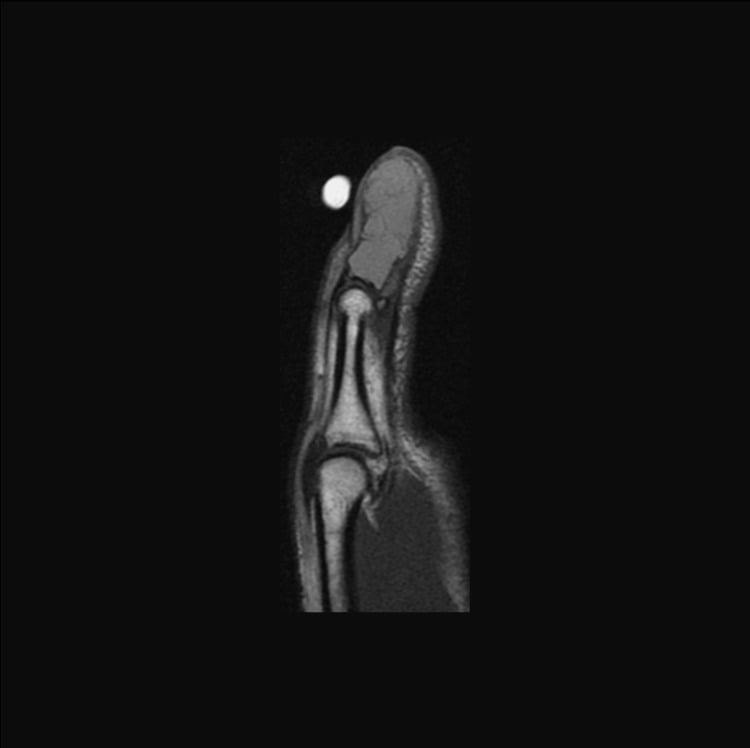
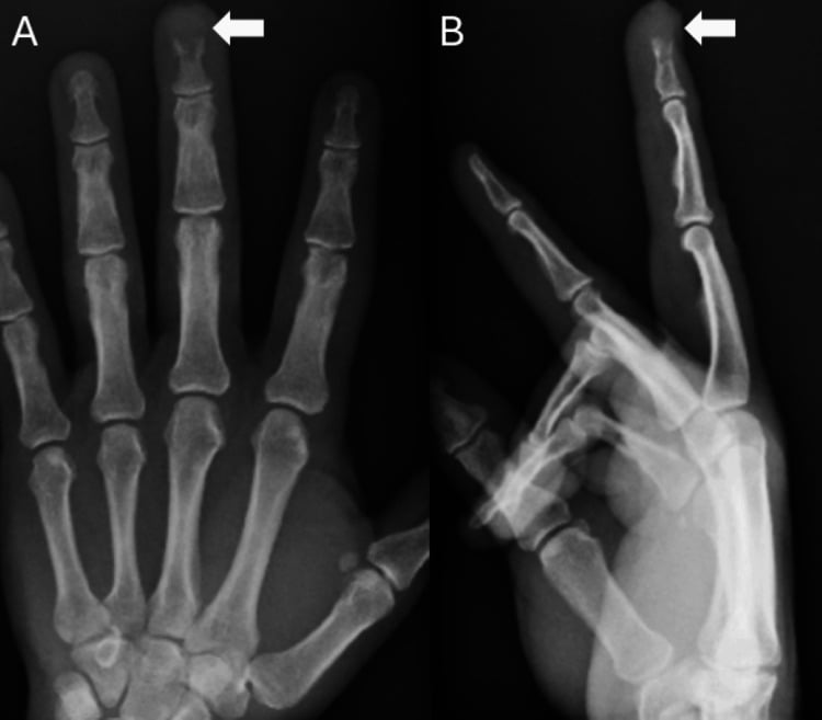
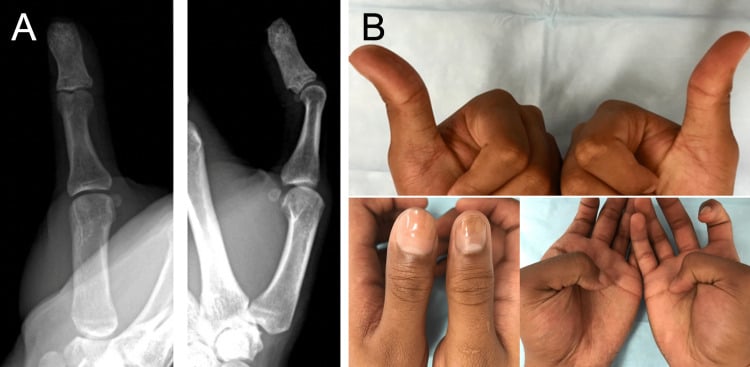
Presentation. Pain, swelling and sometimes a pathological fracture are typical. On imaging it is a lytic, often expansile lesion of a small tubular bone with cortical thinning, and it can look locally aggressive although it is benign.
Histology and the Giant Cell Tumour Question
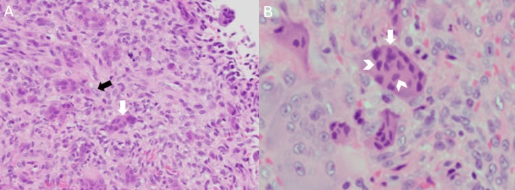
Histology. A fibrous, cellular stroma holds osteoclast-type giant cells distributed unevenly, in clusters, often around foci of haemorrhage, together with reactive woven bone and haemosiderin. In giant cell tumour of bone the giant cells are large and evenly distributed throughout a mononuclear stroma, and it is this uneven, clustered pattern with reactive bone that helps separate the two.
The differential that matters. Giant cell tumour (GCT) of bone is locally aggressive and rare in the small bones of the hand; it favours the meta-epiphysis of long bones around the knee. The morphology overlaps, and a giant-cell-rich lesion of the distal phalanx first diagnosed as GCRG recurred at 8 months in a reported case, proved to be a giant cell tumour and ended in amputation of the fingertip. The diagnosis is genuinely challenging and warrants experienced pathological review.
- Giant cell reparative granuloma
- Benign, reactive
- Giant cell tumour of bone
- Benign but locally aggressive neoplasm
- Giant cell reparative granuloma
- Small bones of hands/feet; jaws
- Giant cell tumour of bone
- Meta-epiphysis of long bones (around knee); rare in hand
- Giant cell reparative granuloma
- Uneven/clustered (around haemorrhage)
- Giant cell tumour of bone
- Large, evenly distributed throughout stroma
- Giant cell reparative granuloma
- Fibrous stroma, reactive woven bone, haemosiderin
- Giant cell tumour of bone
- Mononuclear stromal cells (the neoplastic component)
- Giant cell reparative granuloma
- Curettage (+/- grafting); can recur
- Giant cell tumour of bone
- Extended curettage +/- adjuvants; denosumab; recurrence
Why the call matters. Giant cell tumour of bone metastasises to the lung in about 3% of cases, and this has been reported from a giant cell tumour of the hand, where the tumour is supposedly rare. Calling an aggressive giant-cell lesion of a phalanx a reparative granuloma is therefore not a harmless naming error. It changes whether the excision is adequate, whether the chest is imaged, and the surveillance the patient receives.
Differential Diagnosis
Each lesion that a giant-cell-rich lytic lesion of a small bone must be separated from has its own page, and the separation is what the work-up is for. Correlating the imaging with the histology is part of that work.
- Giant cell tumour of bone - the aggressive neoplasm at the other end, discussed above
- Brown tumour of hyperparathyroidism - the metabolic mimic, which is why calcium and PTH are checked
- Aneurysmal bone cyst - shares both the morphology and part of the spectrum
- Chondroblastoma - epiphyseal, in a child, with chondroid matrix
- Non-ossifying fibroma - metaphyseal, cortical, with a sclerotic rim, and self-limiting
- Rarely, more aggressive lesions - malignancy to exclude includes chondrosarcoma or other lesions; a giant-cell-rich osteosarcoma, with atypical stroma and malignant osteoid, is the one not to miss
Molecular Discriminators
When the morphology overlaps, the markers help:
- H3F3A (H3.3) G34W - the giant cell tumour mutation. The H3.3 G34W immunostain is positive in GCT and negative in GCRG, brown tumour, aneurysmal bone cyst and chondroblastoma, which makes it the single most useful marker when GCRG-versus-GCT is in doubt.
- H3F3B K36M - the chondroblastoma mutation, detected as H3K36M positivity.
- USP6 rearrangement - present in most primary aneurysmal bone cysts and in 8 of 9 (89%) giant cell reparative granulomas of the hands and feet (Agaram 2014), but in none of 8 gnathic central giant cell granulomas.
USP6 does not separate ABC from GCRG. It is not the clean separator it was once taught as. The rearrangement in most small-bone lesions is the molecular evidence that they are solid aneurysmal bone cysts, the same entity. In a hand or foot lesion, USP6 positivity places it on the solid ABC/GCRG spectrum and argues against giant cell tumour, but it cannot tell ABC from GCRG.
The Jaw Form, Syndromic Associations and Medical Treatment
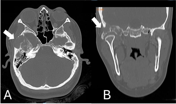
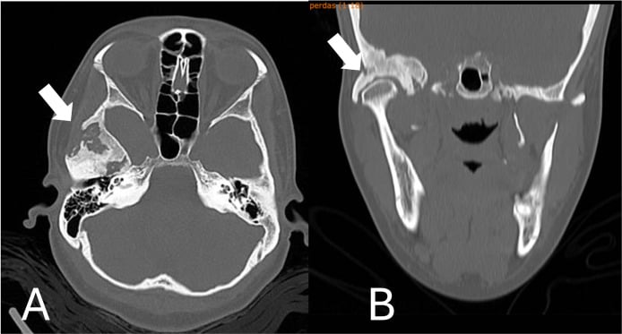
Central giant cell granuloma. The intraosseous giant-cell lesion of the mandible and maxilla is commonest in the anterior mandible, in patients under 30, with a female predominance. The peripheral giant cell granuloma is a different lesion, a soft-tissue lesion of the gum.
Aggressive and non-aggressive forms. The jaw lesion may be:
- Non-aggressive - slow-growing and asymptomatic
- Aggressive - pain, rapid growth, cortical perforation, root resorption and a high recurrence rate
Syndromic associations. Multiple or syndromic giant-cell lesions occur in Noonan syndrome, neurofibromatosis type 1 and cherubism, part of the RASopathy spectrum, so multiple lesions warrant a genetics work-up. Cherubism (SH3BP2) produces bilateral, symmetric mandibular lesions in a child and is self-limiting after puberty.
Medical treatment. Beyond curettage, aggressive or jaw lesions can be treated with intralesional corticosteroid injections, calcitonin, interferon-alpha or denosumab, sparing a more morbid resection. Denosumab is anti-RANKL and shrinks the osteoclastic component of aggressive or unresectable lesions.
Management
Work-up. The work-up has three parts:
- Imaging of the lytic, expansile small-bone lesion
- Calcium and PTH, to exclude a brown tumour
- Biopsy and histology at a centre experienced in bone tumours
Treatment. Curettage with bone grafting treats most lesions and is usually effective. Recurrent or aggressive lesions may need more extensive excision, occasionally amputation of a digit.
Counselling about recurrence. Local recurrence after curettage is well recognised, so the patient needs follow-up. The literature on giant-cell lesions of the small bones is case reports and small series, and no cohort supplies a dependable recurrence rate. Counsel that the lesion can come back rather than quoting a percentage, and if a number is quoted in an exam, attribute it to the series it came from.
Treat a recurrence as a diagnostic event, not just a surgical one. Send fresh tissue for experienced pathological review, ask specifically for H3.3 G34W immunohistochemistry, and check calcium and PTH if that was not done initially. A lesion that behaves aggressively is telling you something the first report may have missed: a "recurrent GCRG" may prove to be a giant cell tumour of bone, which needs different, more aggressive management.
Mnemonics & Memory Aids
REPAIR
Hook:REPAIR: Reactive benign, Extremity small bones/jaws, Pattern (uneven/clustered + reactive bone), vs GCT (even/aggressive), Investigate Ca/PTH, Resect/curette + Recurrence (re-confirm).
STAINThe Molecular Panel - STAIN
Hook:STAIN the ambiguous giant-cell lesion: G34W for GCT, K36M for chondroblastoma, USP6 for the ABC/small-bone-GCRG spectrum - and none of these marks the jaw form.
JAWThe Jaw Form - JAW
Hook:JAW: the gnathic lesion is its own entity - USP6-negative, syndromic when multiple, and answerable to medical therapy when aggressive.
Clinical Decision Scenarios
Practise clinical reasoning and management decisions out loud
“A lytic, expansile lesion of a phalanx shows a giant-cell-rich histology. How do you approach the diagnosis and management?”
What it is
- Benign, reactive giant-cell-rich lesion (not a true neoplasm)
- Small bones of hands/feet and the jaws (central giant cell granuloma)
- Part of the giant-cell-lesion-of-small-bones / solid ABC spectrum
Histology
- Fibrous/cellular stroma; unevenly distributed/clustered giant cells
- Often around foci of haemorrhage; reactive woven bone + haemosiderin
- Contrast: GCT of bone = large, evenly distributed giant cells
Differential & work-up
- Giant cell tumour of bone (locally aggressive; rare in hand)
- Brown tumour (check calcium/PTH); aneurysmal bone cyst
- Imaging + biochemistry + experienced histology review
Management
- Curettage (+/- grafting) - usually effective
- Recurrence recognised but no dependable published rate for the small bones - follow up
- The stakes: GCT metastasises to lung in ~3 percent, reported even from a hand lesion
- Recurrent/aggressive: re-confirm diagnosis (may be GCT) + more extensive surgery
Evidence & Key Studies
Giant-cell lesion of the distal phalanx initially diagnosed as reparative granuloma, recurring as giant cell tumour
- Giant cell tumours of bone are locally aggressive neoplasms that typically occur in the distal femur or proximal tibia and infrequently in the bones of the hand, including the distal phalanx.
- A distal-phalanx lesion underwent curettage with a working diagnosis of giant cell reparative granuloma (with focal fracture callus), but recurred at 8 months and, on histopathology, was confirmed to be a giant cell tumour of bone (requiring amputation of the distal phalanx tip).
- In rare and difficult-to-diagnose giant-cell lesions, consultation with experienced pathologists improves diagnostic accuracy and prevents treatment delays - underscoring the GCRG/GCT diagnostic overlap.
Multiple pulmonary metastases from giant cell tumor of a hand
- Giant cell tumour of bone classically involves the epiphysis of long bones and only rarely the small bones of the hands and feet, but despite being benign it destroys bone, recurs locally and can metastasise.
- Approximately 3 percent of giant cell tumours metastasise to the lung. This report describes multiple pulmonary metastases from a giant cell tumour of the hand in a 54-year-old man, found incidentally as asymptomatic nodules on a routine chest radiograph.
- He achieved near-complete remission with adriamycin and cisplatin. As a single case report it establishes that the event can occur from a hand lesion, not how often - the 3 percent figure refers to giant cell tumours at all sites.
USP6 rearrangements in giant cell reparative granulomas of the hands and feet
- 49 samples from 48 patients: 9 giant-cell-rich lesions of the hands/feet compared with 8 gnathic GCRGs, 22 primary ABCs, 8 giant cell tumours of bone and 2 brown tumours.
- 8 of 9 (89%) giant-cell reparative granulomas of the hands and feet carried USP6 gene rearrangements - as did 13 of 22 (59%) primary aneurysmal bone cysts - while ALL 8 gnathic GCRGs, 8 GCTs and 2 brown tumours were USP6-negative.
- The authors' conclusion: most GCRGs of the hands and feet represent true (solid) aneurysmal bone cysts and should be classified as such, with the GCRG terminology largely reserved for the gnathic lesions.
Distinct H3F3A and H3F3B driver mutations define chondroblastoma and giant cell tumour of bone
- 73 of 77 chondroblastomas (95%) carried p.Lys36Met alterations, predominantly in H3F3B (one of the two histone H3.3 genes).
- 49 of 53 giant cell tumours of bone (92%) carried H3F3A alterations - p.Gly34Trp in all but one (a single p.Gly34Leu) - with exquisite tumour-type specificity.
- The mutations were restricted to the stromal cell population, identifying the mononuclear stromal cell - not the osteoclast-like giant cell - as the neoplastic component of GCT.
The diagnostic overlap and challenge between giant cell reparative granuloma and giant cell tumour of bone in the small bones of the hand (a phalangeal lesion diagnosed as GCRG recurring and proving to be a giant cell tumour), the rarity of giant cell tumour in the hand, and the value of experienced pathological review come from the cited DeFrancisis report. The benign reactive nature of GCRG, its location in the small bones of the hands/feet and jaws, the histological distinction (uneven/clustered giant cells with reactive bone vs even distribution in GCT), the brown-tumour/ABC differentials, and curettage-with-recurrence management are standard, well-established teaching. The roughly 3 percent pulmonary-metastasis figure for giant cell tumour of bone, and its occurrence from a hand lesion, come from the Moon report - a single case, which establishes that it can happen rather than how often. The USP6 profile (rearranged in 8 of 9 small-bone GCRGs, absent in all 8 gnathic ones) comes from the cited Agaram study, and the H3.3 driver mutations (G34W in 92% of GCTs, K36M in 95% of chondroblastomas, stromal-cell restricted) from the cited Behjati study. No cohort gives a dependable local-recurrence rate for giant-cell reparative granuloma of the small bones; the literature is case reports and small series, so no percentage is quoted here. Nor is there a validated follow-up interval, a size or site threshold that mandates wider excision, or a randomised comparison of curettage against any medical adjuvant in this lesion.