Massive Osteolysis / Vanishing Bone Disease
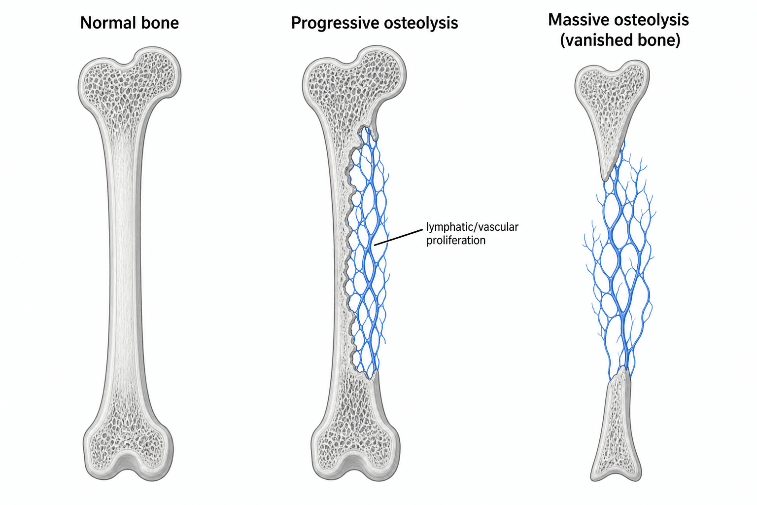
- Gorham-Stout disease (GSD) - massive osteolysis, 'vanishing bone' or 'phantom bone' disease - is a RARE disorder (only a few hundred cases reported) of PROGRESSIVE, SPONTANEOUS, idiopathic OSTEOLYSIS, in which an abnormal proliferation of LYMPHATIC and vascular channels within bone resorbs it and replaces it with fibrous/angiomatous tissue, so the bone literally 'disappears' on serial radiographs.
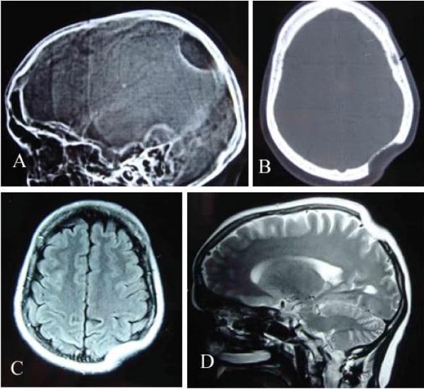
- It can occur at any age but predominantly affects CHILDREN and young adults (with a slight male predominance), and although any bone may be involved it has a predilection for the AXIAL and girdle skeleton - the SPINE, RIBS, pelvis/hip, shoulder girdle, MANDIBLE and skull - frequently with MULTIPLE lesions that can cross joints.
- PAIN is the commonest symptom, with progressive swelling, deformity and PATHOLOGICAL FRACTURE; the hallmark is the documented PROGRESSIVE LOSS OF BONE on serial imaging, often with tapering/pointed bone ends ('sucked candy' appearance).
- The FEARED complication is CHYLOTHORAX / pleural effusion when the thoracic skeleton (ribs/spine) is involved: the proliferating lymphatics leak chyle into the pleural space, causing respiratory compromise - PLEURAL EFFUSION is a recognised RISK FACTOR FOR MORTALITY and demands close monitoring.
- Diagnosis is one of EXCLUSION: there are no agreed criteria, so it rests on demonstrating PROGRESSIVE osteolysis on serial imaging together with characteristic HISTOLOGY (proliferating thin-walled vascular/lymphatic channels with fibrosis, NO malignant cells and NO osteoblastic/reparative response), after EXCLUDING infection, malignancy (metastasis, myeloma), metabolic and other causes of osteolysis.
- There is NO consensus treatment: options include ANTIRESORPTIVES (bisphosphonates), the mTOR inhibitor SIROLIMUS (which targets the lymphatic proliferation and is increasingly used), interferon-alpha, RADIOTHERAPY, and SURGERY (resection, reconstruction/allograft, stabilisation, fracture management); chylothorax is managed with thoracic-duct ligation/embolisation, octreotide, a low-fat/MCT diet and pleurodesis. The course is unpredictable - some stabilise spontaneously.
- “Gorham-Stout = progressive spontaneous massive osteolysis ('vanishing bone') from intraosseous LYMPHATIC/vascular proliferation.
- “Axial/girdle predilection (spine, ribs, pelvis, shoulder, mandible); CHYLOTHORAX (thoracic involvement) is the feared, mortality-associated complication.
- “Diagnosis of EXCLUSION (progressive osteolysis + benign vascular/lymphatic histology, no malignancy). Treat with bisphosphonates / SIROLIMUS / radiotherapy / surgery; no consensus.
Progressive, spontaneous osteolysis on serial imaging - bone 'vanishes' and is replaced by lymphatic/vascular tissue. A diagnosis of exclusion.
Chylothorax when ribs/spine are involved (lymphatic leak into the pleura) - a recognised cause of mortality; monitor and treat aggressively.
Pathology & Presentation
In Gorham-Stout disease, an abnormal proliferation of lymphatic and vascular channels invades bone and drives its progressive resorption, replacing the bone with fibrous/angiomatous tissue - hence 'vanishing' or 'phantom' bone. It predominantly affects children and young adults, with a slight male predominance, and although any bone can be involved it favours the axial and girdle skeleton (spine, ribs, pelvis, shoulder, mandible, skull), often with multiple lesions that may cross joints. Patients present with pain (the commonest symptom), swelling, deformity and pathological fracture, and the diagnostic hallmark is documented progressive bone loss on serial radiographs, classically with tapered/pointed bone ends.
Diagnosis & Management
There are no universally agreed diagnostic criteria, so GSD is a diagnosis of exclusion requiring:
- Progressive osteolysis demonstrated on serial imaging (radiographs/CT/MRI; bone scintigraphy).
- Characteristic HISTOLOGY: proliferating thin-walled vascular/lymphatic channels with fibrous replacement, NO malignant cells and NO osteoblastic/reparative response.
- EXCLUSION of other causes of osteolysis - infection (osteomyelitis), malignancy (metastasis, myeloma, primary bone tumour), metabolic and endocrine disease, and other rare osteolyses.
- Assess for thoracic involvement and chylothorax (chest imaging) given its prognostic importance.
- Medical: bisphosphonates (antiresorptive), the mTOR inhibitor SIROLIMUS (targets the lymphatic proliferation - increasingly first-line), and interferon-alpha.
- Radiotherapy: can arrest progression in selected lesions.
- Surgery: resection of affected bone with reconstruction (allograft/prosthesis), stabilisation and management of pathological fractures - though osteolysis can recur in or around constructs.
- Chylothorax: thoracic-duct ligation/embolisation, octreotide, a low-fat/MCT diet, drainage and pleurodesis; multidisciplinary care.
- The course is unpredictable - some lesions stabilise spontaneously while others progress relentlessly.
Because GSD favours the axial skeleton, thoracic (rib/spine) involvement can cause chylothorax, which is the leading cause of death in this disease through respiratory compromise and nutritional/immune depletion from chyle loss. Any patient with thoracic GSD and a pleural effusion needs urgent multidisciplinary management. Equally, because GSD is rare and mimics infection or malignancy, do NOT diagnose it without excluding those treatable/serious alternatives with biopsy and the appropriate workup.
Evidence & Key Studies
Clinical features and current management of Gorham-Stout disease: a systematic review
- Gorham-Stout disease is a rare complex lymphatic malformation (~400 cases reported); among 206 reviewed patients it most often presented in childhood with a slight male predominance.
- Osteolysis predominantly affected the axial skeleton (spine 46%, ribs 29%, hip 23%, femur 18%, mandible 16%), usually with multiple lesions, and pain was the commonest symptom.
- Surgery (67%) and bisphosphonates (57%) remained mainstream with sirolimus in 18%; PLEURAL EFFUSION was a significant risk factor for mortality.
Management of refractory chylothorax in Gorham-Stout disease: a case report
- GSD is a rare lymphatic-origin disease with progressive osteolysis that commonly causes chylothorax from leakage of lymph from dissolved bone.
- Refractory chylothorax was managed with thoracic-duct embolisation, octreotide, a low-fat diet and surgery, and disease progression was halted after introducing SIROLIMUS.
- Illustrates the chylothorax complication and the role of sirolimus and multidisciplinary care.
According to PubMed, the lymphatic-malformation basis, the childhood/axial predominance, the treatment patterns (surgery, bisphosphonates, sirolimus) and pleural effusion as a mortality risk factor come from the cited Zhou systematic review, and the chylothorax complication and sirolimus/multidisciplinary management from the cited Yamaki case report. The 'vanishing bone' radiographic hallmark, the diagnosis-of-exclusion approach and the characteristic benign vascular histology are standard, well-established teaching. (See also our Metastatic Bone Disease, Osteomyelitis and Bone Tumour Imaging topics.)
Clinical Decision Scenarios
Practise clinical reasoning and management decisions out loud
“A young patient has progressive, spontaneous disappearance of bone on serial radiographs. What is the diagnosis, and how is it confirmed?”
“How is Gorham-Stout disease treated, and why is the chest important?”
Mnemonics & Memory Aids
VANISH
Hook:The bone VANISHes - lymphatic-driven osteolysis; watch for chylothorax.
EXCLUDE
Hook:GSD is a diagnosis you EXCLUDE others to reach.
What it is
- Rare progressive spontaneous massive osteolysis ('vanishing/phantom bone')
- Intraosseous lymphatic/vascular proliferation -> bone resorbed, replaced by fibrous/angiomatous tissue
- Children/young adults; slight male predominance; axial/girdle predilection
Presentation
- Pain (commonest), swelling, deformity, pathological fracture
- Progressive bone loss on serial imaging (tapered ends)
- Chylothorax with thoracic (rib/spine) involvement - mortality risk
Diagnosis (exclusion)
- Progressive osteolysis on serial imaging
- Histology: thin-walled vascular/lymphatic channels, no malignancy, no osteoblastic response
- Exclude infection, malignancy (metastasis/myeloma), metabolic causes
Treatment (no consensus)
- Bisphosphonates, sirolimus (mTOR inhibitor), interferon-alpha
- Radiotherapy; surgery (resection/reconstruction/stabilisation)
- Chylothorax: duct ligation/embolisation, octreotide, low-fat/MCT diet, pleurodesis