Bilateral Pars Fracture | Levine-Edwards Classification | Type IIA - No Traction
- Type IIA: Traction is CONTRAINDICATED - flexion-distraction injury
- Auto-decompression explains low neurological injury rate (canal expands)
- Measure translation AND angulation on lateral radiograph
- Type IIA: angulation out of proportion to translation (greater than 11 degrees, less than 3mm)
- Most treated non-operatively (collar or halo) - surgery for Type III or failed conservative
- “Type IIA recognition: angulation greater than translation (greater than 11 degrees, less than 3mm)
- “All types can have severe injury but neurological deficit rare (canal expands)
- “Associated head/facial trauma common (forehead impact mechanism)
Overview and Clinical Significance
What it is. A hangman's fracture, traumatic spondylolisthesis of C2, is a bilateral fracture through the pars interarticularis of the axis. The name comes from the similar fracture pattern produced by judicial hanging; modern cases are typically from motor vehicle accidents.
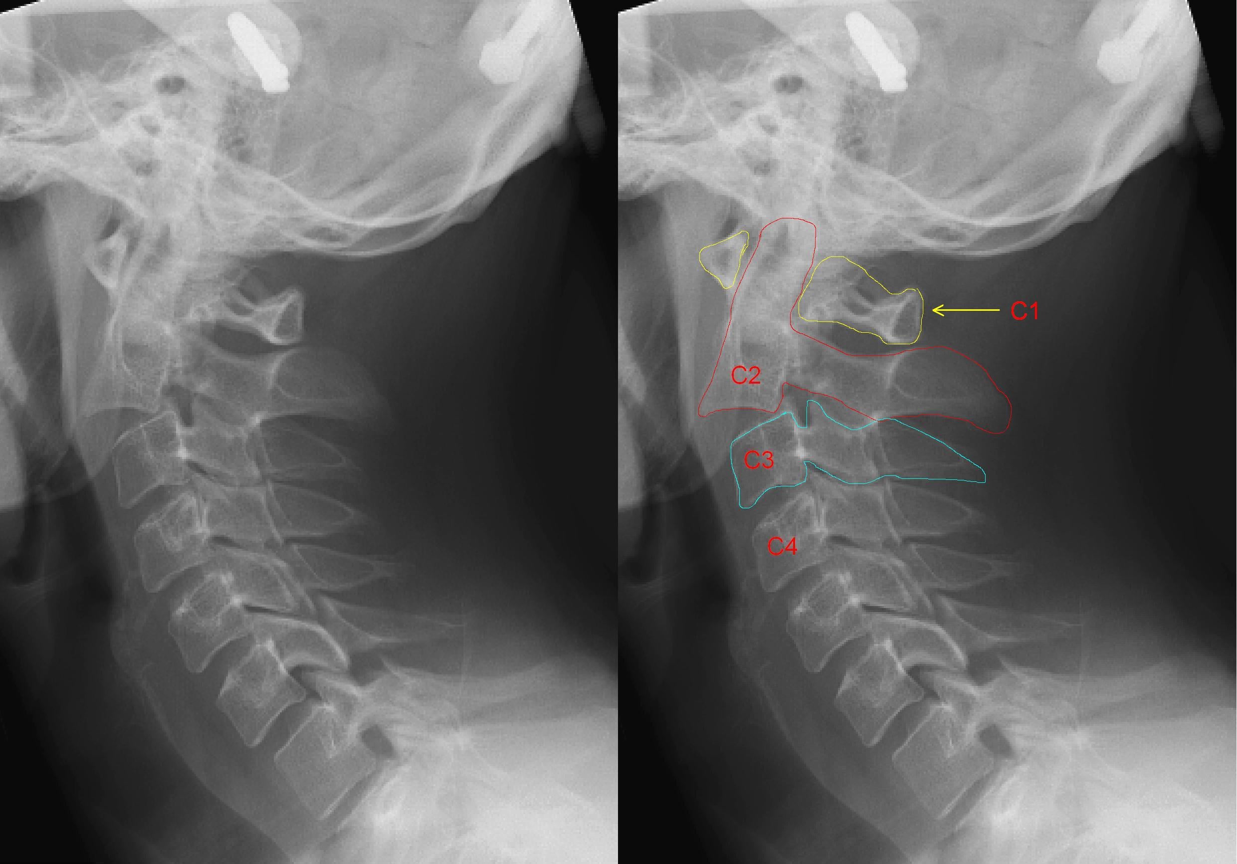
How common. Hangman's fractures make up 4-7% of all cervical fractures and are the most common C2 fracture once the odontoid is excluded. There is a male predominance and two peaks: young adults in motor vehicle accidents and the elderly in falls.
Mechanism. Hyperextension with an axial load is the most common mechanism, and the Type IIA injury is the exception, a flexion-distraction injury.
- Motor vehicle accident, the forehead striking the dashboard
- Falls from height
- Diving accidents
- Type IIA: flexion-distraction
Why the cord is spared. Neurological injury occurs in only about 6%. The bilateral pars fracture separates the posterior ring from the anterior elements, so as the body translates the canal diameter increases rather than narrows, an "auto-decompression". Contrast a burst fracture, where bone retropulses into the canal: "Jefferson expands, Hangman's translates but opens."
The name. Sir Geoffrey Jefferson first described the mechanism in 1927, relating it to judicial hanging. There the drop causes hyperextension and distraction, the pars fractures bilaterally, the cord is transected at C2 and death follows from respiratory arrest, the phrenic nerve arising from C3-5. Modern hangman's fractures differ: lower energy, variable mechanisms, and they rarely cause death.
Pathophysiology and Mechanisms
The axis. C2 has unique anatomy. The odontoid process projects superiorly to articulate with C1, and its fractures are classified separately (Anderson-D'Alonzo). The pars interarticularis is the thin isthmus connecting the superior and inferior articular processes, and it is where a hangman's fracture runs.
- The superior articular facets are large and flat, face superolaterally, and articulate with the C1 lateral masses
- The inferior articular facets face anteroinferiorly and articulate with C3; these are the facets that dislocate in a Type III
- The vertebral artery courses through the transverse foramen and is at risk with displaced fractures
- The body is large, bears the weight from above, and translates forward once both pars have fractured
The classic mechanism (Types I, II and III). Hyperextension with an axial load, in sequence:
- The forehead strikes the dashboard or another object
- The neck is forced into hyperextension
- The axial load is transmitted through the cervical spine
- The pars interarticularis, the weakest point, fractures bilaterally
- The C2 body translates on C3 by a variable amount
The Type IIA mechanism. A different injury altogether: flexion rather than extension, with distraction forces rather than compression. The result is minimal translation but severe angulation, with disruption of the disc between C2 and C3. This mechanism is the reason traction is contraindicated, which the Management section returns to.
Classification Systems
Levine-Edwards. The most widely used classification system, based on mechanism and radiographic parameters. Know what the numbers are before you defend them. The 3 mm translation and 11 degree angulation cut-offs are textbook convention rather than validated thresholds - neither appears in the retrievable abstract of Levine and Edwards, no measurement protocol is specified with them, and interobserver agreement on axis ring fractures has repeatedly proved only moderate. More importantly, Type IIA in the original series was three patients out of 52. The mechanism is sound and the no-traction rule is universally accepted on that mechanism, but it has never been tested. Use the classification for what it reliably does - map mechanism to reduction strategy - and treat the millimetres as a guide to which pattern you are looking at, not as a boundary that decides treatment by itself.
- Translation
- Less than 3mm
- Angulation
- Minimal
- Mechanism
- Axial load + extension
- C2-C3 Disc
- Intact
- Stability
- Stable
- Translation
- Greater than 3mm
- Angulation
- Significant
- Mechanism
- Axial load + extension then flexion
- C2-C3 Disc
- Disrupted
- Stability
- Unstable
- Translation
- Minimal (less than 3mm)
- Angulation
- Severe (greater than 11 degrees)
- Mechanism
- Flexion-distraction
- C2-C3 Disc
- Disrupted
- Stability
- Unstable
- Translation
- Variable
- Angulation
- Variable
- Mechanism
- Flexion-compression
- C2-C3 Disc
- Disrupted + facet dislocation
- Stability
- Very unstable
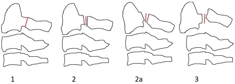
Type I. Translation under 3 mm with minimal angulation. The C2-C3 disc and the posterior longitudinal ligament are intact, the pattern is stable, and it is the most common type.
Type II. Translation over 3 mm with significant angulation. The C2-C3 disc is disrupted and the posterior longitudinal ligament torn, so the pattern is unstable.
Type IIA. Minimal translation, under 3 mm, with severe angulation over 11 degrees. The anterior annulus is intact and the posterior part of the disc disrupted, with the posterior ligaments torn; the mechanism is flexion-distraction.
Type III. Bilateral pars fractures plus a C2-C3 facet dislocation, with complete disc disruption and all ligaments torn. It is the most unstable pattern and carries a high risk of neurological injury.
Type IIA recognition: angulation out of proportion to translation. More than 11 degrees of angulation with less than 3 mm of translation is a Type IIA, and traction will make it worse by further opening the posterior disc.
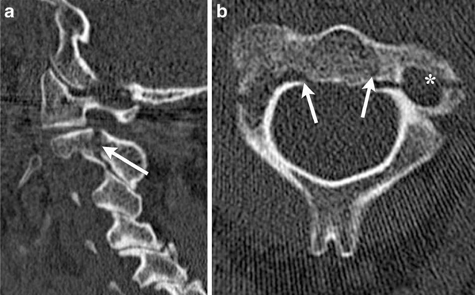
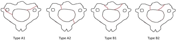
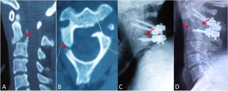
Atypical fractures. Not every C2 ring fracture runs cleanly through both pars. The atypical pattern is covered with the differential diagnosis below, and the drawings here show what to look for on the axial CT.
Clinical Assessment
History. The mechanism is the first clue. A dashboard injury with a forehead strike, a fall onto the head or face, a diving impact with the head, or a contact sport or gymnastics injury are the mechanisms to ask about; a history of a flexion mechanism is the clue to a Type IIA. Patients describe posterior neck pain radiating to the occiput, limited range of motion and headache. Neurological symptoms are rare.
Maintain cervical spine immobilisation until cleared. There is a high association with other cervical and head injuries in motor vehicle accident patients. Complete the ATLS primary survey before the focused spine examination.
Examination. The collar stays on. Look for facial or forehead trauma, palpate for posterior midline tenderness at the C2 level, and complete a full motor and sensory examination, which is usually normal. Head trauma and facial fractures are common companions.
When the cord is at risk. Neurological injury is rare, but it occurs with:
- A complete cord injury from the judicial hanging mechanism
- Type III with cord compression
- Associated injuries: disc herniation, facet locking
- Vertebral artery injury
Differential diagnosis. A C2 ring lucency on imaging is not always a classic hangman's fracture; the distinction changes management and neurological risk.
- Key Distinguishing Feature
- Bilateral pars interarticularis fracture, body translates anteriorly
- Canal Effect
- Canal widens (auto-decompression)
- Management Difference
- Levine-Edwards directed (collar/halo/surgery)
- Key Distinguishing Feature
- Fracture through posterior vertebral body with cortical/pedicle continuity
- Canal Effect
- Canal NARROWS rather than widens - the anatomy is the reason for vigilance. Starr and Eismont reported paralysis in 33%, but that was 2 of only 6 atypical cases in 1993, and neither larger modern series on this page reproduces it: Cai found no higher SCI incidence in atypical fractures (p=0.31), and Al-Mahfoudh had one permanent deficit in 28 atypical fractures, from associated injuries. Treat the canal-narrowing pattern as the warning; do not quote 33% as an incidence
- Management Difference
- Higher surgical/neurological vigilance, CTA
- Key Distinguishing Feature
- Fracture line at the dens, not the pars
- Canal Effect
- Variable
- Management Difference
- Anderson-D'Alonzo classification; often anterior screw or fusion
- Key Distinguishing Feature
- Smooth, corticated, bilateral, symmetric line in a child
- Canal Effect
- None
- Management Difference
- Normal variant - no immobilisation needed
- Key Distinguishing Feature
- Corticated, non-traumatic defect, no soft-tissue swelling
- Canal Effect
- None
- Management Difference
- Incidental; no acute treatment
- Key Distinguishing Feature
- Bilateral pars fracture PLUS dislocated C2-C3 facets
- Canal Effect
- May compress cord
- Management Difference
- Reduction and surgical fusion
A separate C2 fracture that the differential above omits, and a classic exam trap, is the extension teardrop fracture of C2.
What it is. A hyperextension avulsion of the antero-inferior corner of the C2 body by the anterior longitudinal ligament or annulus: a small triangular "teardrop" fragment at the front of C2. It is typically seen in older patients after a hyperextension fall, often striking the chin or forehead, the opposite mechanism context to a flexion teardrop.
Why it matters. It is usually stable. It is a bony avulsion of the anterior column only, with the posterior elements and pars intact, so it is generally a stable injury treated in a collar, unlike a hangman's (bilateral pars) fracture or the dreaded subaxial flexion teardrop.
The two teardrops. The flexion teardrop fracture, classically lower cervical (C5, for example) and from flexion-compression, is a highly unstable three-column injury with a high rate of cord injury (anterior cord syndrome): the polar opposite in stability and prognosis to the extension teardrop of C2.
Clues to stability. A small avulsed fragment wider than it is tall, no or minimal C2-C3 translation, intact pars and posterior elements, and no posterior ligamentous injury on MRI point to the benign extension teardrop. Significant translation, posterior injury, or a tall fragment with retropulsion should raise concern for an unstable pattern.
Imaging
The lateral radiograph. This is the critical view. Measure the translation of C2 on C3 and the angulation at C2-C3, assess the prevertebral soft-tissue swelling, and look for associated injuries. Both measurements are needed, because the type depends on both.
- Translation: from the posterior margin of the C2 body to C3
- Angulation: the angle between the inferior endplate of C2 and the superior endplate of C3
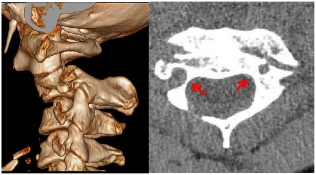
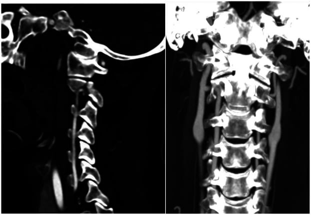
CT. CT shows the bilateral pars fractures, the fracture pattern and any comminution, the facet alignment that defines a Type III, and associated fractures of C1 and C3; its translation measurement is more accurate than the radiograph's. Multiplanar and three-dimensional reconstructions help operative planning, subtle fractures missed on the radiograph are detected, and the vertebral artery foramina can be assessed.
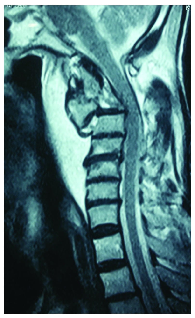
MRI. Indicated for a neurological deficit, for Type II, IIA and III fractures, for surgical planning, and to assess the disc, the ligaments and the cord. The cord is normal in Type I, usually normal in Types II and IIA, and may be compressed in Type III.
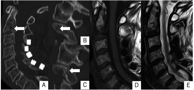
Synchondroses at C2 can mimic fracture lines in a child. A synchondrosis has smooth, corticated edges and is bilateral and symmetric; a true fracture has irregular, non-corticated edges.
Management Algorithm
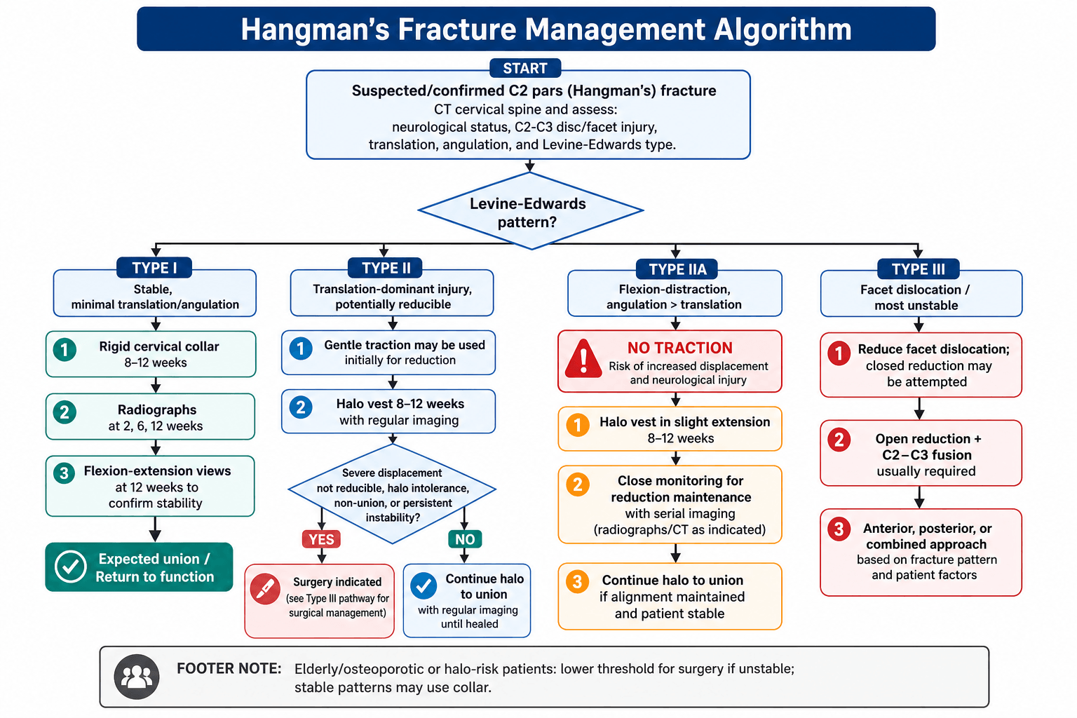
Type I: the collar. A rigid cervical collar (Philadelphia, Miami J or Aspen) for 8-12 weeks; no halo is needed. Radiographs at 2, 6 and 12 weeks, with flexion-extension views at 12 weeks to confirm stability.
Type II: reduce, then halo. Gentle traction may be applied initially to reduce the translation, after which the patient is transitioned to a halo vest for 8-12 weeks with regular imaging. The complication rate is higher than in Type I. Surgery is considered for:
- Severe displacement that cannot be reduced
- A patient unable to tolerate the halo
- Non-union after halo treatment
- Persistent instability
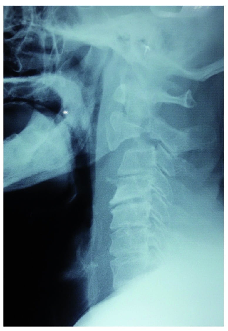
Type IIA is a flexion-distraction injury and traction is absolutely contraindicated: it worsens the angulation and the disc disruption by opening the posterior disc further. The halo goes on in slight extension.
Type IIA: halo in extension. Recognise it by angulation greater than translation, do not apply cervical traction, and apply the halo vest with the neck in slight extension. Extension and compression close the posterior disc disruption and reduce the fracture. Immobilise for 8-12 weeks and monitor the reduction closely.
Type III: surgery. The most unstable pattern, and the facet dislocation must be reduced. Closed reduction may be attempted, but open reduction with C2-C3 fusion is usually required, through an anterior or posterior approach chosen on the fracture pattern.
- Technique
- C2-C3 ACDF
- Indications
- Type III, failed conservative
- Considerations
- Direct disc access, anterior column support
- Technique
- C2 pars screw + C3 lateral mass screw
- Indications
- Type II/III with posterior instability
- Considerations
- Direct fracture fixation possible
- Technique
- Anterior fusion + posterior fixation
- Indications
- Severe instability, Type III
- Considerations
- Most rigid construct
- Technique
- Lag screws across fracture
- Indications
- Select Type II
- Considerations
- Preserves C2-C3 motion (controversial)
The elderly. A fall is more often the mechanism and osteoporosis may be present. Halo complications, pin loosening and infection among them, are higher, so consider a collar for stable patterns and keep a lower threshold for surgery if the fracture is unstable.
Associated injuries. Head trauma is common from the forehead impact, as are facial fractures, and other cervical fractures (a C1 Jefferson fracture, C3) travel with it. Assess the entire cervical spine with CT.
Surgical Technique
Indication. Type III, failed non-operative management, and the irreducible Type II. It is the most common surgical approach for a hangman's fracture that needs surgery.
Technique.
- Patient supine, slight neck extension
- Standard Smith-Robinson right-sided approach
- Identify the C2-C3 level with fluoroscopy
- Complete discectomy and endplate preparation
- Interbody graft or cage placement (PEEK or allograft)
- Anterior plate fixation with screws into the C2 and C3 bodies
- Avoid extending the plate above C2-C3 (adjacent segment issues)
- Intraoperative fluoroscopy confirmation
Why it is chosen. Direct access to the disrupted disc, anterior column support and fusion rates over 90%. Posterior supplementation may be needed for severe instability or a Type III injury.
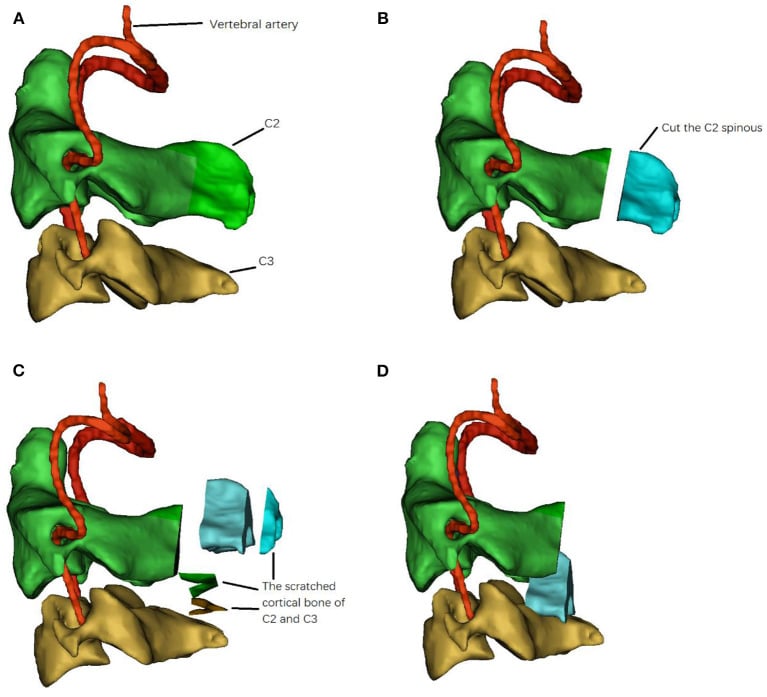
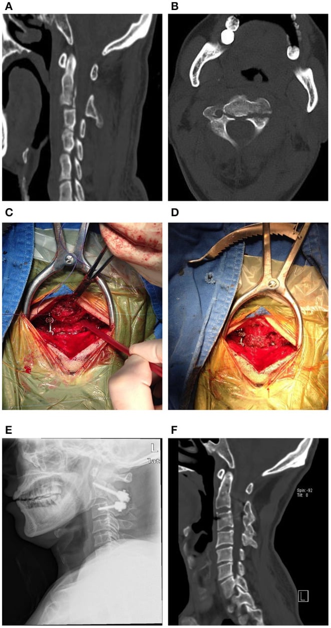
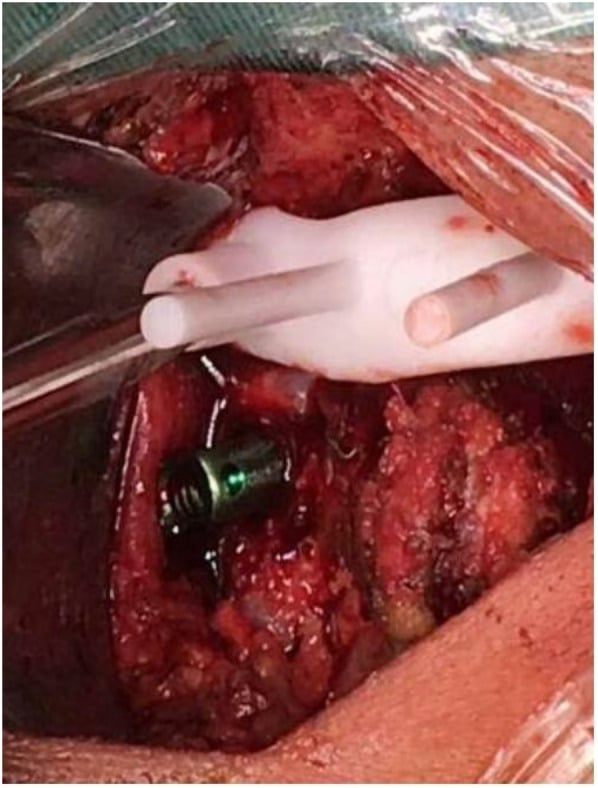
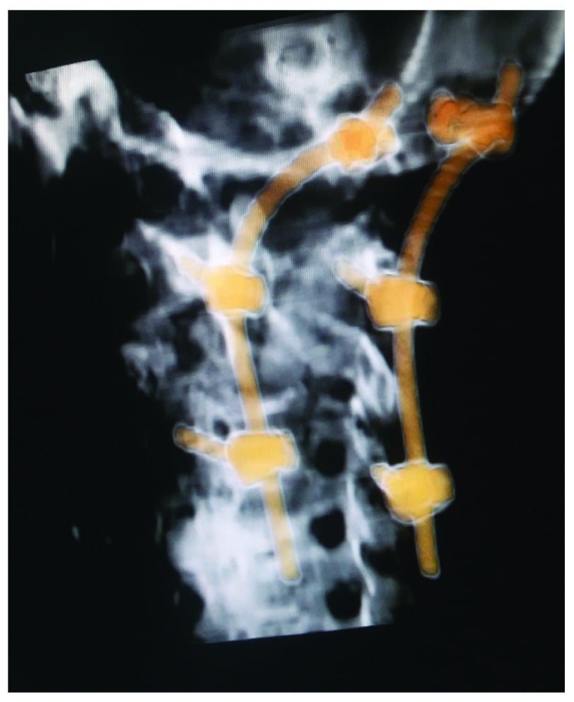
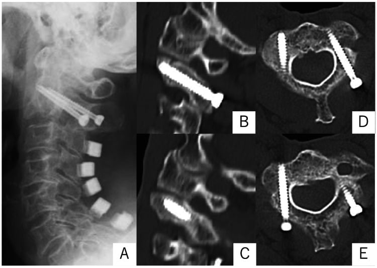
Direct C2 pars (Judet) osteosynthesis is the motion-sparing operative choice for a reducible Type II hangman's fracture: it repairs the ring without fusing C2-C3, but needs a safe pedicle (check the vertebral artery on CT) and is unsuitable for Type III.
Complications
Hangman's fractures have relatively low complication rates compared with other cervical spine injuries, primarily because of the auto-decompression effect. The specific complications depend on fracture type and treatment method.
Non-union. 5-10% overall, higher in Types II and III and rare in Type I. The risk factors are inadequate immobilisation duration, poor patient compliance, smoking and diabetes, and severe initial displacement. An asymptomatic non-union is observed; a symptomatic one needs extended immobilisation or C2-C3 fusion, anterior or posterior.
Malunion. The incidence is variable. The patterns are kyphotic deformity, residual translation and the Type IIA treated in flexion, with poor reduction the risk factor. Asymptomatic malunion is managed conservatively; severe deformity needs corrective osteotomy and fusion.
Vertebral artery injury. Rare, under 5%, and usually asymptomatic; the risk is a displaced fracture through the foramen. CT angiography of the cervical spine screens high-energy injuries. Management is observation and antiplatelet therapy, rarely endovascular intervention.
Neurological complications. Highest in Type III, at 10-15%. Deficit is usually present at the time of injury and delayed deterioration is very rare, under 2%; the risk factors are Type III and cord compression, and it needs urgent surgical decompression. Otherwise the alignment is maintained and decompression offered if indicated.
Halo complications. 10-30%, higher in the elderly, with osteoporosis and with poor compliance: pin-site infection (10-20%), pin loosening (5-15%), respiratory complications, skin breakdown and dysphagia. Pin care and early recognition are the defence; the halo may need early removal and conversion to a collar.
Postoperative Care
The first days. From day 0 to 3: ICU or high-dependency monitoring if neurologically intact, the cervical collar or halo vest kept on, neurological checks every 2-4 hours, multimodal analgesia, mechanical and chemical DVT prophylaxis, and early mobilisation when stable.
Immobilisation after fusion. A hard cervical collar for 6-8 weeks after an anterior fusion alone; a halo vest for 8-12 weeks after a posterior or combined fusion. Serial radiographs at 2, 6 and 12 weeks, and a CT at 12 weeks to assess fusion.
- Assessment
- Wound check, neurology
- Imaging
- Lateral radiograph
- Activity
- Collar/halo wear
- Assessment
- Pain, ROM assessment
- Imaging
- AP + Lateral radiographs
- Activity
- Light activities only
- Assessment
- Fusion assessment
- Imaging
- CT + flexion-extension
- Activity
- Wean immobilisation
- Assessment
- Final fusion check
- Imaging
- CT if non-union concern
- Activity
- Return to full activity
- Assessment
- Discharge if healed
- Imaging
- Only if symptomatic
- Activity
- No restrictions
Rehabilitation. Three phases, each building on the last.
- Phase 1 (0-6 weeks): maintain immobilisation; upper extremity exercises (shoulder, elbow); core strengthening with no neck movement; gait training in the collar or halo
- Phase 2 (6-12 weeks): gradual collar or halo weaning; gentle active range-of-motion exercises; progressive strengthening; proprioceptive training
- Phase 3 (3-6 months): full range-of-motion restoration; advanced strengthening; sport-specific training if appropriate; return-to-work assessment
- Type I
- When comfortable in collar
- Type II/IIA
- When comfortable in halo
- Type III (Surgical)
- 6-8 weeks post-surgery
- Type I
- After collar weaned, full ROM
- Type II/IIA
- After halo removed, ROM restored
- Type III (Surgical)
- 3 months post-surgery minimum
- Type I
- 3 months, confirmed union
- Type II/IIA
- 4-6 months, confirmed union
- Type III (Surgical)
- 6 months post-fusion
- Type I
- 6 months if union confirmed
- Type II/IIA
- Case-by-case, often restricted
- Type III (Surgical)
- Generally not recommended
- Type I
- Patient decision after counselling
- Type II/IIA
- Counsel on risk
- Type III (Surgical)
- Not recommended
Outcomes and Prognosis
Hangman's fractures have generally excellent outcomes when appropriately treated, and the type predicts the shape of the recovery.
- Type I
- Greater than 95%
- Type II/IIA
- 90-95%
- Type III
- 85-90% (surgical)
- Type I
- 95% full duty
- Type II/IIA
- 85% full duty
- Type III
- 70% full duty
- Type I
- Less than 10%
- Type II/IIA
- 20-30%
- Type III
- 30-40%
- Type I
- Minimal
- Type II/IIA
- 10-20 degrees
- Type III
- Complete (if fused)
- Type I
- Excellent (90%)
- Type II/IIA
- Good (80%)
- Type III
- Fair-Good (70%)
Type I. Near 100% union with a collar, full return to function expected, minimal long-term neck pain and minimal long-term sequelae: an excellent prognosis.
Type II. 90-95% union with a halo and good functional outcomes, though some patients have mild chronic neck pain. The non-union that occurs may require late surgery.
Type IIA. Good outcomes if recognised and treated correctly, and the key is avoiding traction. Union rates are similar to Type II with correct management, though there may be more neck stiffness than after a Type I.
Type III. Variable outcomes and the highest rate of neurological injury. Surgery usually achieves a stable fusion, over 85%, at the cost of the C2-C3 motion segment, and there may be persistent neck pain.
Predictors of a poor outcome.
- Type III injuries (highest complication rate)
- Elderly patients (over 65 years)
- Neurological injury at presentation
- Delayed diagnosis or treatment
- Non-compliance with the immobilisation protocol
- High-energy mechanism
- Smoking and comorbidities
Guidelines, Registries & Global Practice
Global Epidemiology
- Traumatic spondylolisthesis of the axis ("hangman's fracture") accounts for approximately 4-7% of all cervical spine fractures and is the second most common axis fracture after the odontoid (Francis et al., J Bone Joint Surg Br 1981, PMID 7263740; Cai et al., Emerg Radiol 2022, PMID 35543854).
- Bimodal age distribution: high-energy motor vehicle collisions in younger adults and low-energy falls in older adults. Recent level-1 trauma-centre data report a mean age of 62.7 years, reflecting an ageing injured population (Cai et al., 2022, PMID 35543854).
- Neurological injury is uncommon (historically approximately 6%) because the bilateral pars fracture tends to decompress rather than narrow the canal; the main exceptions are Type III, atypical posterior-body fractures, and high-energy distraction (Starr & Eismont, Spine 1993, PMID 8272942).
Guideline & Classification Comparison
- Framework
- Evidence-based US spine-trauma guideline
- Core Guidance
- External immobilisation initially for most; surgery for severe C2-C3 angulation, disc disruption, or failure to maintain alignment
- Evidence Level
- Practice option (insufficient evidence for a standard)
- Framework
- Mechanistic 4-type classification
- Core Guidance
- I collar; II reduce then halo; IIA halo in extension (NO traction); III surgery
- Evidence Level
- Level IV defining series
- Framework
- Displacement-and-stability grading
- Core Guidance
- Stable/undisplaced - external brace; displaced or facet dislocation - consider surgery
- Evidence Level
- Level IV
- Framework
- Morphology + facet + modifiers
- Core Guidance
- Escalate to fixation with disco-ligamentous disruption or dislocation
- Evidence Level
- Expert consensus
Registry & Synthesised Evidence
- The largest systematic review (25 studies, 548 fractures) found non-operative union of 94.1% versus 99.4% surgically, with no mortality difference and both anterior and posterior approaches achieving high fusion (Murphy et al., J Orthop Trauma 2017, PMID 28816880).
- For low-grade fractures, a rigid collar matched halo immobilisation for union, with no demonstrable advantage of the halo and lower morbidity - particularly relevant for older patients (Al-Mahfoudh et al., Global Spine J 2016, PMID 27099816).
- There are no national hangman's-fracture–specific arthroplasty/registry datasets (unlike joint replacement); evidence is dominated by retrospective series and pooled reviews, so guidance remains at the level of practice options rather than standards (Hadley et al., 2002, PMID 12431297).
Practice Variation
- Immobilisation device: Halo use has declined in many high-income centres in favour of rigid collars for stable patterns, driven by halo morbidity (pin-site infection, loosening, pneumonia) especially in the elderly.
- Operative threshold: Surgeons increasingly favour early fixation for Type II with marked translation/angulation and for Type III, citing higher union and faster mobilisation; halo-based non-operative care remains standard for Type I and many Type II/IIA fractures.
- Vascular screening: High-grade fractures (Type IIA/III) warrant CT angiography for blunt cerebrovascular injury and brain imaging, given the markedly higher rates of vertebral artery injury and traumatic brain injury (Cai et al., 2022, PMID 35543854).
Viva Scenarios
Clinical Decision Scenarios
Practise clinical reasoning and management decisions out loud
“A 35-year-old male is in an MVA. Lateral cervical radiograph shows a bilateral C2 pars fracture with 5mm translation and 8 degrees angulation. What is your classification and management?”
“A Hangman's fracture is identified with 2mm translation but 15 degrees of angulation. What type is this and how would you manage it differently?”
“Explain why neurological injury is rare in Hangman's fractures despite the unstable nature of the injury.”
MCQ Practice Points
High-Yield Facts for MCQs
Classification:
- Type I: less than 3mm translation, minimal angulation, collar treatment
- Type II: greater than 3mm translation, significant angulation, halo treatment
- Type IIA: minimal translation (less than 3mm) but severe angulation (greater than 11 degrees), NO TRACTION
- Type III: facet dislocation, surgical treatment
Q: How are Type I, II, and IIA distinguished by measurement? A: The Levine-Edwards classification uses 3mm translation and 11 degrees angulation thresholds. Type I is less than 3mm translation. Type II is greater than 3mm translation. Type IIA is less than 3mm translation but greater than 11 degrees angulation.
Key Measurements:
- 3mm: threshold between Type I and Type II translation
- 11 degrees: threshold for Type IIA angulation
- 8-12 weeks: typical immobilization duration
Q: What is the classic exam trap regarding Type IIA fractures? A: Type IIA is a FLEXION-DISTRACTION injury (unlike others). Traction is CONTRAINDICATED as it worsens displacement. Treatment is halo in extension.
Mechanism:
- Type I, II, III: hyperextension with axial loading
- Type IIA: flexion-distraction (opposite mechanism)
- Judicial hanging: hyperextension + distraction (usually fatal)
Neurological Injury:
- Overall rate: approximately 6%
- Explained by "auto-decompression" - canal expands
- Type III has highest risk
- Bilateral pars fractures allow posterior elements to separate
Q: Why is neurological injury rare (6%) in Hangman's fractures? A: "Auto-decompression" - the bilateral pars fractures allow the posterior elements to separate from the anterior body, expanding the spinal canal diameter rather than narrowing it.
Type IIA Critical Points:
- TRACTION IS CONTRAINDICATED
- Halo applied in EXTENSION position
- Flexion-distraction mechanism
- Angulation out of proportion to translation
- Most commonly tested pitfall in exams
Surgical Indications:
- Type III (facet dislocation)
- Failed non-operative management
- Irreducible Type II
- Patient unable to tolerate halo
- Non-union after appropriate immobilization
Q: Which Hangman's fracture requires surgical stabilization? A: Type III (with facet dislocation). It is highly unstable. Type I and II are usually managed with collar or halo respectively.
Imaging:
- Lateral cervical radiograph: key view
- Measure both translation AND angulation
- CT: fracture pattern, facet alignment
- MRI: disc disruption, cord, ligaments
Q: What specific measurements must be taken on lateral X-ray? A: Both translation AND angulation. Measuring only translation will miss Type IIA (minimal translation, severe angulation).
Common Exam Traps
- Type IIA traction: Most common mistake - traction worsens injury
- Neurological injury rate: Not high despite "unstable" fracture
- Measurement confusion: Must measure BOTH translation and angulation
- Mechanism confusion: Type IIA is flexion, not extension
- Surgical timing: Type III needs surgery, not all Hangman's fractures
- Collar vs halo: Type I gets collar, Type II gets halo
- Union rates: Generally excellent with appropriate treatment
MCQ Stems to Expect
- "A patient has C2 pars fracture with 2mm translation and 15-degree angulation. What is the classification and management?"
- "What explains the low neurological injury rate in Hangman's fractures?"
- "What is contraindicated in Type IIA Hangman's fracture?"
- "A Type III Hangman's fracture is characterized by..."
- "What is the most appropriate initial immobilization for a Type II Hangman's fracture?"
Exam Cheat Sheet
Classification
- Type I: less than 3mm translation, minimal angulation = COLLAR
- Type II: greater than 3mm translation, angulation = HALO
- Type IIA: less than 3mm translation BUT greater than 11 degrees angulation = HALO IN EXTENSION, NO TRACTION!
- Type III: facet dislocation → SURGERY
Key Concepts
- Bilateral C2 pars fracture (traumatic spondylolisthesis)
- Neurological injury rare (6%) - canal expands (auto-decompression)
- Type IIA is flexion-distraction - traction worsens injury
- Measure translation AND angulation on lateral cervical radiograph
Critical Pitfalls
- Applying traction to Type IIA (CONTRAINDICATED)
- Missing Type IIA - look for angulation greater than translation
- Treating Type III non-operatively (needs surgery)
- Missing associated C1 or C3 fractures
Exam Tips
- Type IIA recognition is the classic exam trap
- minimal translation + severe angulation = Type IIA
- Explain auto-decompression for why cord spared
- Know surgical options for Type III
Evidence and Guidelines
Levine-Edwards Classification (defining series)
- 52 patients with traumatic spondylolisthesis of the axis (15 Type I, 29 Type II, 3 Type IIA, 5 Type III). Associated neurological deficit in only 4 patients. All fractures healed.
- Type IIA injuries showed INCREASED displacement when placed in traction and were reduced instead with gentle extension and compression in a halo vest - the origin of the 'no traction' rule.
- Fracture type correlated with mechanism: Type I hyperextension-axial loading; Type II hyperextension-axial loading then severe flexion; Type IIA flexion-distraction; Type III flexion-compression.
Francis Grading & Non-operative Outcomes
- Series of 123 patients with traumatic spondylolisthesis of the axis. High incidence of associated face/scalp injuries and upper cervical fractures, but LOW incidence of neurological injury despite apparent gross instability.
- Union was usual regardless of displacement; protection from extremes of flexion and extension was adequate treatment. Early mobilisation in a halo reduced hospital stay without jeopardising the result.
- Surgery (anterior C2-C3 interbody fusion to preserve atlanto-axial rotation) needed only for chronic instability with or without pain.
Atypical Hangman's Fractures (canal-narrowing pattern)
- 19 cases of traumatic spondylolisthesis: 13 standard hangman's fractures and 6 'atypical' fractures running through the posterior vertebral body with unilateral/bilateral continuity of the posterior cortex or pedicle.
- Unlike standard fractures, the atypical pattern NARROWS the spinal canal - this anatomical point is the paper's durable contribution and is not disputed.
- Paralysis was reported in 33% of the atypical group. Note the denominator: that is 2 patients out of 6, and the authors themselves close by asking for larger series to clarify the true incidence.
- Neither larger series cited on this page reproduces that rate - Cai found no higher SCI incidence in atypical fractures (p=0.31), and Al-Mahfoudh reported one permanent deficit across 28 atypical fractures, attributable to associated injuries.
- These atypical C2 injuries must still be recognised as distinct from Levine-Edwards/Effendi types, because the fracture plane and its effect on canal diameter differ.
Effendi Classification (axis ring fractures)
- Retrospective analysis of 131 fractures of the ring of the axis, classified into three types by radiological displacement and stability (Type I undisplaced/stable; Type II displaced with abnormal C2-C3 disc; Type III displaced with C2-C3 facet dislocation).
- Provided the original three-type scheme later modified by Levine and Edwards (who added Type IIA) and refined by Francis (I-V grading).
Surgical vs Non-operative Treatment - Systematic Review
- Pooled union rate was 94.1% for 131 non-surgically treated fractures versus 99.4% for 417 surgically treated fractures; non-union was less likely with surgery (OR 0.12, 95% CI 0.02-0.71).
- No significant difference in mortality between surgical (0.16%) and non-surgical (1.04%) groups; treatment failure was lower in the surgical group.
- Both anterior and posterior approaches achieved high fusion rates with neither clearly superior.
Collar vs Halo for Low-Grade Fractures
- 41 hangman's fractures (mean age 59) from 105 axis fractures; 31.7% typical and 68.2% atypical. Most were low-grade (Type I/II).
- Bony union was achieved in ALL patients; there was NO difference in outcome between rigid collar and halo immobilisation for low-grade fractures, and only 7.3% needed surgery.
- Permanent neurological deficit occurred in just one patient (from associated injuries).
Modern Epidemiology & Vascular/Brain Injury Risk
- Traumatic spondylolisthesis of the axis accounts for 4-5% of all cervical fractures; mean age 62.7 years, motor vehicle collision the commonest cause at this level-1 trauma centre.
- Type I injuries healed well with conservative management, while Type IIA and III were significantly more likely to be managed surgically.
- Type IIA and III injuries carried significantly higher-grade vertebral artery injuries (Biffl III-IV) and an 11-fold increased risk of traumatic brain injury versus Type I/II - mandating CT angiography and TBI screening.
- THE PAPER'S OWN CONCLUSIONS ARE TWO NEGATIVES, AND THEY CUT AGAINST THE CLASSIFICATION THIS PAGE IS BUILT AROUND: fracture type was NOT associated with any clinical outcome, and the authors state that Levine-Edwards typing 'is not enough to guide the treatment plan and does not account for associated injuries'.
- Atypical fractures were NOT found to carry a higher incidence of spinal cord injury in this series (p=0.31) - which does not reproduce the 33% paralysis figure from Starr and Eismont's much smaller 1993 report.