Thompson-Epstein Classification | Pipkin Fractures | Urgent Reduction
- Reduce as an emergency - AVN odds are 5.6 times higher when reduction is delayed beyond 12 hours; pooled AVN ranges 11-43% for posterior dislocations
- Thompson-Epstein classification - Type I (simple), Type II-V (with fractures)
- Pipkin classification for femoral head fractures - Type I (below fovea), Type II (above fovea), Type III (+ neck), Type IV (+ acetabulum)
- Sciatic nerve at risk in posterior dislocations (10-20%) - peroneal division most vulnerable
- CT post-reduction mandatory - assess concentric reduction, loose bodies, fractures
- “Hip fracture-dislocation = true orthopaedic emergency - reduce within 6 hours
- “Thompson-Epstein Type V = Pipkin fracture - use Pipkin classification to guide treatment
- “Posterior wall over 40% = unstable - requires ORIF
- “Pipkin III (head + neck) = disaster - double blood supply insult, high AVN risk
Overview and Epidemiology
Hip fracture-dislocations are high-energy injuries in which the hip dislocates and something breaks with it: the femoral head, the acetabulum or the femoral neck. They are true orthopaedic emergencies, and the Thompson-Epstein classification, which sorts them by the associated fracture, guides treatment.
Reduce as soon as possible, even before CT if needed. The pooled evidence gives an odds ratio of 5.6 for AVN when reduction is delayed beyond 12 hours, and AVN of 11-43% for posterior dislocations overall, so even prompt reduction does not make this a low-risk injury. The conventional 6-hour target is best practice, not a validated cut-off.
Mechanism. Direction decides the pattern.
- Posterior - 90%. High energy: a motor vehicle accident, where the knee strikes the dashboard with the hip flexed, or a fall from height. The associated fractures are of the femoral head (Pipkin), the posterior wall and the femoral neck.
- Anterior - 10%. The hip is forced into abduction and external rotation, usually with lower energy, and associated fractures are less common.
- Central. The head displaces medially through the acetabulum. This is high energy and usually part of a complex acetabular fracture pattern.
Who. The peak is at 20-40 years, the injury of high-energy trauma, with a male predominance of 3:1. It is usually unilateral, and 5-10% of hip dislocations have associated fractures.
Anatomy and Pathophysiology
The joint. The hip is a ball-and-socket joint, the femoral head seated in the acetabulum, and its stability is both bony and ligamentous: labrum, capsule and ligaments. The sciatic nerve lies posterior to it and the femoral nerve anterior.
The blood supply. It comes from the medial femoral circumflex artery (80%), the retinacular vessels and the ligamentum teres (under 10%). Displacement disrupts the retinacular vessels and may stretch or tear the MFCA, and AVN risk increases with time to reduction.
The sciatic nerve. Lying directly behind the hip, it is the nerve at risk in a posterior dislocation. The peroneal division is the most vulnerable, in its lateral position; the tibial division, placed medially, is less so.
How the fractures happen. A posterior force on the flexed hip drives the head out posteriorly, and fractures may come with it:
- Femoral head (Pipkin) - by impaction or shear
- Posterior wall - a shear fracture from the dislocation
- Acetabular floor - the central dislocation pattern
- Femoral neck - rare but devastating (Pipkin III)
Classification Systems
Thompson-Epstein. A classification of posterior dislocations by the fracture that comes with them, and the type points to the treatment. Type V hands over to Pipkin for the head fracture.
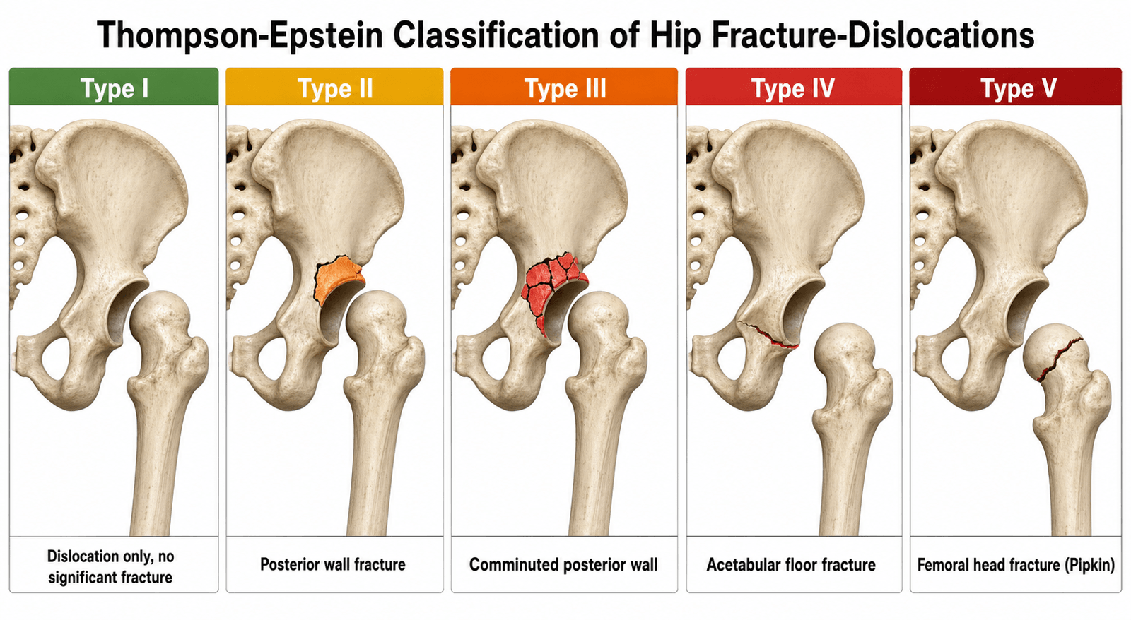
- Associated Fracture
- None, or insignificant
- Treatment
- Urgent closed reduction
- AVN Risk
- Pooled 11-43% across posterior dislocations, rising with Thompson-Epstein grade; odds x5.6 if delayed past 12h
- Associated Fracture
- Single large posterior wall fragment
- Treatment
- Reduce, assess wall size, ORIF if over 40%
- AVN Risk
- 10-15%
- Associated Fracture
- Comminuted posterior wall
- Treatment
- Reduce, ORIF posterior wall
- AVN Risk
- 15-20%
- Associated Fracture
- Acetabular floor (unstable)
- Treatment
- Reduce, ORIF acetabulum
- AVN Risk
- 20-25%
- Associated Fracture
- Femoral head
- Treatment
- Reduce, Pipkin classification guides
- AVN Risk
- 20-40%
Pipkin. The femoral head fractures, and the fovea is the landmark. Below it the fragment is non-weight-bearing and may be excised if small; above it the fragment carries load and is fixed.
- Pattern
- Head fracture inferior to the fovea (non-weight-bearing)
- Treatment
- Excise if small, ORIF if large
- AVN Risk
- 10-15%
- Pattern
- Head fracture superior to the fovea (weight-bearing)
- Treatment
- ORIF required (headless screws)
- AVN Risk
- 20-25%
- Pattern
- Pipkin I or II + femoral neck fracture: double blood supply insult
- Treatment
- THA (elderly) or fix neck + head (young)
- AVN Risk
- The dominant risk; no cited rate (see Complications)
- Pattern
- Pipkin I, II or III + acetabular fracture: complex pattern
- Treatment
- Fix acetabulum + address head
- AVN Risk
- 30-50%
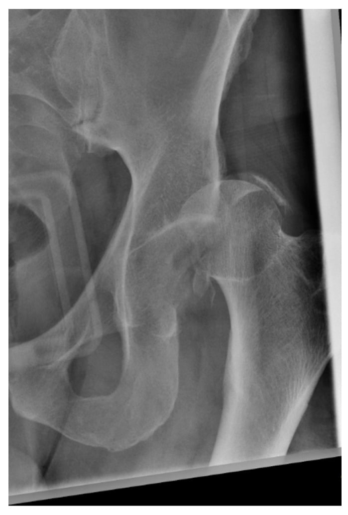
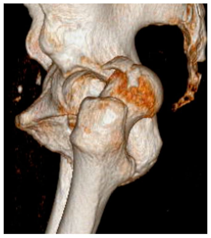
Timing. Injuries are also described by the time to reduction, and every group is still reduced:
- Acute (under 6 hours) - the optimal time; urgent closed reduction
- Subacute (6-12 hours) - still reducible; urgent reduction is still indicated
- Delayed (over 12 hours) - high AVN risk; still reduce, and open reduction may be needed
Clinical Assessment
History. The mechanism is high-energy trauma: a dashboard injury in a motor vehicle accident (the posterior dislocation), a fall from height, or contact and high-impact sport. The patient has immediate severe pain, cannot bear weight and has a deformed leg, shortened and rotated, with numbness or weakness if the nerve is injured.
Inspection. The deformity is obvious and tells you the direction. A posterior dislocation leaves the leg shortened, adducted and internally rotated; an anterior dislocation leaves it externally rotated and abducted. Swelling may be minimal at first.
Palpation and movement. The hip is tender, the femoral head may be palpable posteriorly or anteriorly, and the greater trochanter sits in the wrong place. Movement is severely limited by pain and mechanical block, and the deformity is fixed.
Neurovascular status. For the sciatic nerve, test dorsiflexion, plantarflexion and sensation in the first web space and the lateral foot; for the circulation, the distal pulses and capillary refill.
Document sciatic nerve function before and after reduction; the pre-reduction record is critical for clinical and medicolegal reasons. The peroneal division is the most vulnerable, so dorsiflexion and first web space sensation are the tests that matter.
Associated injuries. The fractures and injuries that accompany the dislocation:
- Femoral head fracture (Pipkin) - 10-15%
- Posterior wall fracture - 20-30%
- Acetabular fracture - 10-15%
- Femoral neck fracture - 5-10% (Pipkin III)
- Knee injuries (PCL, patella) - 10-15%
- Head injury - 10-15%
- Other fractures - 20-30%
Investigations
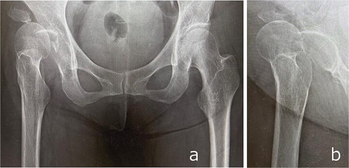
Radiographs. An AP pelvis, and a lateral hip if possible. They show the head out of the acetabulum and the associated fractures of the head, acetabulum and neck. The pre-reduction film is essential because a femoral neck fracture must be excluded before reduction is attempted: traction risks displacing it.
Always scrutinise the femoral neck on the pre-reduction radiograph. Attempting a vigorous closed reduction across an undiagnosed femoral neck fracture can convert it into a displaced fracture and devastate the blood supply. If a neck fracture coexists (Pipkin III), the strategy changes fundamentally.
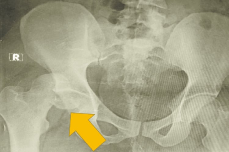
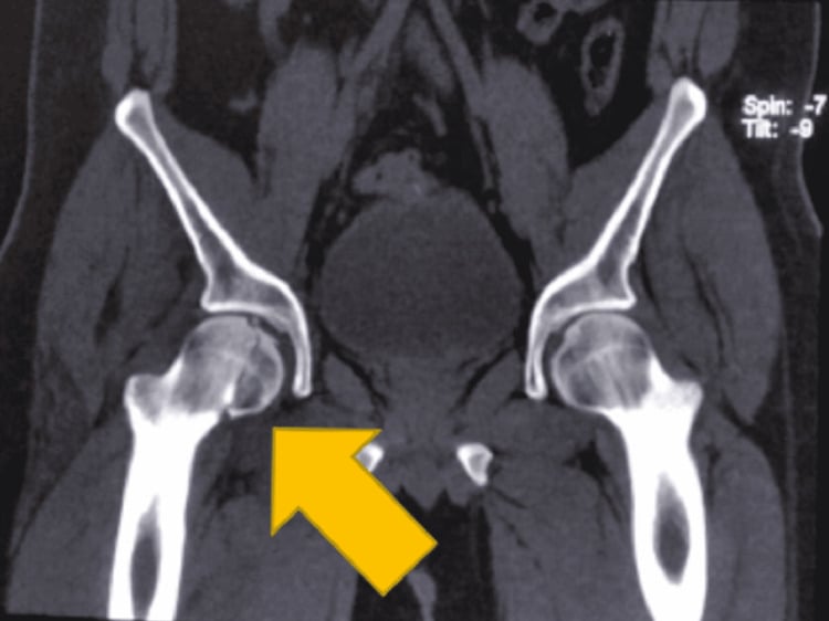
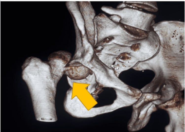
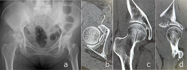
CT. Post-reduction CT is mandatory, even when the joint appears located. It confirms a concentric reduction, classifies a femoral head fracture by Pipkin, measures the size of the posterior wall fragment, finds loose bodies in the joint, and plans the surgical approach. Pre-reduction CT is usually not needed: reduce first, then scan.
MRI. Rarely needed acutely and not routine. It is indicated for specific concerns: soft-tissue injury, and AVN assessment later, at 6-12 weeks.
Differential Diagnosis
The acutely painful, deformed hip after trauma has a focused differential. The priority is to tell a true dislocation, which is reduced now, from an isolated fracture, which follows a different pathway.
- Typical Limb Position
- Shortened, adducted, internally rotated
- Key Discriminator
- Femoral head out of acetabulum on AP; wall/head fragment
- Action
- Emergent reduction, then CT
- Typical Limb Position
- Abducted, externally rotated, often extended
- Key Discriminator
- Head medial/inferior to acetabulum
- Action
- Emergent reduction, exclude vascular injury
- Typical Limb Position
- Shortened, externally rotated
- Key Discriminator
- Lucency across neck, head IN acetabulum
- Action
- Do NOT attempt dislocation-style reduction; fix neck
- Typical Limb Position
- Shortened, markedly externally rotated
- Key Discriminator
- Fracture line trochanter to trochanter
- Action
- Fixation pathway (DHS/IM nail)
- Typical Limb Position
- Variable, may be subtle
- Key Discriminator
- Head migrated medially through floor
- Action
- Judet views/CT, acetabular pathway
- Typical Limb Position
- Pattern depends on direction
- Key Discriminator
- History of THA, low-energy event
- Action
- Closed reduction, assess components/instability
Management Algorithm
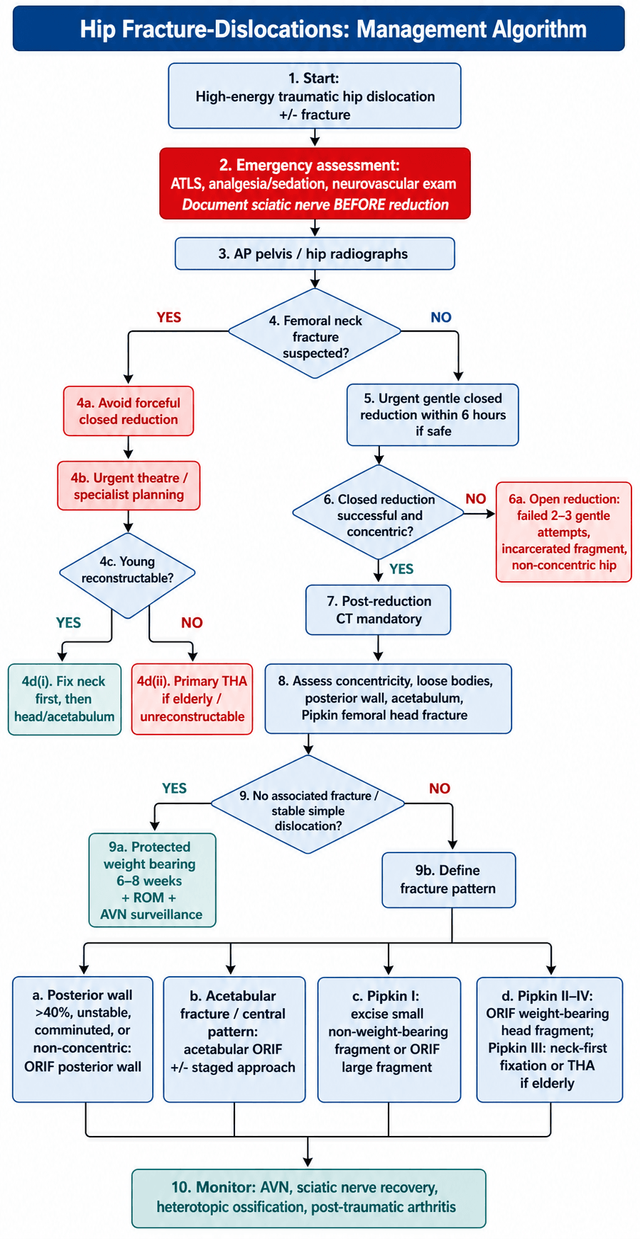
The pathway. Reduce, scan, then treat the fracture.
- Emergency. Pre-reduction radiograph to exclude a femoral neck fracture, nerve function documented, then closed reduction in the emergency department or theatre, within the conventional 6-hour window.
- Post-reduction CT. Concentricity, loose bodies and the fractures (Pipkin, acetabular, neck), and the plan for definitive treatment.
- Type I, no fracture. Observe, with protected weight bearing for 6-8 weeks and radiographs for AVN at 6 and 12 weeks and 1 year.
- Types II-V. Treat the fracture as the classification directs.
Closed reduction. The Allis manoeuvre for a posterior dislocation:
- Anaesthesia, sedation or general
- An assistant stabilises the pelvis
- Traction in line with the deformity
- Flexion and internal rotation to disimpact
- Then traction, external rotation and extension
- Confirm reduction with fluoroscopy
The Stimson manoeuvre is the alternative: the patient prone with the leg hanging, and gentle traction and rotation. Make no more than 2-3 gentle attempts; if they fail, open reduction is required.
Open reduction. The approach is Kocher-Langenbeck (posterior) or anterior (Smith-Petersen), depending on the pattern.
- Absolute - failed closed reduction after 2-3 gentle attempts, a non-concentric reduction, incarcerated fragments
- Relative - associated fractures that need fixation, delayed presentation (over 12 hours)
The Irreducible and Non-Concentric Hip
Confirming concentricity. Take an immediate post-reduction AP, then the mandatory CT, and compare the joint space with the normal side. A medial or superior clear space greater than 2 mm wider than the contralateral hip means the hip only looks reduced: there is interposed tissue or a retained fragment until CT proves otherwise, and it is an indication for CT and urgent operative removal, never for observation. A hip that reduces then re-subluxates, or clunks through range, signals mechanical incongruity or gross instability rather than a stable reduction.
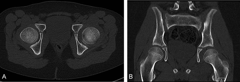
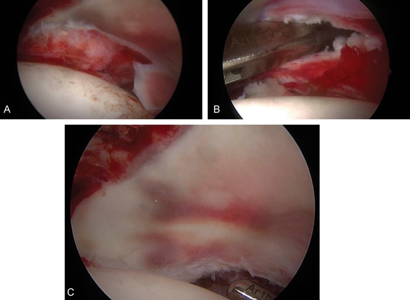
Why a closed reduction fails. A small minority of posterior dislocations cannot be reduced closed. The recognised blocks:
- Buttonholing of the femoral head through a rent in the posterior capsule or the short external rotators
- An inverted or infolded labrum trapped between head and acetabulum
- An incarcerated osteochondral or bony fragment, a posterior-wall or Pipkin head fragment lodged in the joint: the block specific to the fracture-dislocation
- Interposed piriformis or gluteal tendon
- An ipsilateral femoral neck fracture absorbing the reduction force, where forcing traction is dangerous
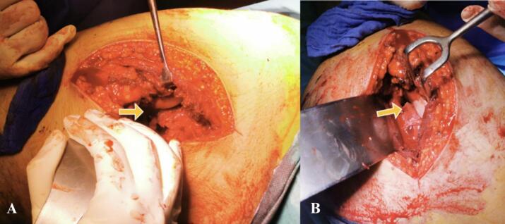
Why a reduced hip stays non-concentric. Even after a reduction that looks successful the joint may stay incongruent, most commonly from an incarcerated intra-articular fragment, and otherwise from an infolded labrum, a retained ligamentum teres remnant or interposed capsule.
What to do. Stop pulling. An irreducible or non-concentric hip after 2-3 gentle attempts under adequate relaxation needs open reduction, by the approach the pathology dictates, usually Kocher-Langenbeck for a posterior fragment: remove the loose bodies, reduce and fix the wall or head fragment, and confirm concentric congruity. Retained fragments are not benign; they cause third-body wear, chondral abrasion and accelerated post-traumatic arthritis.
Surgical Technique
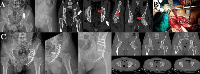
Posterior wall. Fix a wall over 40% (unstable), a comminuted wall (Type III) or a non-concentric reduction. Through a Kocher-Langenbeck approach, identify and protect the sciatic nerve, reduce the wall fragments, fix them with a spring plate or buttress plate, and check stability with fluoroscopy. The 40% figure is a guide, and Controversies sets out its limits.
The sciatic nerve lies directly behind the hip and is at risk in the posterior approach. Identify and protect it before any dissection, retract it with vessel loops, avoid excessive retraction, and document nerve function before and after surgery.
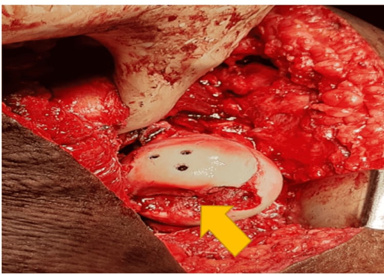
Femoral head (Pipkin). The head is approached from the front, anterior approach (Smith-Petersen or Watson-Jones), to preserve its blood supply.
- Pipkin I - excise a small fragment; fix a large one with headless screws
- Pipkin II - the weight-bearing portion, so ORIF with headless screws, countersunk
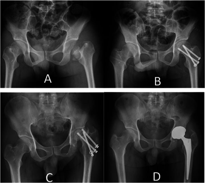
- Pipkin III - fix the neck first and urgently, then address the head; the elderly may need THA
Acetabulum (Type IV). A complex pattern that needs acetabular ORIF, perhaps as a staged procedure, with the femoral head addressed separately. The approach, posterior, anterior or combined, depends on the fracture.
Complications
- Incidence
- 11-43% (pooled, posterior)
- Risk Factors
- Time to reduction, displacement, Pipkin type
- Prevention/Management
- Reduce as soon as possible; odds x5.6 if past 12h
- Incidence
- 10-20%
- Risk Factors
- Posterior dislocation, surgical approach
- Prevention/Management
- Protect nerve, document function
- Incidence
- 20-30%
- Risk Factors
- Surgical approach, delayed reduction
- Prevention/Management
- Prophylaxis (indomethacin, radiation)
- Incidence
- 20-30%
- Risk Factors
- Cartilage injury, AVN, malreduction
- Prevention/Management
- Anatomic reduction, early reduction
- Incidence
- 5-10%
- Risk Factors
- Instability, inadequate fixation
- Prevention/Management
- Secure fixation, assess stability
AVN. It follows disruption of the blood supply, delayed reduction and displacement, and it is prevented by urgent, anatomic reduction. Read the evidence carefully (Kellam and Ostrum): the pooled rate for posterior dislocations, 10.6-43%, rises with Thompson-Epstein grade, and the odds ratio of 5.6 for reduction after 12 hours is a relative increase, not an absolute 40% rate. No validated sub-6-hour stratum exists in the cited evidence. The dominant risk is Pipkin III, an associated displaced femoral neck fracture: in Yoon's series both AVN cases were Pipkin III, and no cited rate exists, so do not quote one. Monitor with radiographs at 6 and 12 weeks and 1 year, and offer THA if it becomes symptomatic.
Sciatic nerve injury. Caused by the dislocation itself or by the surgical approach, and prevented by protecting the nerve at operation and documenting its function. Observe it: most injuries recover by 2 years, and a nerve with no recovery by 6 months is explored. The Cornwall review puts the incidence at about 10% in adults and recovery, at least partial, at 60-70%, and complete recovery is less common than that.
Heterotopic Ossification Prophylaxis
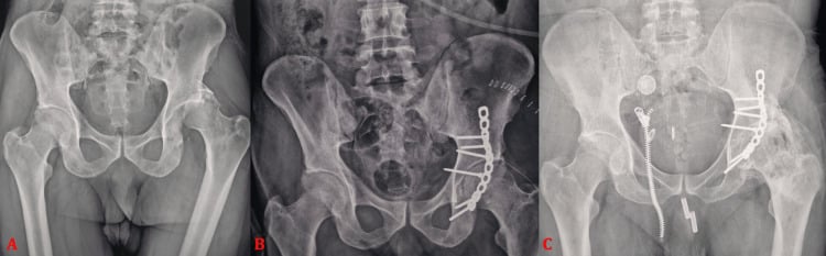
Heterotopic ossification occurs in roughly 20-30% after these injuries (25% in the surgical-hip-dislocation meta-analysis), and prophylaxis with indomethacin or radiation is standard advice. When, how much and to whom is the recurring viva point.
Who is at risk. HO is the price of operative posterior exposure rather than of the dislocation itself. Risk is highest with the Kocher-Langenbeck approach, extensive muscle stripping, retained devitalised gluteus minimus, delayed surgery, an associated head injury, and male sex. A simple closed reduction with no operative wound carries a low risk and needs no prophylaxis; reserve indomethacin or single-dose radiotherapy for high-risk operative cases.
Grading: Brooker. Only the higher grades (III-IV) usually become clinically relevant, through loss of motion.
- Brooker I - islands of bone within the soft tissues
- Brooker II - bone spurs from pelvis or femur with at least 1 cm between opposing surfaces
- Brooker III - opposing bone surfaces less than 1 cm apart
- Brooker IV - apparent bony ankylosis of the hip
- Regimen
- 25 mg three times daily (75 mg/day) for about 4-6 weeks
- Caveats
- GI and renal effects; theoretical impairment of fracture/osteotomy union; benefit debated
- Regimen
- About 700-800 cGy within roughly 24-72 hours of surgery
- Caveats
- Logistics/resource-dependent; avoided in the very young over malignancy concerns; shield un-united fracture lines
- Regimen
- Excise devitalised muscle at the time of fixation
- Caveats
- Reduces the osteogenic burden; universally applicable adjunct
Established HO. Excise only symptomatic HO and only once it is mature, shown by a quiescent bone scan and a normalised alkaline phosphatase, usually beyond 6-12 months. Cover the excision with peri-operative single-dose radiotherapy or indomethacin to limit recurrence.
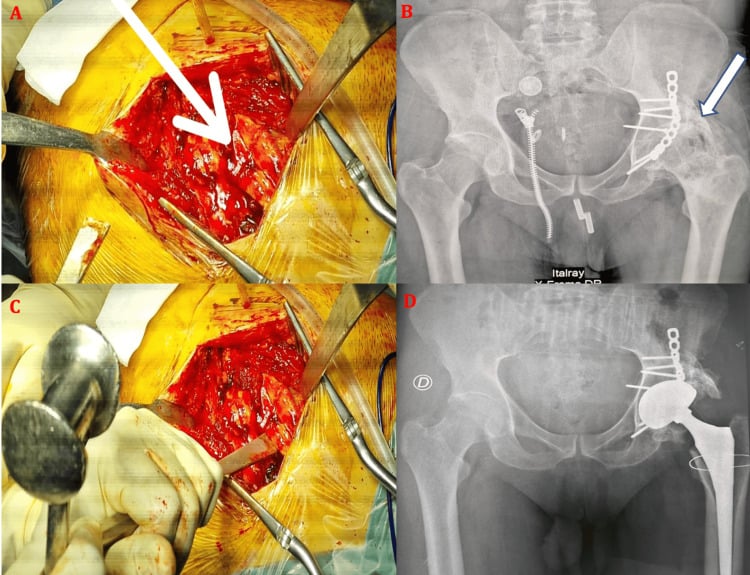
Postoperative Care
Early. No immobilisation: mobilise early and start hip range of motion immediately, with physiotherapy for range and strengthening. Weight bearing depends on the fractures. A simple dislocation is touch-down weight bearing for 6-8 weeks; with fractures the patient is non-weight bearing until they heal.
Rehabilitation protocol. It runs in four stages:
- Weeks 0-2 - touch-down or non-weight bearing depending on the fractures; hip range of motion; quadriceps and hip strengthening; ice and elevation
- Weeks 2-6 - protected weight bearing continues; progressive range and strengthening; balance and proprioception
- Weeks 6-12 - progressive weight bearing if the fractures have healed; full range; progressive activity
- Weeks 12 and beyond - return to sport when strength and range are normal; continued monitoring for AVN
Outcomes and Prognosis
The success-rate and return-to-function percentages in this section are conventional teaching ranges, not measurements from a cited cohort; the verified outcome data on this page are Kellam & Ostrum (AVN 10.6-43%, OR 5.6 beyond 12 hours), Upadhyay (24% osteoarthritis at a mean 14.6 years, occupation-driven), Khalifa (85% satisfactory, 74% anatomic, AVN 12%/HO 25%/OA 16%) and Yoon (AVN confined to Pipkin III).
- Success Rate
- 85-90% (with urgent reduction)
- Return to Pre-Injury Level
- 80-85%
- Complications
- 10-15% (AVN, HO, arthritis)
- Success Rate
- 70-85% (depends on fracture pattern)
- Return to Pre-Injury Level
- 70-80%
- Complications
- 20-30% (AVN, HO, arthritis, nerve injury)
Post-traumatic arthritis. At 10 years it affects 20-30% after a Type I injury and 30-40% after Types II-V, driven by cartilage injury, AVN and malreduction.
What moves the outcome. Urgent reduction (under 6 hours), a simple Type I dislocation with no associated fracture, and complete rehabilitation favour a good result. Reduction delayed over 12 hours, associated fractures (Types II-V), Pipkin III and incomplete rehabilitation count against it.
Prevention and Return to Sport
Prevention. Primary prevention is seatbelt use, which prevents the dashboard injury, airbag deployment, safe driving practices and protective equipment in sport. After the injury, complete rehabilitation before returning to sport, keep up strength and conditioning, and return to activity gradually.
Return to sport. Usually at 6-12 months after surgery, depending on associated injuries and AVN risk, and 70-85% return to their pre-injury level; associated injuries, AVN and compliance with rehabilitation decide who does. The criteria:
- Full range of motion, equal to the other side
- Strength greater than 90% of the other side
- No pain or instability
- No AVN on imaging
- Single-leg hop greater than 90% of the other side
- Agility testing passed and sport-specific drills completed
Guidelines, Registries & Global Practice
Global Epidemiology
- Hip dislocation overall is uncommon; roughly 5-15% of all traumatic dislocations and the great majority follow high-energy mechanisms (road traffic collisions, falls from height).
- Posterior pattern accounts for around 90% of cases; associated fractures (femoral head, posterior wall, neck) are present in a substantial minority and define the fracture-dislocation subgroup.
- Strong male predominance (roughly 3:1) with a young peak (20-40 years), mirroring high-energy trauma demographics worldwide.
- In low- and middle-income settings the same injury skews toward motorcycle and pedestrian trauma, and pre-hospital delay is the dominant modifiable risk factor for AVN.
Side-by-Side Guidance
- Emphasis
- Emergency reduction + anatomic articular restoration
- Practical Recommendation
- Urgent closed reduction, post-reduction CT, ORIF of displaced head/wall fragments via appropriate approach
- Emphasis
- Polytrauma pathway and timely senior decision-making
- Practical Recommendation
- Treat within an ATLS framework, escalate to pelvic/acetabular network for column or complex wall fractures
- Emphasis
- Evidence-informed individualised care
- Practical Recommendation
- Reduce emergently, document neurovascular status, CT all fracture-dislocations before definitive fixation
- Emphasis
- Centralisation of acetabular surgery
- Practical Recommendation
- Refer complex acetabular/Pipkin patterns to high-volume units; consider surgical hip dislocation for selected head fractures
Registries and Practice Variation
- Dedicated dislocation registries are limited, but national arthroplasty registries (NJR UK, AJRR US, AOANJRR Australia, SHAR Sweden) capture the downstream burden: post-traumatic arthritis and AVN are recognised indications for conversion to total hip arthroplasty in young adults.
- High-resource centres: 24/7 trauma access, immediate CT, fellowship-trained pelvic/acetabular surgeons and surgical-hip-dislocation capability.
- Limited-resource centres: emphasis on rapid closed reduction (the single highest-value intervention), selective transfer for complex fixation, and pragmatic non-operative management of small fragments.
Hip fracture-dislocations are a common viva topic. Know that this is a true orthopaedic emergency (urgent reduction, AVN risk rises sharply after 12 hours), the Thompson-Epstein classification (Type I-V), the Pipkin classification for Type V (I-IV), the posterior wall stability question (around 40%, confirmed by EUA), sciatic nerve at risk (about 10%), and urgent reduction technique. Be prepared to discuss surgical approaches and complications.
Controversies and Areas of Uncertainty
The 6-hour rule. Widely taught, but not a sharp biological threshold. The statistically robust step-up in AVN in the meta-analysis comes around the 12-hour mark, and the pragmatic message is unchanged: reduce as soon as safely possible.
The posterior wall threshold. "40% = unstable" oversimplifies. Static CT poorly predicts stability in the 20-50% indeterminate zone, and examination under anaesthesia remains the reference standard for the borderline wall.
Which approach for the femoral head. For displaced femoral head fractures the debate continues between anterior approaches, posterior approaches and the Ganz surgical hip dislocation. Surgical hip dislocation gives the best articular view but adds a trochanteric osteotomy, and its reported AVN rates are comparable rather than clearly superior.
Pipkin I: excise or fix? Small infra-foveal fragments may be excised, but excising larger fragments risks instability and degenerative change. There is no consensus fragment-size cut-off; the decision is individualised to fragment size, comminution and joint congruity.
State the conventional teaching, then show you understand its limits: "The classic teaching is reduction within 6 hours, although the best evidence shows the major rise in AVN is after 12 hours, so my principle is emergent reduction as soon as the patient is safe." This demonstrates consultant-level nuance.
MCQ Practice Points
Q: What is the golden window for hip dislocation reduction? A: As soon as possible. The only quantified threshold in the pooled evidence is 12 hours - AVN odds are 5.6 times higher beyond it - and the traditional 6-hour target is best practice rather than a validated cut-off. Pooled AVN for posterior dislocations is 11-43%, so prompt reduction reduces risk but does not remove it.
Q: What does Thompson-Epstein Type V indicate? A: Femoral head fracture (Pipkin) - Type V = dislocation with femoral head fracture. Use Pipkin classification (I-IV) to guide treatment of the head fracture.
Q: What is the treatment for Pipkin II femoral head fracture? A: ORIF with headless screws - Pipkin II = fragment above fovea (weight-bearing surface). Must fix to restore articular surface. Anterior approach to avoid posterior blood supply.
Q: What posterior wall size requires ORIF? A: Over 40% - Posterior wall over 40% = unstable, requires ORIF. Wall 20-40% = may be stable (assess with EUA). Wall under 20% = usually stable.
Q: What is the incidence of sciatic nerve injury in posterior hip dislocations? A: 10-20% - Peroneal division most vulnerable (lateral position). Most injuries recover by 2 years. Document nerve function before and after reduction.
Q: Why is Pipkin III (head + neck fracture) considered a disaster? A: Double blood supply insult - Both femoral head and neck blood supply disrupted. Very high AVN risk. Often requires THA, even in young patients.
Exam Viva Scenarios
Practise clinical reasoning and management decisions out loud
“A 30-year-old man presents to ED after a motor vehicle accident. He was the driver and his knee struck the dashboard. He has a painful hip and cannot move his leg. Examination shows leg shortened, adducted, and internally rotated. X-ray shows posterior hip dislocation. After urgent closed reduction, CT shows a Pipkin II femoral head fracture (fragment above fovea, weight-bearing surface).”
“A 35-year-old athlete presents after a high-energy fall. He has a posterior hip dislocation with a large posterior wall fracture. After urgent closed reduction, CT shows posterior wall fracture involving 45% of the wall. Examination under anesthesia shows instability.”
“A 45-year-old man is brought in after a head-on collision. AP pelvis shows a posterior hip dislocation, and on close inspection there is a lucency across the femoral neck. Two gentle closed reduction attempts under sedation fail. How do you proceed and how does the neck fracture change your plan?”
Key Anatomy
- Hip joint: Ball-and-socket, femoral head in acetabulum
- Sciatic nerve: Posterior to hip, vulnerable in posterior dislocation
- Blood supply: MFCA 80%, retinacular vessels, ligamentum teres under 10%
- Fovea: Key landmark for Pipkin classification (above vs below)
Classification
- Thompson-Epstein: Type I (simple), Type II (wall), Type III (comminuted), Type IV (floor), Type V (Pipkin)
- Pipkin: I (below fovea), II (above fovea), III (+ neck), IV (+ acetabulum)
- By timing: Acute (under 6h), Subacute (6-12h), Delayed (over 12h)
- Posterior wall threshold: over 40% = unstable = ORIF required
Treatment Algorithm
- Urgent closed reduction within 6 hours (golden window)
- Post-reduction CT mandatory (assess reduction, fractures, loose bodies)
- Type I: Observe, protected weight bearing 6-8 weeks
- Type II-V: Address fractures (ORIF wall, Pipkin, acetabulum)
Surgical Pearls
- Kocher-Langenbeck approach for posterior, protect sciatic nerve
- Anterior approach for Pipkin (preserves blood supply)
- Posterior wall over 40% = unstable = ORIF required
- Pipkin II = weight-bearing = ORIF required
Complications
- AVN: 11-43% pooled for posterior dislocation; odds x5.6 if reduced after 12h
- Sciatic nerve injury: 10-20% (most recover by 2 years)
- Heterotopic ossification: 20-30% (prophylaxis with indomethacin)
- Post-traumatic arthritis: 20-30% (at 10 years)
Evidence Base
Time to Reduction and AVN Risk
- Posterior dislocation AVN event rate 10.6-43%, rising with Thompson-Epstein grade
- Odds ratio of AVN 5.6 for reduction after 12 hours versus before 12 hours
- Injury severity correlates with both AVN and post-traumatic arthritis
Long-term Outcome of Simple Dislocation
- 24% developed osteoarthritis at mean 14.6-year follow-up
- Higher OA rate in manual workers (up to 45% in pit-accident miners)
- Even Thompson-Epstein Type I carries meaningful long-term arthritis risk
Pipkin Classification (Original)
- Defines the four femoral-head fracture subtypes still used today
- Fovea is the landmark separating type I (below) from type II (above)
- Type III (with neck fracture) recognised as the worst prognostic group
Sciatic Nerve Injury in Hip Dislocation
- Sciatic nerve injury approximately 10% in adults, peroneal branch most affected
- At least partial recovery in 60-70% of patients
- Prompt reduction relieves nerve distortion; document function before and after
Posterior Wall Stability: CT vs EUA
- Walls under 20% usually stable, over 40-50% usually unstable
- Static CT unreliable in the indeterminate 20-50% range
- Dynamic EUA is the preferred determinant of hip stability
Surgical Hip Dislocation for Femoral Head Fractures
- Satisfactory outcome 85%, anatomic reduction 74%
- AVN 12%, heterotopic ossification 25%, osteoarthritis 16%
- Surgical hip dislocation gives controlled head exposure with acceptable vascular safety
Femoral Neck Fracture Drives AVN in FHFD
- AVN occurred only in patients with displaced femoral neck fractures (Pipkin III)
- Good or excellent outcome in the majority at 5.1 years
- Confirms Pipkin III as the dominant AVN risk subtype